
Biology Trending 4e Eli Minkoff and Jennifer HoodDeGrenier The mind and the body interact The immune system maintains health Cells of the immune system and the lymphatic circulation Innate immunity ID: 1039502
Download Presentation The PPT/PDF document "Chapter 14: Mind, Body and Immunity" is the property of its rightful owner. Permission is granted to download and print the materials on this web site for personal, non-commercial use only, and to display it on your personal computer provided you do not modify the materials and that you retain all copyright notices contained in the materials. By downloading content from our website, you accept the terms of this agreement.


1. Chapter 14: Mind, Body and ImmunityBiology Trending, 4eEli Minkoff and Jennifer Hood-DeGrenier
2. The mind and the body interactThe immune system maintains healthCells of the immune system and the lymphatic circulationInnate immunitySpecific immunityImmunological memoryPassive immunityHarmful immune responses and immunosuppression
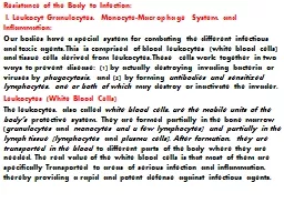
3. Figure 14.1 White blood cells that provide innate and specific immunity. A. Three types of white blood cells (left to right: monocyte, neutrophil, lymphocyte), surrounded by smaller red blood cells (erythrocytes). Monocytes and neutrophils are phagocytic, functioning in innate immunity. Lymphocytes, which function in specific immunity, are of two types that cannot be distinguished by appearance alone: the B-lymphocytes secrete antibodies, while T-lymphotcytes kill virally infected cells and cancer cells. B. Phagocytic leukocyte engulfing a yeast cell.
4. Figure 14.2 Macroscopically visible parts of the immune system. The parts of the lymphatic circulation are in blue, and the major lymphatic tissues (thymus, bone marrow, tonsils, etc.) are colored orange.
5. Figure 14.3 The stages of inflammation.
6. Figure 14.4 Ways in which the immune system can eliminate non-self antigens.
7. Figure 14.5 Immune selection. Developing B and T lymphocytes, each with a unique antigen receptor on its cell membrane, encounter characteristic 'self' antigens on accessory cells. Any developing lymphocyte whose antigen receptor binds to a self antigen on an accessory cell dies. Lymphocytes whose receptors do not bind develop to maturity and are available to bind to any foreign antigens that match their antigen receptors at a later time.
8. Figure 14.6 B cell activation. When a B cell binds to an antigen, it becomes activated and starts to divide repeatedly, forming a clone of genetically identical B cells. Some of these cells become antibody-secreting cells (A), while others become memory cells (M). Both types of B cell have receptors that bind to the same antigen that was originally encountered, and this is the basis of antigen-specific immunity.
9. Figure 14.7 The activity of the immune system changes in response to a second exposure to an infectious microorganism.
10. Figure 14.8 Passive immunity transferred from mother to child.
11. Figure 14.9 Allergic release of histamine.
12. The neuroendocrine system consists of neurons and endocrine glandsThe autonomic nervous systemThe stress and relaxation responses
13. Figure 14.10 Functions of the sympathetic and parasympathetic nervous systems.
14. Figure 14.11 The phases of the stress response. Stressors induce a variety of physiological changes, including immunological changes, which are mediated by the sympathetic nervous system and adrenal hormones. CRH is corticotrophin releasing hormone.
15. The neuroendocrine system interacts with the immune systemEvidence for one interconnecting networkThe placebo effectEffects of stress on healthConditioned learning in the immune systemVoluntary control of the immune system
16. Figure 14.12 Immunohistochemical staining showing the presence of the neurotransmitter norepinephrine in the rat spleen. Large black arrows: blood vessels. Small red arrows: nerve fibers synthesizing norepinephrine.
17. Figure 14.13 The placebo effect.
18. Figure 14.14 Classical conditioning of the immune response.