
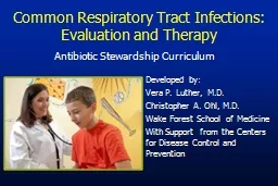
Antibiotic Stewardship Curriculum Developed by Vera P Luther MD Christopher A Ohl MD Wake Forest School of Medicine With Support from the Centers for Disease Control and Prevention Objectives ID: 911347
Download Presentation The PPT/PDF document "Common Respiratory Tract Infections: Eva..." is the property of its rightful owner. Permission is granted to download and print the materials on this web site for personal, non-commercial use only, and to display it on your personal computer provided you do not modify the materials and that you retain all copyright notices contained in the materials. By downloading content from our website, you accept the terms of this agreement.


Slide1
Common Respiratory Tract Infections: Evaluation and Therapy
Antibiotic Stewardship Curriculum
Developed by:
Vera P. Luther, M.D.
Christopher A. Ohl, M.D.
Wake Forest School of Medicine
With Support from the Centers for Disease Control and Prevention
Slide2Objectives
Review the etiology, diagnosis and therapy of 5 common respiratory tract infections: community-acquired pneumonia, acute bronchitis, rhinosinusitis, pharyngitis, and acute otitis media (AOM)
List criteria for symptomatic therapyList criteria for each of the 5 conditions that indicate antibiotic therapy is the most appropriate treatment
List the first line antibiotic therapy for each of the 5 conditions when indicated
Slide3Outline
IntroductionEvaluation and therapyCommunity-acquired pneumonia
Acute bronchitisRhinosinusitis
Acute pharyngitis
AOM
Conclusion
Slide4Common Respiratory Tract Infections
Community-acquired pneumoniaAcute bronchitis
PharyngitisRhinosinusitisAOM
Slide5Respiratory Infections are the Most Common Reason for Office Visits
IMS America NDTI (National Disease Therapeutics Index)
2001.Mehrotra A. Health Affairs 2008 Sep-Oct;27(5):1272-82.
Number of Office Visits (millions)
Respiratory
Hypertension Gastrointestinal Diabetes Depression
Infections Disorders
180
100
80
60
40
20
0
161
73
55
35
26
Slide6Over half of Antibiotic Use in Adults is for Respiratory Tract Infections
2004-2005 Physician Drug & Diagnosis Audit (PDDA)
Slide7Slide8Burden of Acute Respiratory Tract Infections
Significant time away from school and workSignificant healthcare expenditures for clinic visits, hospitalization and medicationsMortality rare except for community-acquired pneumonia in persons with comorbidities
Slide9Pathogens
Respiratory viruses account for the majority of infectionsBacterial infections are more prominent in acute otitis media and pneumonia
Antibiotic resistance is common among
S. pneumoniae, H. influenzae,
and
M.
catarrhalis
isolates
Streptococcus pneumoniae
Haemophilus influenzae
Moraxella catarrhalis
Streptococcus pyogenes
Mycoplasma sp
.
Chlamydiophila sp.
Slide10Proportion of Resistant Invasive Streptococcus
pneumoniae spp.,
1992-2008
Percent Fully Resistant
Source: CDC Active Bacterial Core Surveillance and Sentinel Surveillance Network.
Erythromycin resistance data not available
Slide11Outline
IntroductionEvaluation and therapyCommunity-acquired pneumonia
Acute bronchitisRhinosinusitis
Acute pharyngitis
Acute otitis media
Conclusion
Slide12Community- Acquired
Pneumonia
Slide13Community-Acquired Pneumonia
Overview3-4 million cases/year
10 million patient visits/yearApproximately 80% are mild to moderate in severity and treated as outpatients
500,000 hospitalizations and 45,000 deaths/year
(8
th
leading cause of death)
Mortality
1% in outpatients
5% in inpatients
25-50% in patients admitted to ICU
File TM, Marrie TJ
Postgrad Med
2010;122(2):130.
Slide14Community-Acquired Pneumonia
SymptomsCough
FeverPleuritic chest pain Dyspnea
Sputum production
Slide15Community-Acquired Pneumonia
Diagnosis
Common physical examination findings
Fever
Respiratory rate > 24 breaths/minute
Heart rate > 100 beats/minute
Crackles/râles usually present on auscultation
Evidence of consolidation on exam
Peripheral white blood cell count (WBC) usually elevated
Chest x-ray (CXR) should be used to confirm diagnosis
Slide16Community-Acquired Pneumonia
Microbiology and Proportion of Deaths in Adults
Microbial Agent
S. pneumoniae
H. influenzae
S. aureus
Gram Negative Rods
Miscellaneous Bacteria
“Atypical” Bacteria
Legionella
spp.
Mycoplasma
spp.
C. pneumoniae
Viral (including influenza)
Aspiration
Proportion of Hospital Admissions20-60%
3-10%3-5%
3-10%3-5%10-20%
2-8%1-6%4-6%2-15%6-10%Deaths
66%7%6%
3%9%6%
5%1%<1%<1%
ND
Slide17Antibiotic Considerations
Therapy is almost always empiric initiallyMost important pathogen to target is S. pneumoniae
based on its frequency and associated morbidity and mortalityLocal prevalence of macrolide- resistant
S.
pneumoniae
influences antibiotic
choice
“Atypical pathogens” more common among older children and adults
Sputum gram-stain showing the typical lancet-shaped gram positive diplococci of
S.
pneumoniae
If an etiology is identified
,
therapy should be de-escalated and directed at that pathogen
Slide18Community-Acquired Pneumonia
Treatment Recommendations for Outpatients
Clinical CharacteristicTreatment Regimen
Previously healthy and no risk factors
for drug-resistant
S. pneumoniae
Macrolide*
Doxycycline
Risk factors for drug resistant
S. pneumoniae
Presence of comorbidities
or immunocompromised
Use of antimicrobials within the
previous 3 months
Regions with a high rate (>25%) of macrolide-resistant
S. pneumoniae
Respiratory fluoroquinolone**
High dose amoxicillin
plus macrolide*
Amoxicillin/
clavulanate
plus macrolide*
Alternative: Ceftriaxone, cefpodoxime or cefuroxime plus macrolide*
* Azithromycin, Clarithromycin or Erythromycin** Gemifloxacin, Levofloxacin or Moxifloxacin
Mandell et al. Clin Infect Dis 2007. 44: S27-S72
Slide19Community-Acquired Pneumonia
Treatment Recommendations for InpatientsClinical Characteristic
Treatment Regimen
Non-ICU Admission
Respiratory
fluoroquinolone
**
Cefotaxime or ceftriaxone
plus
macrolide
*
Ampicillin plus
macrolide
*
Ertapenem plus
macrolide
*
ICU Admission
Cefotaxime or ceftriaxone
or
ampicillin-sulbactam
PLUSAzithromycin
or fluoroquinolone
* Azithromycin, Clarithromycin or Erythromycin
** Gemifloxacin, Levofloxacin or MoxifloxacinMandell et al. Clin Infect Dis 2007. 44: S27-S72
Slide20Community-Acquired Pneumonia: Reasons for Overtreatment
Community-acquired pneumonia is commonly misdiagnosedAbnormal findings on chest radiographs often lead to “cannot rule out pneumonia”
e.g. atelectasis, malignancy, hemorrhage, pulmonary edema, heart failure, pulmonary embolism, effusions, fibrosisEmergency department protocols are designed to expedite therapy
Pines, et. al.
J Emerg Med. 2009 Oct;37(3):335-40.
Slide21Acute
Bronchitis
Slide22Acute Bronchitis
Definition: An acute respiratory tract infection that may last up to 3 weeks in which cough, with or without phlegm, is a predominant feature and alveolar inflammation is not present (normal
chest radiograph)
Occurs predominately in the late fall, winter and early spring
Common: Up to 5% of adults self report an episode each year
Gonzales et al. Annals of Int Med. 2001;134(6):521
Brahman. Chest 2006;129:95S-103S
Slide23Acute Bronchitis
Almost Always a Viral EtiologyLess than 10% due to bacterial causes
Etiologic diagnosis not usually attempted unless influenza suspectedAntibiotic therapy not indicated and should not be offered
Exception: some episodes of prolonged paroxysmal cough are due to
Bordetella pertussis
Viral Causes of Bronchitis
Respiratory Syncytial Virus
Adenovirus
Parainfluenza
virus
Rhinovirus
Influenza virus
Gonzales et al. Annals of Int Med. 2001;134(6):521
Brahman. Chest 2006;129:95S-103S
Slide24Patient Management
Some patients may expect an antibiotic based on past experience or expectationsExplain to the patient why an antibiotic is not necessary and that these drugs may have unwanted side-effects
Use terms like “chest cold” rather than bronchitis or infectionSuggestions for symptom relief
Humidified air
Over-the-counter pain relievers
Some recommend cough suppressants
No role for bronchodilators in absence of asthma or chronic obstructive pulmonary disease (
COPD
)
Slide25Acute
Rhinosinusitis (ARS)
Slide26Acute Rhinosinusitis
Broad term describing multiple disease processes affecting the nasal cavity and sinuses with a duration of <4 weeksAllergy
Infection (viral, bacterial, fungal)Polyps
Frequent: 1 of 7 adults per year seeks medical attention for acute rhinosinusitis (ARS)
Chow et al. Clin Infect Dis. 2012; 54(8):e72-112
Slide27Acute Viral Rhinosinusitis (Common Cold)
Pathogens: Viruses similar to acute bronchitisCommon symptoms: Nasal congestion and mucous discharge, facial pressure, post-nasal dischargeUsually symptoms peak at 2-3 days and resolve by day 7-10
Diagnosis relies on exam: radiographs not sensitive or specificTreat with topical and oral decongestants, nasal irrigation, +/- topical corticosteroids
No indication for antibiotics
Meltzer et. al. Mayo Clin Proc. 2011 86: 427
Chow et al. Clin Infect Dis. 2012; 54(8):e72-112
Slide28Acute Bacterial Rhinosinusitis (ABRS)
Pathogens: S. pneumoniae, H. influenzae, M. catarrhalis, Streptococcus sp, S. aureus
, anaerobes
Much less frequent than viral ARS
Follows <2.0% of viral
ARS
cases
Important to attempt to differentiate from viral ARS
CT imaging indicated for severe infection with suspected orbital or intracranial extension
Symptoms Suggesting Bacterial Infection
Symptoms > 10 days
Unilateral maxillary face pain
Maxillary tooth
ache
Unilateral maxillary
sinus tenderness
Unilateral purulent nasal discharge
Double sickening
(symptoms improve then worsen)
Green or colored nasal discharge and cough do not predict
ABRS
.
Meltzer et. al. Mayo Clin Proc. 2011 86: 427
Chow et al. Clin Infect Dis. 2012; 54(8):e72-112
Slide29ABRS treatment
First-line antibiotic therapy:
Amoxicillin-clavulanatePenicillin allergy: doxycycline, levofloxacin or moxifloxacin
Adjunctive treatment
Hydration, analgesics, antipyretics
Irrigation with physiologic or hypertonic saline
Intranasal corticosteroids for those with concurrent allergic rhinitis
Topical or oral decongestants or antihistamines not indicated due to lack of effect
Meltzer et. al. Mayo Clin Proc. 2011; 86: 427, Young J et al. Lancet. 2008; 371:908,
Chow et al. Clin Infect Dis. 2012;
54(8):e72-112
Slide30Acute Pharyngitis
Slide31Acute Pharyngitis
Classically the triad of fever, sore throat and pharyngeal inflammationPathogens:Viruses: Epstein-Barr, Cytomegalovirus, respiratory viruses, enteroviruses, Herpes simplex type I
Bacteria: Group A Streptococcus
(GAS), Non-group A
Streptococcus
,
Arcanobacterium hemolyticum,
and
Fusobacterium
spp.
Pharyngitis in 85-95% of adults and 80-85% of children is due to viruses
For uncomplicated pharyngitis, antibacterial therapy is reserved for GAS infection
Slide32Clinical Features of Pharyngitis
Features suggestive
of GAS etiology
Sudden onset
sore throat
Fever
Headache
Tonsillopharyngeal inflammation
Tonsillopharyngeal
exudate
Palatal petechiae
Tender
anterior cervical adenopathy
Winter-early
spring presentation
Age 5-15 years
History of exposure to GAS
pharyngitis
Features suggestive
of viral etiology
Absence of fever
Conjunctivitis
Coryza
Cough
Hoarseness
Ulcerative mouth
lesions
Viral
type rash
Overlap between GAS and viral pharyngitis may be considerable
McIsaac et al. JAMA. 2004; 291:1587, Bisno et al. Clin Infect Dis. 2002; 35:113
Slide33Acute Pharyngitis Diagnosis
For adults and children with features that strongly suggest a viral etiology, testing is not indicatedIn persons with findings suggestive of GAS infection, confirmation with a rapid antigen detection test (
RADT) or culture is neededIn children and adolescents a negative RADT has a low negative predictive value and should be backed up with a throat culture for GAS
McIsaac et al. JAMA. 2004; 291:1587, Bisno et al. Clin Infect Dis. 2002; 35:113
Slide34Acute Pharyngitis Treatment
Antibiotics for those with confirmed GASPenicillin or amoxicillinPenicillin allergic: first generation cephalosporin for minor allergy and clindamycin or
macrolide if anaphylaxis
No GAS resistance to penicillin has been reported
Symptomatic treatment:
Over-the-counter pain relievers/antipyretic
Throat lozenges or sprays
Adequate oral hydration
Corticosteroids not recommended
Slide35Acute Otitis Media
Slide36Acute Otitis Media (AOM)
Acute illness with fluid and mucosal inflammation of the middle ear spaceExtremely common in young children: By age 3, two-thirds have had at least one episodeMuch less common in adults
Increased risk with some ethnic groups, exposure to polluted air (including tobacco smoke), and with children who attend daycare
Slide37Acute Otitis
MediaPathogenesis: Anatomic and physiologic disruption of eustachian tube drainage of the middle ear with subsequent fluid accumulation and bacterial infection
Often follows viral respiratory infectionIncidence due to
S. pneumoniae
decreasing due to vaccination of children starting in 2000
Pathogen
Proportion of cultures (2001-2003) (%)
S.
pneumoniae
23
H.
influenzae
36
M.
catarrhalis
3
Group A
Streptococcus
1.3
None
41
Adapted from Casey et. al. Pediatr Infect Dis J. 2004; 23:824
Slide38Acute Otitis
MediaSymptoms/signs
Fever, chills, ear pain, ear drainage, hearing loss, lethargy, irritability, pulling on earExam
Tympanic membrane erythema, loss of landmarks and bulge
Presence of middle ear fluid on pneumatic otoscopy or tympanometry, or
otorrhea
Slide39Acute Otitis Media: Treatment
Many cases of AOM (~25%) are due to viruses and will not respond to antibioticsA significant number of cases due to bacteria will spontaneously resolve without antibiotics
If antibiotics are indicated, use high dose amoxicillin
Severe illness: Amoxicillin-
clavulanate
Penicillin allergy: 2
nd
or 3
rd
generation cephalosporin, azithromycin or clarithromycin
AAP. Pediatrics. 2004; 113:1451
Slide40Acute Otitis Media Treatment
Age
Certain
Diagnosis
Uncertain Diagnosis
<6 mo
Antibacterial therapy
Antibacterial therapy
6 mo -2 yr
Antibacterial therapy
Antibacterial therapy if severe
illness; observation option if
non-severe illness
≥ 2 yr
Antibacterial therapy if severe illness; observation option if non-severe illness
Observation option
AAP. Pediatrics. 2004; 113:1451
Slide41Acute Otitis Media
Symptom reliefOral analgesicsTopical analgesic spray/drops
Warm, moist cloths over earAvoid narcotics
Prevention
Conjugate
pneumococcal and
H
aemophilus
vaccination
Influenza vaccination
Rarely
antibiotic prophylaxis for frequent recurrences
Slide42Outline
IntroductionEvaluation and therapyCAP
Acute bronchitisRhinosinusitisAcute pharyngitis
Acute otitis media
Conclusion
Slide43Conclusion
Antibiotics are frequently given for respiratory tract infections in outpatient and inpatient settingsInappropriate antibiotic use is common for these diagnosesMisdiagnosis of pneumonia is common
Most upper respiratory infections are viral and do not need antibiotic treatmentObservation without antibiotics is an option for children with acute otitis media
Guidelines exist for the appropriate treatment of respiratory tract infections
Slide44Treatment Guidelines and Resources
Centers for Disease Control and Prevention (CDC)http://www.cdc.gov/getsmart/
Get Smart: Know When Antibiotics Work
Adult Guideline Summaries
Pediatric Guideline
Summaries
Infectious Diseases Society of America (IDSA)
http://www.idsociety.org/IDSA_Practice_Guidelines/
American Academy of Pediatrics (AAP)
American Academy of Family Physicians(AAFP)