
Proteins are major components of all cellular systems Proteins consist of one or more linear polymers called polypeptides Proteins are linear and never branched Different AAs are linked together via ID: 912016
Download Presentation The PPT/PDF document "Structure of Protein Protein" is the property of its rightful owner. Permission is granted to download and print the materials on this web site for personal, non-commercial use only, and to display it on your personal computer provided you do not modify the materials and that you retain all copyright notices contained in the materials. By downloading content from our website, you accept the terms of this agreement.


Slide1
Structure of Protein
Slide2ProteinProteins are major components of all cellular systemsProteins consist of one or more linear polymers called polypeptidesProteins are linear and never branchedDifferent AA’s are linked together via PEPTIDE bondsThe individual amino acids within a protein are known as RESIDUESThe smallest known Protein is just nine residues long - OXYTOCINThe largest is over 35,000 residues - the structural protein
TITIN
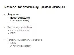
Slide3ProteinIn the absence of stabilizing forces a minimum of 40 residues is needed to adopt a stable 3D structure in water.Protein sequence can be determined by systematically removing the AA’s one at a time from the amino end - Edman degradationSequence the gene or cDNA for any protein and use the genetic code to determine the AA sequence
Slide4structural organization of proteinsPrimary StructureSecondary StructureTertiary StructureQuaternary Structure
Important Forces determine structure
Covalent bond
Weak forces
Slide5Structure organization
Slide6Primary StructureThe sequence of amino acids present in the polypeptide chainCovalently linked by peptide bonds- Peptide bondBy convention, the primary structure of a protein starts from the amino- terminal (N) end and ends in the carboxyl-terminal (C) endLargely responsible for its function
Slide7peptide bond has some double bond like character (40%) due to resonance C – N bond length of peptide is 10% shorter than that found in usual C – N As a consequence of resonance, peptide bonds are almost planar
Slide8Cis conformation and Trans conformationTrans conformation is normally present in the residues as Cis conformation leads to the steric clashCis conformation is possible for peptide bond next to the proline residue
Slide9IMPORTANCE OF PRIMARY STRUCTURETo predict secondary and tertiary structures from sequence homologies with related proteins. (Structure prediction)Many genetic diseases result from abnormal amino acid sequencesTo understand the molecular mechanism of action of proteinsTo trace evolutionary paths
Slide10METHODS OF Primary structure DETERMINATION1. Amino Acid Composition2. Degradation of protein into smaller fraction3. Determination of amino acid sequence
Slide11Amino Acid Compositionunordered amino acid composition of a protein prior to attempting to find the ordered sequenceKnowledge of the frequency of certain amino acids may also be used to choose which protease to use for digestion of the proteinMisincorporation of low levels of non-standard amino acids2 stepsHydrolyse a known quantity of protein into its constituent amino acids- by acid or alkali treatmentSeparate and quantify the amino acids- by chromatography
Slide12N-and C-terminal amino acid analysisReagents used which can label N-terminal amino acidsSanger's reagent (1-fluoro-2,4-dinitrobenzene) and dansyl derivatives such as dansyl chloride are used
Slide13Generation of small fragmentsUrea or Guainidine hydrochloride- disrupts weak forces and dissociates the protein into polypeptide unitsNo of polypeptide chains than identified with dansyl chloridePolypeptide is broken down (i) Enzymatic cleavage- trypsin, chymotrypsin, pepsin etc
(ii) Chemical clevage- cyanogen bromide
Slide14Amino acid sequence determinationEdman degradation-
Slide15SequentorProtein or peptide is immobilized in the reaction vesselEdman degradation is performedEach cycle releases and derivatizes one amino acid from the protein or peptide's N-terminusThe released amino-acid derivative is then identified by HPLC
Process is done repetitively for the whole polypeptide
Slide16POLYPEPTIDE CHAIN CONFORMATIONS
The only reasonably free movements are rotations around the C α-N bond (measured as ϕ ) and the C α-C bond (measured as Ѱ).The conformation of the backbone can therefore be described by the torsion angles (also called dihedral angles or rotation angles)Two torsion angles in the polypeptide chain, also called Ramachandran angles
Slide17RAMACHANDRAN PLOTTo visualize the backbone of amino acid residuesDeveloped in 1963 by G. N. RamachandranEach dot on the Ramachandran plot shows the angles for amino acids.The regions on the plot with highest density are most favorable combinations of phi-psi values and called “allowed” regions, also called “low – energy” regions.
Some values of φ and ψ are forbidden since the involved atoms will come too close to each other, resulting in a steric clash. Such regions are called “disallowed” regions