
or intestinal obstruction is a mechanical or functional obstruction of the intestines preventing the normal transit of the products of digestion It can occur at any level distal to the stomach and it is a surgical ID: 929055
Download Presentation The PPT/PDF document "Intestinal obstruction Bowel obstructio..." is the property of its rightful owner. Permission is granted to download and print the materials on this web site for personal, non-commercial use only, and to display it on your personal computer provided you do not modify the materials and that you retain all copyright notices contained in the materials. By downloading content from our website, you accept the terms of this agreement.


Slide1
Intestinal obstruction
Slide2Bowel obstruction
(or
intestinal obstruction
) is a mechanical or functional obstruction of the intestines, preventing the normal transit of the products of digestion. It can occur at any level distal to the stomach and it is a surgical
emergency
. The condition is often treated conservatively over a period of 2-5 days with the patient's progress regularly monitored by an assigned physician. It’s a common cause of acute abdominal pain and represents 5%-20% of acute surgical admission.
Slide3Epidemiology
1% of all hospitalization
3% of emergency surgical
admissions
More frequent in female patients because of gynecological-obstetric and pelvic surgical operations are important etiologies for post operative adhesions
Adhesion is the most common cause of intestinal obstruction
80% of bowel obstruction due to small bowel obstruction and the most common causes are adhesion—hernia---neoplasm while 20% due to colon obstruction and the most common cause is CR-cancer 60-70% while 30% are
diverticular
disease and
volvulus
Mortality rate range between 3% for simple bowel obstruction to 30% when there is strangulation or perforation
Recurrent rate vary according to method of treatment if conservative 12% while the operation treatment recurrent rate 8-32%
Slide4The Questions that should be answered in patient with IO:
-Is this bowel obstruction or
ileus
?
-Is this a small or large bowel obstruction?
-Is this proximal or distal obstruction?
-What is the cause of this obstruction?
-Is this a complex or simple obstruction?
-How should we start investigation the patient?
-What is the immediate/ intermediate treatment plan?
-What are the indications for surgery?
-
Slide5Types
On the basis of presence or absence of bowel activity
Dyanamic
A mechanical blockage acting as a barrier to the progression of gut contents
.
Adynamic
:
is a paralytic or functional variety of obstruction
Slide6Classification of intestinal obstruction
1-Small bowel obstruction &large bowel obstruction.
2-Mechanical obstruction & functional obstruction.
3-Simple obstruction & complicated obstruction.
4-Partial obstruction& complete obstruction .
5-Acute obstruction
-Sub acute obstruction
-Acute on chronic obstruction .
-Chronic obstruction.
6-Congenital &acquired
Slide78L of isotonic fluid received by the small intestines (saliva, stomach, duodenum, pancreas and
hepatobiliary
)
7L absorbed
2L enter the large intestine and 200 ml excreted in the faeces
Air in the bowel results from swallowed air ( O
2
& N
2
) and bacterial fermentation in the colon ( H
2
, Methane & CO
2), 600 ml of flatus is releasedEnteric bacteria consist of coliforms, anaerobes and strep.faecalis.Normal intestinal mucosa has a significant immune roleDistension results from gas and/ or fluid and can exert hydrostatic pressure.In case of BO Bacterial overgrowth can be rapidIf mucosal barrier is breached it may result in translocation of bacteria and toxins resulting in bactaeremia, septaecemia and toxaemia.
Patho
-physiology I
Slide8Patho-physiology II
Obstruction results in:
Initial overcoming of the obstruction by increased
paristalsis
Increased
intraluminal
pressure by fluid and gas
Vomiting
sequestration of fluid into the lumen from the surrounding circulation
Lymphatic and venous congestion resulting in oedematous tissues
Factors 3,4,5 result in
hypovolaemia
and electrolyte imbalanceFurther: localised anoxia, mucosal depletion necrosis and perforation and peritonitis.Bacterial over growth with translocation of bacteria and it’s toxins causing bacteraemia and septicaemia.
Slide9Causes of small IO:-
Extraluminal
Mural
Luminal
Adhesions
Hernia
Volvulus
Neoplasms
Crohns
TB
Intussusception
Congenital
F. Body
Bezoars
Gall stone
Food Particles
Ascaris
Slide10Etiology
Mechanical bowel obstruction:
Small bowel obstruction:
Adhesion 60%
Hernia 20%
Neoplasm 5%
Volvulus
5%.
Others: IBD-GALL STONE-FOREIGN BODY-INTUSSUSCEPTION.
Large bowel obstruction :
Cancer 60%.
Diverticular
disease 15%.Volvulus 15%.Others: hernia –fecal impaction-inflammatory.
Slide11Features of obstructions
In high small bowel obstruction ,vomiting occurs early and is profuse with rapid dehydration .Distension is minimal with little evidence of fluid levels on abdominal radiography.
In low small bowel obstruction, pain is predominant with a central distention .Vomiting is delayed. Multiple central fluid level are seen in radiography.
In large bowel obstruction, distension is early and pronounced .Pain is mild and vomiting and dehydration are late .The proximal colon and
caecum
are distended are distended on abdominal radiography .
Slide12Cardinal clinical features of acute obstruction :
Abdominal pain
Distension
Vomiting
Absolute constipation .
Slide13Abdominal pain
Most patients who have small –bowel obstruction experience
crampy
abdominal pain that comes in wave .The pain is around the umbilicus.
Slide14Vomiting
Small bowel obstruction usually cause vomiting The vomit is green if the obstruction is in the upper small intestine and brown if it is in the lower small intestine.
Slide15Constipation
Constipation and inability to pass gas are signs of bowel obstruction .However ,when the bowel partially blocked a person may have stool leak and pass gas. Patient with a complete obstruction may have a bowel movement if there is stool below the obstruction.
Slide16Distension
With the blockage of the lower small intestine ,the
epigastric
area may be
distinded
or bloated.
Slide17Clinical features of strangulation
Constant pain
Tenderness with rigidity
Shock
Discoloration
Constitutional symptom
Slide18Slide19Clinical Findings
Examination
Others
Systemic examination
If deemed necessary.
CNS
Vascular
Gynaecological
musculoskeletal
Abdominal
Abdominal distension and it’s pattern
Hernial orifices
Visible peristalsis
Cecal distension
Tenderness, guarding and rebound
Organomegaly
Bowel sounds
High pitched
Absent
Rectal examination
General
Vital signs:
P, BP, RR, T, Sat
dehydration
Anaemia, jaundice, LN
Assessment of vomitus if possible
Full lung and heart examination
Slide20Initial Management in the ER
Resuscitate:
Air way (O
2
60-100%)
IVF : Crystalloids at least 120 ml/h. (determined by estimated fluid loss and cardiac function).
NPO.
Decompress with
Naso
-gastric tube and secure in position
Insert a urinary catheter (hourly urinary measurements) and start a fluid input / output chart
Intravenous antibiotics (no clear evidence)
If concerns exist about fluid overloading a central line should be insertedFollow-up lab results and correction of electrolyte imbalance“Never let the sun set or rise on an obstructed bowel ”
Slide21How to initially investigate your patient
Lab:
CBC (
leukocytosis
, anaemia,
hematocrit
, platelets)
Clotting profile
Arterial blood gasses
U&
Crt
, Na, K, Amylase, LFT and glucose, LDH
Group and save (x-match if needed) Optional (ESR, CRP, Hepatitis profileRadilogical:Plain x-raysUSS ( free fluid, masses, mucosal folds, pattern of peristalsis, Doppler of mesenteric vasculature, solid organs)CT, MRI, Contrast studies……ECG and other investigations for co-morbid factors
Slide22Diagnostic in 50 %
CXR
: superior to erect
abd
X ray for
pneumo
peritoneum
Abdo
X ray
: Changes appear in
3-5
hrs
if complete obstruction and take days if incomplete obstructionHigh grade SBO : > 3.6 cm diameter of loops ; 2.5 times more in number ; air fluid levels > 2 ; wider than 2.5 cm and differing in height >2 cm String of beads sign : small bubbles of gas trapped in rows between valvulae conniventes . Diagnostic of SBO (sometimes also in IBD and Ileus)Coffee bean sign: closed loop obstr where the arms of the loop dilated with gas separated by thick intestinal
wall
Pseudo tumor sign
: closed loop fill with fluid looks like soft tissue mass
XRAY
Slide23SBO
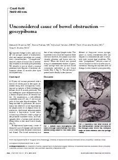
Slide24Fluid levels with gas above; ‘
stepladder pattern’.
Ileal obstruction by adhesions; patient erect.
Supine radiograph from a patient with complete small bowel obstruction shows
distended small bowel loops
in the central abdomen with
prominent valvulae conniventes
(small white arrow)
Figure 3.
Lateral decubitus view of the abdomen, showing air-fluid levels consistent with intestinal obstruction
(arrows).
Slide25Slide26The Difference between small and large bowel obstruction
Small Bowel
Large bowel
Central ( diameter 3 cm max)
Vulvulae
coniventae
Ileum: may appear tubeless
Peripheral ( diameter 6 cm max)
Presence of
haustration
Slide27Coffee bean sign
Slide28Slide29Slide30LARGEBOWEL
SMALL BOWEL
Haustra
present
Absent
Number of loops
Few
Many
Valvulae
conniventes
absent
Present in jejunumDistribution of loopsPeripheral centralRadius of curvature of loopsLarge SmallDiameter5 cm3-5 cmSolid feces +-
Slide31Water soluble contrast gets diluted
Poor mucosal detail
No therapeutic effect in SBO
Follow through
:
500 ml
of
42 %
barium ,
fluroscopic
radiographs at 15-30 min interval till
ileo
cecal valve. When barium reaches cecum put a rectal tube and insufflate air to distend the rt colon and distal iluemEnteroclysis: duodenal intubation , 30-40% barium infusion at 60-90 ml /minDouble contrast enteroclysis: followed by infusion of air / methyl cellulose BARIUM
Slide32Role of barium
gastrografin
studies
As: follow through, enema
Limited use in the acute setting
Gastrografin
is used in acute abdomen but is diluted
Useful in recurrent and chronic obstruction
May able to define the level and mural causes.
Can be used to distinguish
adynamic
and mechanical obstruction
Barium should not be used in a patient with peritonitis
Slide33When CT not available
Operator dependent modality
Obstruction
: lumen
> 3 cm
; length
> 10 cm
; distal
seg
shows to and fro
or whirling motion
( differentiates from ileus )
Cause can be detectedSeverity : free fluid + ; aperistalsis ; bowel wall thickening > 3 mm ( sugg infarction )USG
Slide34Role of CT
Used with iv contrast, oral and rectal contrast (triple contrast).
Able to demonstrate abnormality in the bowel wall, mesentery, mesenteric vessels and peritoneum.
It can
define:
the level of obstruction
The degree of obstruction
The cause: volvulus, hernia, luminal and mural causes
The degree of
ischaemia
Free fluid and gas
Ensure: patient vitally stable with no renal failure and no previous
alergy to iodineFigure: Axial computed tomography scan showing dilated, contrast-filled loops of bowel on the patient’s left (yellow arrows), with decompressed distal small bowel on the patient’s right (red arrows). The cause of obstruction, an incarcerated umbilical hernia, can also be seen (green arrow), with proximally dilated bowel entering the hernia and decompressed bowel exiting the hernia.Source: Jackson, PG. & Raiji M., Evaluation and Management of Intestinal Obstruction, January 2011, American Academy of Family Physicians (AAFP), 83: 2 (160-164)
Slide35Slide36Can detect extra luminal
patho
and detailed info about the small bowel wall.
And does not use
ionised
radiation
MR ENTEROCYLSIS
Slide37Initial Management in the ER
Resuscitate:
Air way (O
2
60-100%)
Insert 2 lines if necessary
IVF : Crytloids at least 120 ml/h. (determined by estimated fluid loss and cardiac function). Add K
+
at 1mmmol/kg
Draw blood for lab investigations
Inform a senior member in the team.
NPO.
Decompress with Naso-gastric tube and secure in positionInsert a urinary catheter (hourly urinary measurements) and start a fluid input / output chartIntravenous antibiotics (no clear evidence)If concerns exist about fluid overloading a central line should be insertedFollow-up lab results and correction of electrolyte imbalanceThe patient should be nursed in intermediate careRectal tubes should only be used in Sigmoid volvulus.
Slide38INDICATIONS FOR SURGERY
Absolute
Generalised peritonitis
Localised peritonitis
Visceral perforation
Irreducible hernia
Relative
Palpable mass lesion
'Virgin' abdomen
Failure to improve
Trial of conservatism
Incomplete obstruction
Previous surgeryAdvanced malignancyDiagnostic doubt - possible ileusSource: http: Surgical Tutor.co.uk
Slide39Ileus
Associated with the following conditions:
Postoperative and bowel resection
Intraperitoneal infection or inflammation
Ischemia
Extra-abdominal: Chest infection, Myocardia infarction
Endocrine: hypothyroidism, diabetes
Spinal and pelvic fractures
Retro-peritoneal haematoma
Metabolic abnormalities:
Hypokalaemia
Hyponatremia
UraemiaHypomagnesemiaBed riddenDrug induced: morphine, tricyclic antidepressants
Slide40Acute Mesenteric Occlusion
Acute ischemic of mesenteric vessel.
Commonly SMA
Causes: AF, mural thrombosis,
atheromatous
plaque from aortic aneurysm and
valave
vegetation from endocarditis
Features: -Sudden onset of severe
abd
. pain in
pt
with AF and atherosclerosis -Persistent vomiting and defecation then passage of altered blood -Hypovolumic shock Investigations: - Neutrophil leukocytosis - Abd Xray: Absence of gas in thickened small intestinesTreatment: - Anti-coagulant - Embolectomy - Revascularization - Colectomy
Slide41PSEUDO-OBSTRUCTION
Obstruction usually colon- occur in the absence of mechanical cause or acute intra-abdominal disease.
Associated with a variety of syndromes in which there is underlying neuropathy and/or a range of other factors
IDIOPATHIC
SEPTICAEMIA
Metabolic
Retroperitoneal irritation
Severe trauma
at lumbar
area
Drugs
ShockSecondary GI involvement
Slide42Upright abdominal X-ray demonstrating a small bowel obstruction. Note multiple air fluid levels
.
Slide43Upright abdominal X-ray of a patient with a large bowel obstruction showing multiple air fluid levels and dilated loops of bowel
.
Slide44Hernia
Slide45Other causes of small
io
.
IBD
Gall stone Ileus
Intussusception
Slide46Sigmoid Volvulus
Colonic Obstruction
Slide47Ileus