

Dr Ruth Heisey Family PhysicianGP Oncologist Womens College HospitalPrincess Margaret Cancer Centre Clinician InvestigatorAssociate Professor University of Toronto Sandy Fawcett RNEC NPAdult ID: 596930
Download Presentation The PPT/PDF document "Breast Cancer Screening and Diagnosis" is the property of its rightful owner. Permission is granted to download and print the materials on this web site for personal, non-commercial use only, and to display it on your personal computer provided you do not modify the materials and that you retain all copyright notices contained in the materials. By downloading content from our website, you accept the terms of this agreement.


Slide1
Breast Cancer Screening and Diagnosis
Dr. Ruth HeiseyFamily Physician/GP OncologistWomen’s College Hospital/Princess Margaret Cancer CentreClinician Investigator/Associate ProfessorUniversity of TorontoSandy Fawcett RN(EC) NP-AdultGattuso Rapid Diagnostic CentreBreast Disease SiteUniversity Health Network- Princess Margaret Cancer Centre Adjunct Lecturer University of TorontoMay 2, 2014Slide2
Canadian Breast Cancer Statistics
In 2013: 23,800 women will be diagnosed 5,000 will dieOne in nine expected to develop breast cancerMortality rates decliningwww.cancer.caSlide3
Objectives:Review current breast screening guidelines
Introduce personalized risk assessment tools Review strategies for timely breast cancer diagnosisSlide4
Question
Which is the gold standard tool used to screen for breast cancer?Breast ultrasoundBreast MRIMammogramClinical breast examSlide5
Breast Cancer Screening Principles
Breast screening aims to detect cancer before palpable (pre-clinical phase)Early detection leads to better outcomeSlide6
ONCOLOGY - Cancer biology
Tumor growth and detection1012
109
time
Diagnostic
threshold
(1cm)
Undetectable
cancer
Detectable
cancer
Limit of
clinical
detection
Host
death
Number of
cancer cellsSlide7
2011: Breast Cancer Screening Guidelines CMAJ 2012 Warner et alSlide8
The Canadian Task Force screening recommendations are for average risk women with no breast symptomsSlide9
Screening Mammography
Canadian Task Force Recommendations:“For women aged 50-74, we recommend routinely screening for breast cancer every two to three years”www.ctfphc.orgSlide10
Screening Mammography
Canadian Task Force Recommendations:“For women aged 40-49, we recommend not routinely screening for breast cancer with mammography”www.ctfphc.orgSlide11
Screening Mammography
Canadian Task Force Recommendations (40-49yo):“this recommendation places a relatively low value on a very small absolute decrease in mortality… clinicians should discuss the benefits and harms with their patients and must help each woman to make a decision consistent with her values and preferences”www.ctfphc.orgSlide12Slide13
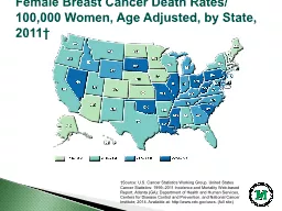
Effect of Mammographic Screening (1976-2008)Early stage breast cancers-2 fold increaseLate stage breast cancers-small decreaseMore than 30% of breast cancers detected were
overdiagnosed (would never have resulted in clinical symptoms if left untreated)NEJM Bleyer and WelchSlide14
Mammographic Screening-Polling Results
NEJM 2013 Feb 368;9, Colbert,AdlerSlide15
Views on mammographic screeningUntil we can determine which cancers will remain indolent we must “ treat all (cancers)as potential killers ”Need to prioritize interventions that increase life expectancy and reduce disease burden
Agreement that women at greater risk need vigilant screeningNEJM 2013 Feb 368;9, Colbert,AdlerSlide16
Clinical Breast Exam
Canadian Task Force Guidelines:“We recommend not routinely performing clinical breast examinations alone or in conjunction with mammography to screen for breast cancer”Slide17
Detection of breast cancer by physical examination versus mammogram for different age groups:
Clinical Breast Cancer 2005;6(4):330-3Slide18
CBEContinue as part of periodic health exam or antenatal visit (opportunistic approach)Slide19
What is average risk?No family history of breast cancerNo previous breast biopsies showing atypical hyperplasia (AH) or lobular carcinoma in situ (LCIS)No history of chest wall radiationSlide20
What is higher than average?Moderate/High:History of breast biopsy showing ADH(atypical ductal hyperplasia), LCIS (lobular carcinoma in situ)
Previous history of breast cancer Family history of breast cancerSlide21
What is higher than average?Very High:BRCA carrier or untested first degree relative of BRCA carrierPrevious chest wall radiation
History of LCIS, ADH and family historySlide22
Role of Screening MRI
Definite role for very high risk patients such as BRCA mutation carriers in conjunction with mammography and CBEMRI more sensitive for detecting breast cancers than mammography, ultrasound or CBE aloneMRI=77-100%Mammography=16-40% JAMA 2004, 292 (11) 1317-25J Clin Oncol 2006, 23:8469-8476Slide23
Magnetic Resonance Imaging ( MRI )
Bilateral breast MRISlide24
American Cancer Society Recommendations for Screening MRIGene mutation (BRCA 1 or 2; Li-
Fraumeni syndrome; Cowden syndrome; Bannayan-Riley-Ruvalcaba syndrome)First-degree relative with one of these mutations (if the woman has not yet been tested)History of radiation therapy to the chest between ages 10 and 30Lifetime risk >20-25% based largely on family history Saslow D, et al. CA Cancer J Clin 2007;57(2):75-89 Slide25
Warner et alSlide26Slide27
OBSP High Risk Screening Program- 2011MRI
in addition to mammogram annuallyfor women ages 30-69: known BRCA carrier untested first degree relative of BRCA carrier chest irradiation before age 30 and at least 8 years previously ≥ 25% lifetime risk of breast cancer (using IBIS or BODICEA risk calc)
www.cancercare.on.caSlide28Slide29
Breast Screening in Clinical Practice
All women should be asked re: family history of breast, ovarian cancer or both If concerns re: mutation carrier discuss implications and referral Consider mammography screening in all women starting at age 40 (no woman should be denied!)Slide30
Breast Screening in Clinical Practice
The 50-74yo asymptomatic woman:Mammogram q 2 years (annual if high risk)Consider OBSPDiscuss breast awarenessOpportunistic CBESlide31
Breast Screening in Clinical Practice
The 40yo asymptomatic woman:Consider mammogram q1-2 years based on risk, density and patient preferenceDiscuss breast awareness Opportunistic CBESlide32
Breast Screening in Clinical Practice
The 75yo asymptomatic woman:Continue to offer mammography until life expectancy is less than 10 yearsSlide33
Breast Screening in Clinical Practice
Moderate/High risk:Annual mammography and CBE starting at age 40Slide34
Breast Screening in Clinical Practice
Very high risk: (e.g. BRCA carrier)Annual mammography, MRI starting at age 30CBE every 6 monthsSlide35
Personalized Risk AssessmentTo determine who should be offered:Referral for consideration of genetic testing
Enhanced screeningPreventive TherapySurgerySlide36
R
B-RST +ve E IBIS > 20-25%P GAIL > 3%
S BRCA carrier
Very high risk
High risk
Average/
M
oderate
risk
Management of Women at Risk for Breast Cancer
Figure 1:Slide37
Why determine candidates for genetic counseling?33yo strong family history breast cancer, start screening digital mammography age 40At a
ge 42 presents with bloating irregular periods- Stage 3c ovarian cancerYou now take a more thorough family history-BRCA1 carrier Slide38
refSlide39
Why Calculate Risk?Risk calculators useful in primary care
B-RST Tool: determine candidates for referral for genetic counselingIBIS: determine candidates for enhanced screeningGail model: determine candidates for preventive therapySlide40
R: Referral (for genetic testing)Two or more first degree relatives same side of family with breast cancer (maternal or paternal)Family members with breast cancer diagnosed before the age of 50 (maternal or paternal)Relative with bilateral breast cancer or breast and ovarian cancer
Multiple relatives with ovarian cancerMale relative with breast cancerAshkenazic Jewish (Eastern European Jewish) ancestryRelative known to be BRCA mutation carrierSlide41
Breast –Referral Screening Tool (B-RST) https://www.breastcancergenescreen.orgSlide42
B-RSTSlide43
E: Enhanced screeningUse IBIS tool to calculate lifetime risk www.ems
-trials.org/riskevaluatorif lifetime risk ≥ 25% refer to OBSP high risk program for MRI screening in addition to mammographic screening www.cancercare.on.ca/common/pages/UserFile.aspx?fileId=99484Slide44
IBIS Risk Calculator: Slide45
IBIS: Calculated RiskSlide46
P: Preventive TherapyConsider for women with strong family history, or history of atypical hyperplasia or LCIS.Use Gail model to assess eligibility for chemoprevention
http://www.cancer.gov/bcrisktool/If 5 year risk ≥3% offer preventive therapySlide47
http://www.cancer.gov/bcrisktool/
Gail model:Slide48
Family Hx
in first degreerelative
Age of first
live birth (or
nulliparity)
Number
of breast
biopsies
Age at
menarche
Age
CP1089285-9
Breast Cancer Risk Assessment Tool
(GAIL MODEL)
5 year risk (>1.66 %)
http://www.cancer.gov.bcrisktool/.comSlide49
Canadian Task Force Recommendations
Fair evidence to recommend counseling about the potential benefits and risks of using tamoxifen to reduce the likelihood of breast cancer in higher risk women (B)Who qualifies?: A woman with >1.7% 5-year risk using Gail modelwww.ctfphc.orgSlide50
S: Prophylactic SurgeryFor highest risk women: known BRCA carriers, or history of LCIS (lobular carcinoma in situ) or AH (atypical hyperplasia) and a significant family historyAlways offer reconstructionSlide51
R
B-RST +ve E IBIS > 20-25%P GAIL > 3%
S BRCA carrier
Very high risk
High risk
Average/
M
oderate
risk
Management of Women at Risk for Breast CancerSlide52
Question
Which is the gold standard tool used to screen for Breast Cancer:Breast ultrasoundBreast MRIMammogramClinical breast examSlide53
Clinical Presentation
Most often breast cancer is first noticed as a painless lump in the breast or armpit (55%)Slide54
Question
What is the most common type of breast cancer?DCIS (Ductal carcinoma in situ)LCIS (Lobular carcinoma in situ)Invasive Ductal CarcinomaInvasive Lobular CarcinomaSlide55
Signs and Symptoms
1. Breast lumpsometimes detected during a screening mammogram or clinical breast examconstantly present and does not fluctuate with menstrual cyclemay feel like it is attached to the skinmay feel hard and irregular may be tender but not usually painfulSlide56
Signs and Symptoms
2. Thickening or lump in the axillaenlarged lymph node – usually means that the lymphatic system is fighting an infection in that areasometimes means that breast cancer has spread to the lymph nodes3. Inverted nipplemay be a normal findingnipples that become inverted should be reportedSlide57
Signs and Symptoms
4. Nipple dischargehas many different causes and should always be reported may be a sign of cancer if it occurs spontaneously, bloody, unilateral, uniductal5. Persistent crusting, ulceration or eczema-type symptoms on the nipplemay be a sign of Paget's disease, a rare form of breast cancerSlide58
Signs and Symptoms
6. Changes in breast size and shapea change in the outline or contour of the breasta change in the size of the breast7. Changes in the skin of the breastpuckering of the skinthickening and dimpling of the skinredness, swelling and increased warmth in the breastSlide59
Mrs. B
77 yr old postmenopausal woman noticed a non-tender mass in the upper outer quadrant (UOQ) of the left breast. PMHx:Hypertension, obesity. Nulliparous. Menarche 11 Menopause 50.Mother and maternal aunt - breast cancer- diagnosed age 48 and 51Clinical breast exam: Breasts are large and in the left breast there was a firm, mobile, 3.5 x 3.5cm mass palpable at 1 o'clock 6 cm FN. No palpable left axillary adenopathy
. Right breast and axilla were unremarkable.Next step:Order diagnostic bilateral
mammogram and left breast ultrasoundSlide60Slide61Slide62Slide63Slide64
Mrs. B
Imaging:Mammography reveals a 3cm mass in the UOQ left breast. Right breast unremarkableLeft breast ultrasound- 3.3 X 3.2 X 2.0cm hypoechoic area 1 o’clock position 6cm FN. Left axilla ultrasound: nodes unremarkableNext step:Core biopsySlide65
Diagnosis- Tissue Confirmation
Core needle biopsy is the preferred method Ultrasound guidedFine needle aspiration for axillary lymph nodesSlide66
Pathophysiology & Anatomy
Slide67
Types of Breast Cancer
Invasive (70%)Ductal LobularIn-situ (30%)Ductal (DCIS)Lobular (LCIS)Other:InflammatoryPaget’s, mucinous, medullary, tubular, pregnancy inducedSlide68
PathophysiologySlide69
Mrs. B
Core biopsy left breast 1 o’clock 6cm FN (3.2cm mass)Pathology:Invasive ductal carcinoma ER+ PR+ HER2-Next steps:Referral to surgeonConsider referral to geneticsHealthy living education/ Survivorship program
Referrals to medical and radiation oncologySlide70
Question
What is the most common type of breast cancer?DCIS (Ductal carcinoma in situ)LCIS (Lobular carcinoma in situ)Invasive Ductal CarcinomaInvasive Lobular CarcinomaSlide71Slide72
Questions?