
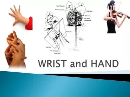
Bone Anatomy Scapoid Lunate Triquetrium Pisiform Trapeziod Trapezium Capitate Hamate Hand and Wrist Anatomy 14 phalanges 2 sesamoid bones thumb 5 metacarpals 8 carpal bones Distal Radius ID: 233882
Download Presentation The PPT/PDF document "Wrist and Hand Anatomy" is the property of its rightful owner. Permission is granted to download and print the materials on this web site for personal, non-commercial use only, and to display it on your personal computer provided you do not modify the materials and that you retain all copyright notices contained in the materials. By downloading content from our website, you accept the terms of this agreement.


Slide1
Wrist and Hand AnatomySlide2
Bone Anatomy
Scapoid
Lunate
TriquetriumPisiformTrapeziodTrapeziumCapitateHamateSlide3
Hand and Wrist Anatomy
14 phalanges
2 sesamoid bones (thumb)
5 metacarpals8 carpal bones
Distal Radius
Forms small ulnar notch to accept the ulnar head
Radial styloid process
Distal Ulna
Ulnar styloid process arises from medial surface
Ulnar headSlide4
Articulations
Distal Radioulnar
Formed by ulnar head and ulnar notch
Allows 1 degree freedom of movementPronation/supinationRadius glides around the ulnaSlide5
Radiocarpal joint
Reinforced by ligamentous thickening
Formed by distal radius articulating with scaphoid, lunate and triangular fibrocartilage disk(TFCC)
Ellipsoid joint (2 degrees freedom)Flexion/extensionRadial/ulnar deviationSlide6
Intercarpal Joints
Palmar/dorsal/interosseous ligaments between each carpal
Very little glidingSlide7
Midcarpal Joints
Proximal/distal carpal row separated by a single joint cavity with small fibrous projections connecting the rows
Limited mobility in flex/ext, radial/ulnar deviationSlide8
Carpometacarpal Joint (CMC)
MC1/trapezium
MC2/trapezoid
MC3/capitateMC4and 5/hamate (forms 1 articulation)Slide9
1
st
CMC (thumb)
Saddle joint2 degrees of freedom(3)Flexion/extensionAbduction/adductionAccessory rotation
Allows for oppositionSlide10
2-4 CMC
Plane/synovial joint
1 degree freedom
Flexion/extension5th CMC2 degree freedomFlexion/extension
Abduction/adductionSlide11
Metacarpophalangeal Joint (MCP)
Two degrees freedom of movement
Flexion/extension
Abduction/adductionThumb can abduct at any point/fingers only when extended
Collateral ligaments
Varus/valgus force
When fingers are in flexion they tighten and limit abduction/adductionSlide12
Interphalangeal Joint
One degree freedom of movement
Flexion/extension
Collateral ligamentsSlide13
Ligament Support
Volar Carpal Ligaments
Volar Radiocarpal Ligament
Three bandsVolar Ulnocarpal LigamentScapholunate Interosseous LigamentLunotriquetral LigamentSlide14
Ligament Support
Dorsal Carpal Ligaments
Dorsal Radiocarpal Ligament
Dorsal Intercarpal Ligament
Radial Collateral Ligament
Ulnar Collateral LigamentSlide15
Carpal Tunnel
Fibro-osseous structure
Floor is proximal carpal bones
Roof is transverse carpal ligamentTunnel contains 10 structuresMedian n., flexor pollicis
longus
tendon, 4 slips of flexor
digitorium
superficialis,
4 flexor
digitorium
profundus
Compression results in
paresthesia
2-4 fingers and decrease gripSlide16Slide17
HandSlide18
Wrist flexors (median n.)
Superficial
Flexor carpi radialis
Palmaris longus
Flexor carpi ulnaris
Flexor digitorium superficialis
Pronator teres
Deep
Flexor digitorium profundus
Flexor pollicis longus
Pronator quadratusSlide19
Palmar (intrinsic)
Thenar
Abductor pollicis brevis
Flexor pollicis brevisOpponens PollicisTendon FPLAdductor pollicisSlide20
Hypothenar
Abductor digiti minimi
Opponens digiti minimi
Flexor digiti minimi brevisSlide21
Central
Tendons FDS/FDP
Superficialis (PIP)
Profundus (DIP)
Lumbricales
Radial side profundus tendon(extensor hood)
Flex MP/ext PIP/DIP
Palmar aponeurosis
Interossei
4 palmar/4 dorsalSlide22
Extrinsic Hand MusclesSlide23
Extensor Indicis
O
Dorsal surface lower ½ body of ulna
Interosseus membraneI Ulnar side of index finger’s EDC tendonNRadial (posterior interosseus)
F
MCP and IP Ext of 2
nd
digitSlide24
Extensor Pollicis Longus
O
Posterior 1/3 ulna
Interosseus membrane
I
Posterior surface of base of thumb distal phalanx
N
Radial (posterior interosseus)
F
CMC, MCP and IP Ext of 1
st
digitSlide25
Extensor Pollicis Brevis
O
Dorsal 2/3 of radius
IDorsal surface of base of proximal 1st phalanxN
Radial (posterior interosseus)
F
CMC & MCP Ext of thumb
CMC ABD of thumbSlide26
Abductor Pollicis Longus
O
Posterior distal 2/3 of ulna
Posterior middle 1/3 of radiusInterosseus membraneIRadial side of base of 1st
metacarpal
N
Radial (posterior interosseus)
F
CMC ABD & Ext of thumbSlide27
Flexor Pollicis Longus
O
Anterior middle ½ of radius
Interosseus membraneI Palmar surface of base of distal 1st phalanx
N
Median (anterior interosseus)
F
IP Flexion of thumbSlide28
Extensor Digiti Minimi
O
Lateral epicondyle of humerus
IExtensor expansion of 5
th
digit
N
Radial (posteior interosseus)
F
MCP and IP extension of 5
th
digitSlide29
Flexor Digitorum Superficialis
O
Medial epicondyle of humerus
Coronoid processMiddle ½ anterior radius
I
Four tendons separating into two parts that insert into sides of bases of middle 2-5 phalanxes
N
Median
F
MCP flexion digits 2-5
PIP flexion digits 2-5Slide30
Flexor Digitorum Profundus
O
Anteriomedial surface of ulna
Interosseus membraneIFour tendons inserting into distal phalanxes of digits 2-5N
Media 2-3 digits
Ulna 4-5 digits
F
DIP flexion of 2-5 digitsSlide31
Intrinsic Hand Muscles
Thenar EminanceSlide32
Abductor Pollicis Brevis
O
Scaphoid tuberosity
Trapezium ridgeTransverse carpal ligamentILateral base f proximal 1st
phalanx
N
Median
F
CMC & MCP ABD of thumbSlide33
Flexor Pollicis Brevis
O
Superficial head – trapezium
Deep head – trapezoid, capitate and palmar ligaments of distal carpal bones
I
Base of prximal 1
st
phalanx on radial side
Extensor expansion
N
Superficial – median
Deep – Ulnar
F
CMC & MCP Flexion of thumbSlide34
Opponens Pollicis
O
Trapezium
Transverse Carpal Ligament
I
Radial side of 1
st
metacarpal shaft
N
Median
F
OppositionSlide35
Intrinsic Hand Muscles
Hypothenar EminenceSlide36
Abductor Digiti Minimi
O
Pisiform
IUlnar side base of 5th proximal phalanxNUlnar
F
MCP ABD of 5
th
digitSlide37
Opponen Digiti Minimi
O
Hook of hamate
Transverse carpal ligamentIUlnar border of entire 5th metacarpal bone
N
Ulnar
F
MCP flexion & rotation of 5
th
digitSlide38
Flexor Digiti Minimi
O
Hamate bone
Transverse carpal ligamentI
Ulnar side of proximal 5
th
phalanx
N
Ulnar
F
MCP Flexion of 5
th
digitSlide39
Other Intrinsic Hand MusclesSlide40
Adductor Pollicis
O
Oblique Head
Capitate boneBases of 2-3 metacarpals
Transverse Head
Proximal 2/3 of palmar surface of 3
rd
metacarpal
I
Ulnar side of base of 1
st
proximal phalanx
N
Ulnar
F
CMC ADD of thumbSlide41
Palmar Interossei
O
1
st – ulnar side base of 1st metacarpal bone
2
nd
– ulnar side of 2
nd
MC bone
3
rd
– radial side of 4
th
MC bone
4
th
– radia side of 5
th
MC bone
I
Extensor expansion of 2,4 and 5
th
digits
N
Ulnar
F
ADD of 1
st
, 2
nd
, 4
th
and 5
th
digits toward midline of handSlide42
Dorsal Interossei
O
1
st lateral head – ulnar side of 1st metacarpal bone
1
st
medial head – radial side of 2
nd
metacarpal bone
2
nd
, 3
rd
, 4
th
space between metacarpal bones
I
1
st
– radial side 2
nd
proximal phalanx
2
nd
– radial side of 3
rd
3
rd
– ilnar side of 3
rd
4
th
– ulnar side of 4
th
N
Ulnar
F
ABD of 2
nd
, 3
rd
, and 5
th
finger from midlineSlide43
Lumbricales
O
Tendons of FDP
IExtensor expansion on dorsal aspect of each digits radial sideN1 and 2 – median
3 and 4 – ulnar
F
MCP flexion 2-5 digits
DIP & PIP ext 2-5 digitsSlide44Slide45
Palmaris Brevis
O
Flexor
retinaculumI
Palmar surface skin on
ulnar
side of hand
N
Ulnar
F
Wrinkles skin of hand on
ulnar
sideSlide46
Cords Give off Branches!!
(in axilla)
Lateral Musculocutaneous
Median
Medial Ulnar
Posterior Radial Axillary (thoracodorsal) (subscapular)Slide47
PUT IT ALL TOGETHER…...
pg 416Slide48
Innervation by Posterior Cord
Radial Nerve (largest branch)
Course:
Through arm, around humerus, around lateral epicondyle, then divides
Innervates: all posterior muscles of arm and forearm
Triceps brachii, anconeus, supinator,
brachioradialis
Divides in forearm
:
Superficial
= skin of arm and dorsolateral surface of hand
Deep
= extensor muscles of forearm (eg E. carpi radialis L + B)
Damage to Radial Nerve = wristdrop
Inability to extend the hand, st inability to fully extend forearmSlide49Slide50Slide51
Innervation by Posterior Cord
(continued)
Axillary Nerve
(runs w/ humeral circumflex a.)
Innervates:
Deltoid and Teres minor (motor inn)
Capsule of shoulder, skin of shoulder (sensory inn)
Subscapular Nerve
{branches of C5 + C6 rami}
Innervates: Subscapularis, Teres major
Thoracodorsal Nerve
(runs w/thoracodorsal a+v)
Innervates: Latissimus dorsiSlide52
Innervation by Lateral Cord
Musculocutaneous
Course: branches to arm, distal to elbow becomes cutaneous for lateral forearm skin
Innervates
Biceps brachii, brachialis, coracobrachialis (motor inn)
Skin distal to elbow (sensory)
Suprascapular
(runs w/suprascapular a+v) {C5, C6}
Innervates: Supraspinatus, InfraspinatusSlide53
Innervation by both Lateral and Medial Cords
Median
Course:
middle of brachial plexus, does not branch in arm, distal to elbow provides many branches to most forearm flexors, passes through carpal tunnel to hand to lateral palmar intrinsics
Innervates: most muscles of anterior forearm
(motor inn)
(eg) most flexors, some intrinsics (thumb)
Innervates: skin of lateral 2/3 hand on palm side, dorsum of fingers 2+3
(sensory inn)
Nerve Damage = “Ape” Hand
Inability to Oppose ThumbSlide54
Innervation by Medial Cord
Ulnar
Course:
runs along medial side of arm, behind medial epicondyle, superficial to carpal tunnel into hand, branches to supply intrinsics and skin
Innervates:
FCU and part of FDP, most intrinsics (motor inn)
Skin of medial 2/3 of hand A+P (sensory inn)
Nerve Damage: Clawhand
Inability to extend fingers at interphalangeal joints, results in permanent flexion = clawSlide55Slide56
Cutaneous Innervation to the HandSlide57
Ulnar Nerve
Brachial Artery
Median Nerve
Ulnar Nerve
Median Nerve
Radial Artery
Musculocutaneous Nerve
UlnarArtery
Where’s Radial Nerve?Slide58Slide59
Thank you…