
and Eye diseases Otitis media is the inflammation of the area between Eardrum Tympanic membrane and the inner ear including Eustachian tube Infections of airfilled cavities of the head occur when ID: 909535
Download Presentation The PPT/PDF document "Otitis media, Otitis externa" is the property of its rightful owner. Permission is granted to download and print the materials on this web site for personal, non-commercial use only, and to display it on your personal computer provided you do not modify the materials and that you retain all copyright notices contained in the materials. By downloading content from our website, you accept the terms of this agreement.


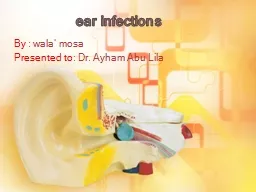


Slide1
Otitis media, Otitis externa , and Eye diseases:
Otitis media
:
is the inflammation of the area between Eardrum (Tympanic membrane) and the inner ear; including Eustachian tube.
-Infections of
air-filled cavities
of the head occur when
normal drainage routes become obstructed
.
-Infection of air-filled cavities of the head results in:
1-Otitis media.
2-Sinusitis.
3-Mastoiditis.
Slide2N
-Because the cavity of the middle ear is contiguous with the
mastoid air cells(spaces of temporal bone); individuals with acute otitis media also have mastoiditis.
Slide3N
-
The majority of cases
occur in children between 6 and 36 months of age.-Children are susceptible to otitis media for several
reasons:
1-The
medial orifice
of the
eustachian tube
is
more open
in
infancy than later in life.
2-
Milk
feeding
(giving a bottle at bedtime) results in
reflux
of
pharyngeal contents
into the
lumen
of
eustachian
tube
.
3-
Eustachian
tube
is
shorter
and more
horizontal
in young
children.
4-The viral infection of upper respiratory tract and
lymphoid
tissue
results in
eustachian tube obstruction
.
Slide4Pathogenesis:
-
Inflammation
of upper respiratory tracts due to: 1-Viral infections; influenza A or B, and adenovirus. 2-Allergy
(Rhinitis).
-Swelling of lymphoid tissue
(Eustachian tonsil)
around
eustachian
tube.
-
Eustachian tube obstruction
.
-Absorption of air of middle ear slowly by surrounding
tissues.
-
Creation of negative pressure
(vacuum) in the middle ear.
-
Accumulation of fluids
; so
normal flora
of upper
respiratory tract could
invade middle ear space
.
Slide5N
-
Colonization
of middle ear cavity lining epithelium.-If the microbe has a polysaccharide
capsule
:
-
P
olyclonal
lymphocyte
activator
; cytokines production;
chemotaxis
of immune cells and
inflammation
.
-Conductive hearing loss.
-If the infection is not treated; otitis media and mastoiditis
could be complicated by:
1-
Facial
nerve paralysis
.
2-
Infection of peripheral nerves; results in
deeper
brain
abscess
.
Slide6N
3-
Infection of veins that bridge
surrounding bony structures and the cerebral cortex; septic thrombophlebitis) results in subdural empyema (in some cases;
related to
epidural
abscesses
).
Acute abscess
is frequently caused by a
mixed bacterial flora
c
onsisting of
obligate and facultative anaerobic bacteria
;
similar
to the
mixture of microbes
infecting middle ear, mastoid, and sinuses.
Slide7N
Treatment of poly-microbial brain abscesses:
Antibiotic combination:
1-Vancomycin or Ceftriaxone: to cover Staphylococci and
other
Gram positive
beta-lactamase producers.
2-
Metronidazole
: to cover
anaerobic bacteria
.
3-
Quinolones
or
Macrolides
working effectively at acidic
pH.
Slide8The Normal flora of upper respiratory tracts:
-
Streptococcus
pneumoniae (Nasopharynx).-Haemophilus influenzae
(non-type b) (Nasopharynx).
-
Moraxella
catarrhalis
(Nasopharynx).
-
Staphylococcus aureus
.
-Coagulase negative
Staphylococcus
species.
-
Diphtheroids
species.
-
Neisseriae
species.
-
Candida
species.
Slide9Causes of Otitis media:
1-
Streptococcus
pneumoniae (the most common cause).2-Non-typeable Haemophilus influenzae
.
(the second common cause).
-Both
Strep.
p
neumoniae
and
H.
influenzae
causes
80%
of
otitis media cases
.
3-
Moraxella
catarrhalis
.
-Gram’s negative non-motile
coccobacilli
in pairs.
-Aerobic fastidious oxidase positive bacteria.
4-Other normal flora of upper respiratory tracts(rare).
Slide10Clinical Classification of Otitis media:
1-Acute suppurative:
-Pus accumulated in middle ear; mainly in infant and children.
2-Chronic:
A-Recurrent OM due to other causes.
B-Secretory OM : Very common persisting middle ear
effusion
after OM in
40% of cases
.
(
Thick fluid consistency
).
Management:
1-Amoxicillin or Ceftriaxone.
2-Amoxicillin/
Clavulanate
for
B
eta-Lactamases strains
.
Slide11Otitis Externa :
Otitis Externa
: is an inflammation of the outer ear and ear canal.
-It is mainly caused by bacterial or fungal agents.-Otitis externa could be established due to:
1-Swimming in polluted water (germs contamination).
2-Impairment in the integrity of the skin (dermatitis).
-Hospital acquired otitis
externa
could be caused by
hospital dwelling bacteria as a post-surgical infection.
Slide12Causes of Otitis Externa:
Exogenous:
-
Pseudomonas aeruginosa (the most common cause).
Endogenous:
Normal flora of outer ear canal
:
-Coagulase negative
Staphylococci
.
-
Staphylococcus aureus
.
-Gram negative bacilli
.
-Fungi:
Candida species. Malassezia furfur.
Slide13Clinical classification of Otitis Externa:
1-Acute localized secondary to folliculitis:
-Painful pustule with local lymph node enlargement; Staphylococcus aureus.2-Acute diffuse (Swimmer’s ear):
-
Pseudomonas aeruginosa
.
-Itchy, painful, edematous, reddened OE with purulent
discharge.
3-Chronic OE secondary to chronic otitis media.
-Fungi, Candida plus other chronic disease
(Immunocompromised Pat. ).
Slide14N
4-Malignant OE
(
Necrotizing OE) Secondary to diabetic microangiopathy; Pseudomonas spp
.
-Serious infection.
-Not-malignant (Non-Cancerous).
-Malignant in its progressive, fatal course.
-Spreading of infection to surrounding bone, blood
vessels, facial nerve and meninges.
-
Complicated by
inflammation of cranial nerves
and their
branches ;
facial
(7th) nerve paralysis. -It is associated with
immunocompromised
patients
.
-Diagnosis: Full investigations: CT Scan, MRI.
Slide15N
Management:
1-Acute
: As OM.2-Acute diffuse: Local Neomycin, Polymyxin.3-Chronic
:
Local imidazole for fungi and treatment of OM.
4-Malignant OE
: I.V Tobramycin and
Ceftazidime
(weeks)
With surgery.
Diagnosis:
Clinical specimen
:
Ear Swab (cotton swab).
Slide16N
Culture:
on Enriched media.
-Blood agar incubated under aerobic conditions. -Chocolate agar: incubated under anaerobic conditions.Isolation of Pseudomonas species:-Encapsulated, motile, Gram negative bacilli.
-Oxidase
positive
,
Exopigments
production.
-Antibiotic resistance
strains.
(
greenish
yellowish
exopigment
production; pyoverdin).
Slide17N
Isolation of
Staphylococci
:Staphylococcus aureus:Gram positive cocci, coagulase positive, novobiocin sensitive, and Mannitol fermenters.
Other
Staphylococci
:
Gram positive
cocci
, coagulase negative,
novobiocin
resistance.
Novobiocin
sensitivity Mannitol fermentation.
Slide18Infectious diseases of the Eye:
Keratitis
:
inflammation of transparent eye’s cornea; the anterior part of the eye (covers
the
iris, and pupil).
Causes
:
1-Amoebic keratitis
:
a serious corneal infection usually affecting contact lens
wearers.
Etiology
:
Acanthamoeba
.
2-Bacterial keratitis
:
Due to injury of wearing
contact lenses.
Etiology: Staphylococcus aureus, &Pseudomonas species.
Slide19N
Staphylococcus aureus
is a major cause of infections of the eyelid and cornea.
Staphylococcus aureus can infect the glands of the eyelid; resulting in the production of a sty.
Sty
is a painful red swelling
on the margin of the eyelid.
Treatment
:
bacitracin ointment.
Slide20N
3-Fungal keratitis:
Keratomycosis
: Etiology: Fusarium species. -Infection
is established due to corneal injury
in agriculture workers or
immunocompromised
patients.
Fusarium
Chlamydospores
.
Fusarium
Macroconidia
.
Slide21N
4-Viral Keratitis:
Etiology: Herpes simplex virus types 1 and 2.
Diseases
:
A-Primary infectious keratitis:
Vesicular eruption
of the eyelid, infection of cornea
leading to
corneal ulcers
.
B-Recurrent herpes keratitis:
-(More common than primary
keratitis).
-In immunocompromised patients. -Mild irritation and photophobia.
Slide22N
5-Onchocercal Keratitis:
Onchocerciasis
: -Parasitic infection of the eye’s cornea (Corneal lesions). -Etiology: Onchocerca
volvulus
.
-Transmitted by the bite
of blackfly
.
-
Disease
:
African River blindness
.
Slide23nChemotherapy for Keratitis and Corneal Ulcers:
1-Bacterial Keratitis:
Local broad-spectrum antibiotics; Vancomycin and tobramycin.2-Viral keratitis: Acyclovir.
3-Fungal Keratitis:
Amphotericin B.
4-Amoebic keratitis:
Propamidine
drops plus oral ketoconazole.
Slide24Conjunctivitis:
Conjunctiva: is a thin, translucent, mucous membrane that lines the eyelid and covers the white portion of the eyeball.
Conjunctivitis is divided according to etiology into:
A-Bacterial conjunctivitis: -Redness, swelling of the eyelid, and
muco
-purulent discharge
.
- Yellowish-greyish discharge:
pyogenic
cocci
infection.
-
Treatment:
Local bacitracin
or neomycin.
Slide25N
Types of bacterial conjunctivitis:
1-Trachoma: Etiology:
Chlamydia trachomatis:Serotypes A, B, Ba, and
C
causes chronic
keratoconjunctivitis
(
Trachoma
) that results in
blindness
.
Trachoma is a leading cause of blindness in endemic areas of northern India, the Middle East, and North Africa.
Transmission:
-Personal contact ; eye-to-eye via droplets by
contaminated
hands (transfer of elementary bodies).
Chemotherapy:
Oral azithromycin or tetracycline.
Slide26N
Chlamydia
trachomatis:-Unicellular obligatory intracellular bacteria that has rigid cell wall. -Infective stage: The elementary body.
-
Inclusion
bodies (Trachoma) infected
conjunctival
epithelial cells
(Reticulate body: diagnostic stage).
Slide27N
2-Ophthalmia
neonatorum
: Etiology:1-Neisseria gonorrhoeae:
-The most severe cause of
hyperacute
bacterial
conjunctivitis of newborn.
-It is acquired during passage of newborn through the
birth canal of a mother infected by gonococci.
-
Neisseria
species are
Gram
negative oxidase positive
diplococci
that ferment glucose only.
Slide28N
2-
Chlamydia
trachomatis:(Ophthalmia neonatorum):
-This type of newborn conjunctivitis is associated with
serotypes
D-K
.
- 50% of Infants born with infection due to passage
through the
birth canal.
-
inclusion conjunctivitis heals without eye damage
-
Treatment of both types 1 and 2
:
(
Prophylactic drug
):
Erythromycin ointment; most strains of Neisseria
gonorrhoeae
are
Beta-Lactam
resistant
.
Slide29N
B-Viral Conjunctivitis
:(Pink Eye):
diffuse pinkness of Conjunctiva. -Adenovirus infection is the most common cause of viral conjunctivitis.
-
Acute conjunctivitis
, and
pharyngoconjunctival
fever
.
-A more serious infection is
epidemic
keratoconjunctivitis
,
which involves
formation
of a
painful ulcer
of the
corneal epithelium
. Electron microscopy: Double-Stranded DNA, Icosahedral
naked virus.
Slide30N
-
Herpes simplex virus
cause serious Herpetic keratoconjunctivitis, which requires treatment with acyclovir.
-
Acute hemorrhagic conjunctivitis
is a highly
contagious disease caused by:
1-Enterovirus 70
.
2-Coxsackievirus A
24
.
Viral hemorrhagic
Conjunctivitis
due to Enterovirus-70
infection.
Slide31Diagnosis of Eye infection:
Clinical specimens:
1-Eye cotton Swab.
2-Conjunctival Scraping. 1-Direct microscopy: A-Swab
for Microbiology (
Gram’s stain
):
detection of
G+ve
and
G-
ve
bacteria
, and
yeast
.
B-
Conjunctival
scrapes
for cytology lab:
detection of Chlamydia diagnostic stage. C-Conjunctival Scrapes for immunohistochemistry:
detection of
viral infections
or
Chlamydia
infection
.
Slide32N
2-Detection of viral genetic material and
Chlamydia
genetic material by molecular methods: 1-Nucleic acid DNA hybridization.(Probe hybridization). 2-PCR :
Primer amplification of genetic material.
3-Cultivation of bacterial agents:
-Eye swab should be inoculated on Enriched media.
-
Blood agar
and
Chocolate agar
should be incubated
under
aerobic
and
anaerobic
conditions
respectively
.
Clinical significance
: isolation of pyogenic cocci and Neisseria gonorrhoeae
.
Slide33Detection of Virus and Chlamydia genetic materials by
Immunofluorescent
Microscopy:
Detection of viral antigen by Localization of specific Adenovirus-
monoclonal antibodies.
Coxsackievirus
receptor on Epithelial cells of conjunctiva.