04
Caecum and Vermiform Appendix
Colon
Rectum
Anus GROSS ANATOMICAL DESCRIPTIONS<br>
05
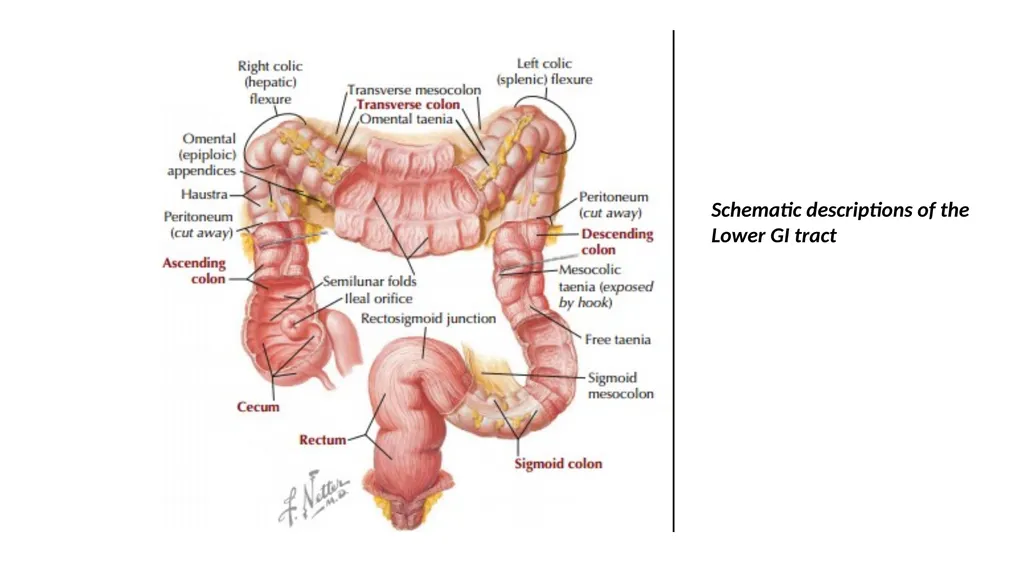
Schematic descriptions of the Lower GI tract<br>
06
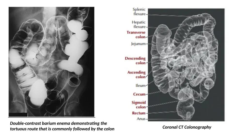
Double-contrast barium enema demonstrating the�tortuous route that is commonly followed by the colon Coronal CT Colonography<br>
07
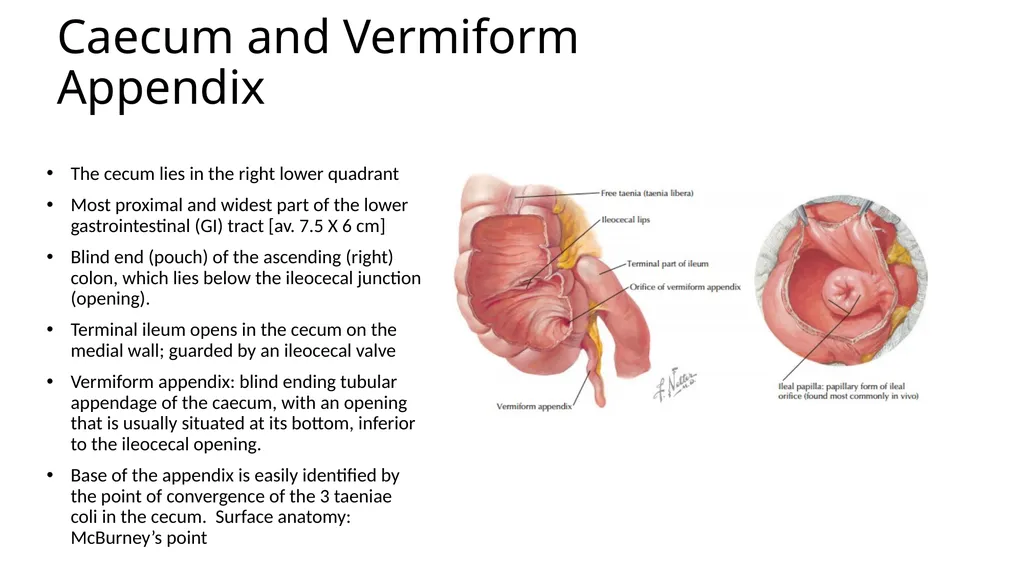
Caecum and Vermiform Appendix The cecum lies in the right lower quadrant
Most proximal and widest part of the lower gastrointestinal (GI) tract [av. 7.5 X 6 cm]
Blind end (pouch) of the ascending (right) colon, which lies below the ileocecal junction (opening).
Terminal ileum opens in the cecum on the medial wall; guarded by an ileocecal valve
Vermiform appendix: blind ending tubular appendage of the caecum, with an opening that is usually situated at its bottom, inferior to the ileocecal opening.
Base of the appendix is easily identified by the point of convergence of the 3 taeniae coli in the cecum. Surface anatomy: McBurney’s point<br>
08
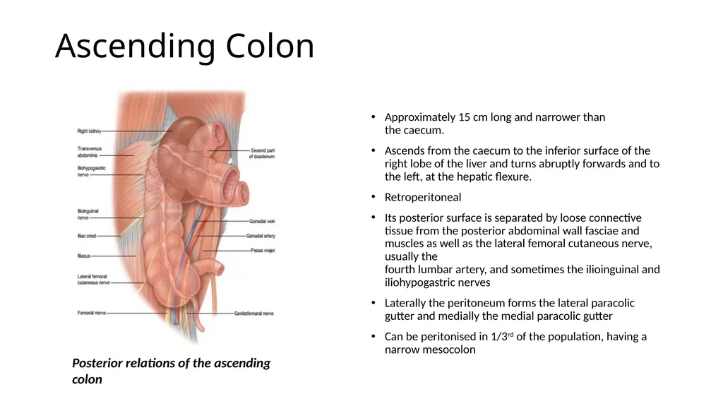
Ascending Colon Posterior relations of the ascending colon Approximately 15 cm long and narrower than�the caecum.
Ascends from the caecum to the inferior surface of the right lobe of the liver and turns abruptly forwards and to the left, at the hepatic flexure.
Retroperitoneal
Its posterior surface is separated by loose connective tissue from the posterior abdominal wall fasciae and muscles as well as the lateral femoral cutaneous nerve, usually the�fourth lumbar artery, and sometimes the ilioinguinal and iliohypogastric nerves
Laterally the peritoneum forms the lateral paracolic gutter and medially the medial paracolic gutter
Can be peritonised in 1/3rd of the population, having a narrow mesocolon<br>
09
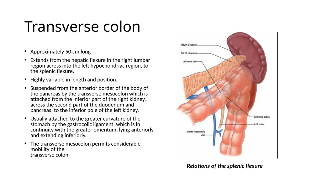
Transverse colon Approximately 50 cm long
Extends from the hepatic flexure in the right lumbar region across into the left hypochondriac region, to the splenic flexure.
Highly variable in length and position.
Suspended from the anterior border of the body of the pancreas by the transverse mesocolon which is attached from the inferior part of the right kidney, across the second part of the duodenum and pancreas, to the inferior pole of the left kidney.
Usually attached to the greater curvature of the stomach by the gastrocolic ligament, which is in continuity with the greater omentum, lying anteriorly and extending inferiorly.
The transverse mesocolon permits considerable mobility of the�transverse colon. Relations of the splenic flexure<br>
10
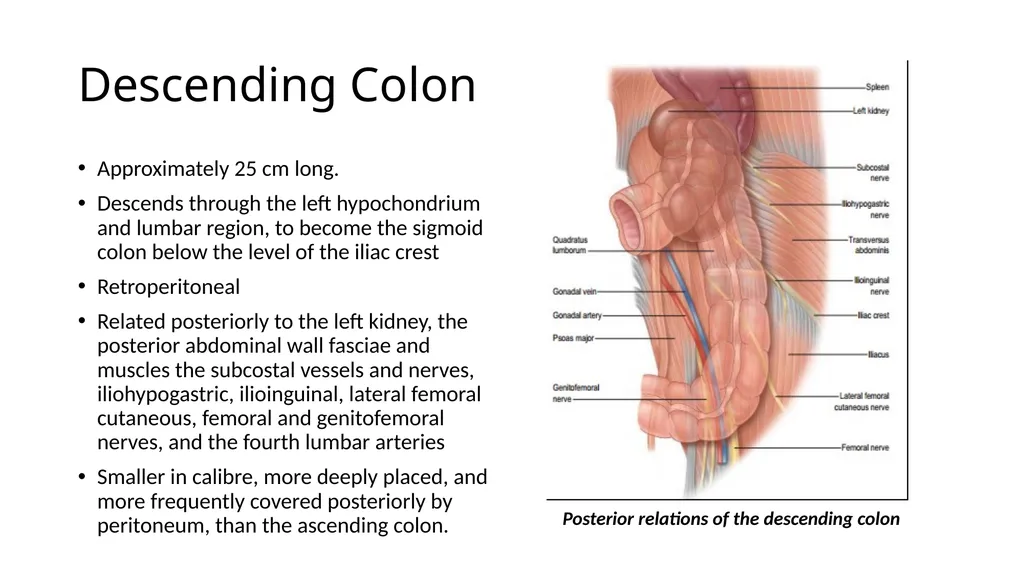
Descending Colon Approximately 25 cm long.
Descends through the left hypochondrium and lumbar region, to become the sigmoid colon below the level of the iliac crest
Retroperitoneal
Related posteriorly to the left kidney, the posterior abdominal wall fasciae and muscles the subcostal vessels and nerves, iliohypogastric, ilioinguinal, lateral femoral cutaneous, femoral and genitofemoral nerves, and the fourth lumbar arteries
Smaller in calibre, more deeply placed, and more frequently covered posteriorly by peritoneum, than the ascending colon. Posterior relations of the descending colon<br>
11
Sigmoid colon Begins below the pelvic inlet and ends at the rectum.
Usually completely peritonised and attached to the posterior abdominal wall by the “V” shaped sigmoid mesocolon.
Most variable in length and form of all colonic segments.
Variable diameter and length in different races
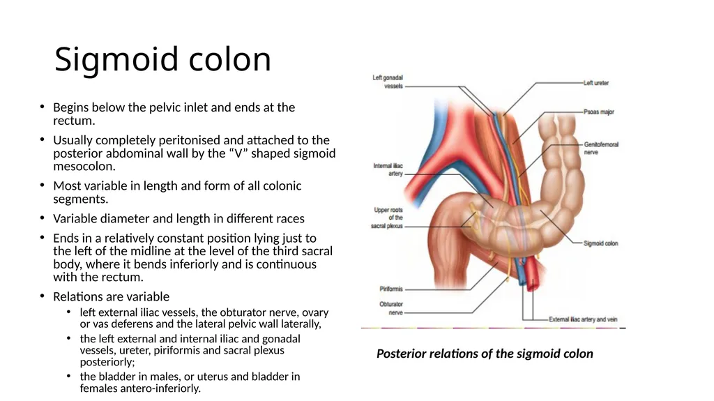
Ends in a relatively constant position lying just to the left of the midline at the level of the third sacral body, where it bends inferiorly and is continuous with the rectum.
Relations are variable
left external iliac vessels, the obturator nerve, ovary or vas deferens and the lateral pelvic wall laterally,
the left external and internal iliac and gonadal vessels, ureter, piriformis and sacral plexus posteriorly;
the bladder in males, or uterus and bladder in females antero-inferiorly. Posterior relations of the sigmoid colon<br>
12
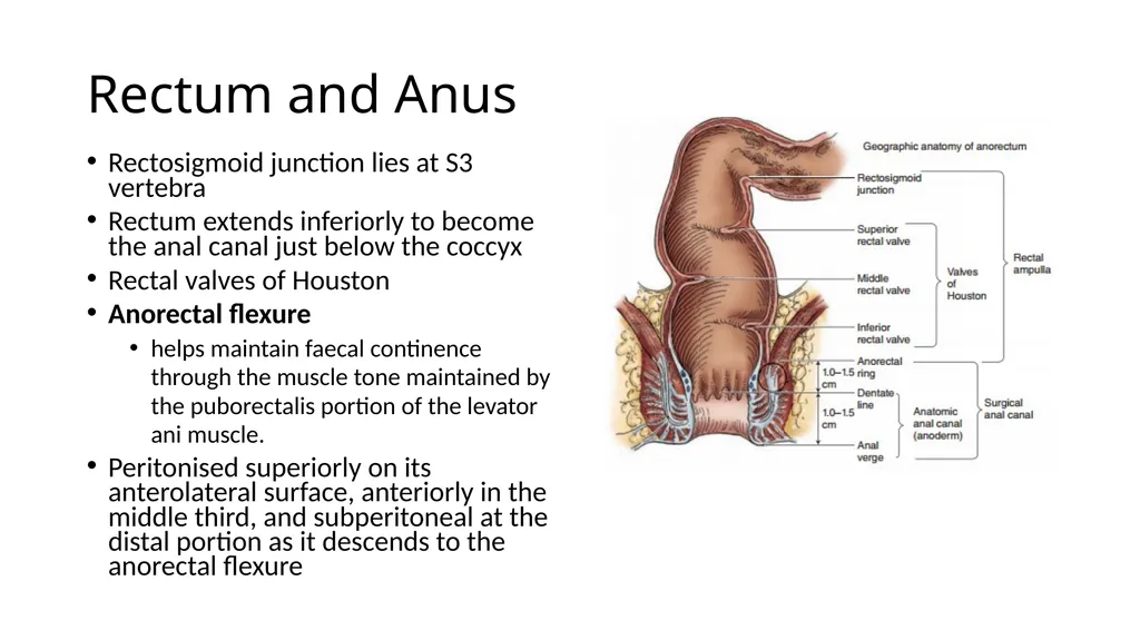
Rectum and Anus Rectosigmoid junction lies at S3 vertebra
Rectum extends inferiorly to become the anal canal just below the coccyx
Rectal valves of Houston
Anorectal flexure
helps maintain faecal continence through the muscle tone maintained by the puborectalis portion of the levator ani muscle.
Peritonised superiorly on its anterolateral surface, anteriorly in the middle third, and subperitoneal at the distal portion as it descends to the anorectal flexure<br>
13
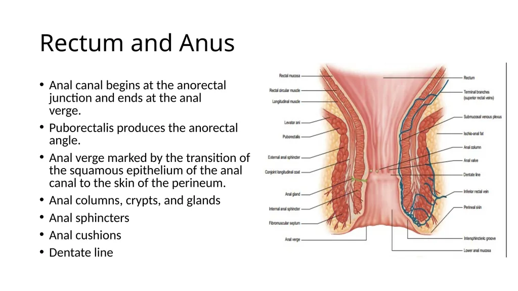
Rectum and Anus Anal canal begins at the anorectal junction and ends at the anal�verge.
Puborectalis produces the anorectal angle.
Anal verge marked by the transition of the squamous epithelium of the anal canal to the skin of the perineum.
Anal columns, crypts, and glands
Anal sphincters
Anal cushions
Dentate line<br>
14
Blood Supply to the Lower GI Tract The normal distribution of supply to the ileum, right colon, and right side of transverse colon from the middle colic artery and ileocolic arteries is shown. The distribution of the arterial supply from the inferior mesenteric artery to the left side of the transverse colon, the descending colon, the sigmoid, and the upper rectum is also demonstrated. The venous drainage of the large bowel and rectum is illustrated; in particular, the drainage of the left colon via the inferior�mesenteric vein to the splenic vein is shown.<br>
15
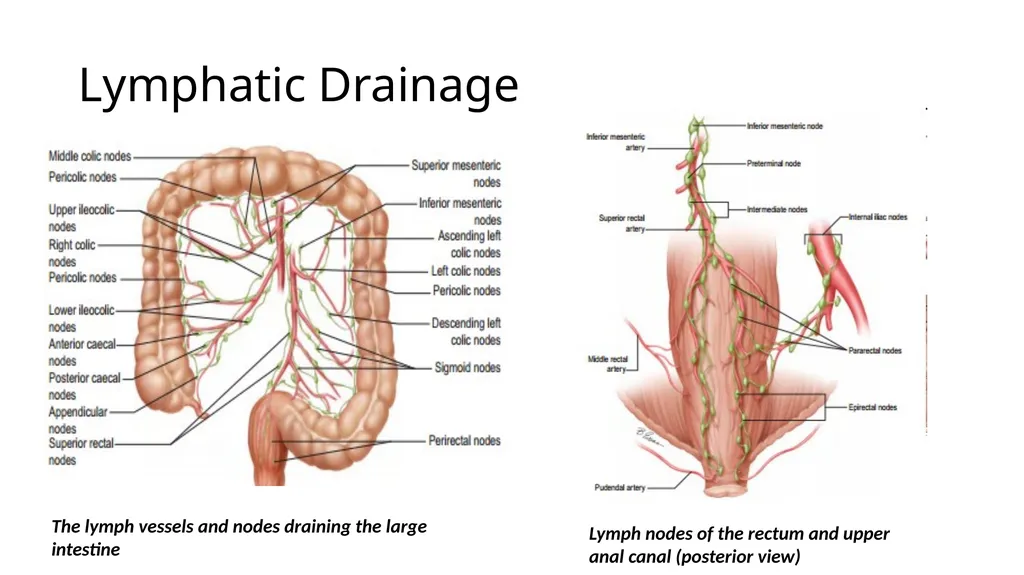
Lymphatic Drainage The lymph vessels and nodes draining the large intestine Lymph nodes of the rectum and upper anal canal (posterior view)<br>
16
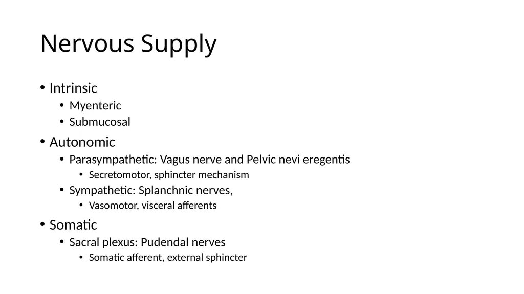
Nervous Supply Intrinsic
Myenteric
Submucosal
Autonomic
Parasympathetic: Vagus nerve and Pelvic nevi eregentis
Secretomotor, sphincter mechanism
Sympathetic: Splanchnic nerves,
Vasomotor, visceral afferents
Somatic
Sacral plexus: Pudendal nerves
Somatic afferent, external sphincter<br>
17
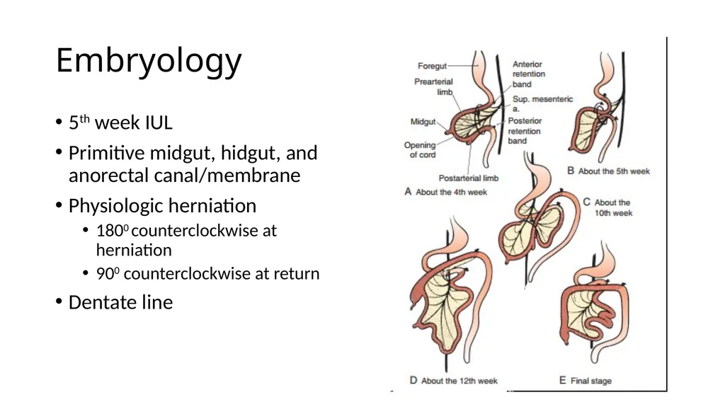
Embryology 5th week IUL
Primitive midgut, hidgut, and anorectal canal/membrane
Physiologic herniation
1800 counterclockwise at herniation
900 counterclockwise at return
Dentate line<br>
18
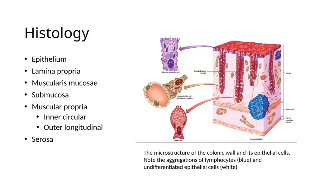
Histology Epithelium
Lamina propria
Muscularis mucosae
Submucosa
Muscular propria
Inner circular
Outer longitudinal
Serosa The microstructure of the colonic wall and its epithelial cells. Note the aggregations of lymphocytes (blue) and undifferentiated epithelial cells (white)<br>
19
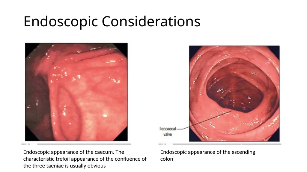
Endoscopic Considerations Endoscopic appearance of the caecum. The characteristic trefoil appearance of the confluence of the three taeniae is usually obvious Endoscopic appearance of the ascending�colon<br>
20
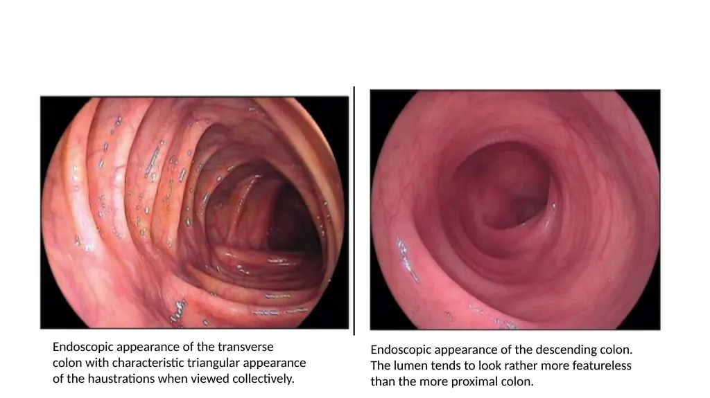
Endoscopic appearance of the transverse�colon with characteristic triangular appearance�of the haustrations when viewed collectively. Endoscopic appearance of the descending colon. The lumen tends to look rather more featureless than the more proximal colon.<br>
21
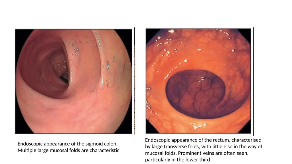
Endoscopic appearance of the sigmoid colon. Multiple large mucosal folds are characteristic Endoscopic appearance of the rectum, characterised by large transverse folds, with little else in the way of mucosal folds, Prominent veins are often seen, particularly in the lower third<br>
22
References Gray’s Anatomy: The Anatomical Basis of Clinical Practice. 40th Edition. 2008.
Shackelford’s Surgery of the Alimentary Canal. 7th Edition 2013
Maingot’s Abdominal Operations. 12th Edition 2013.
Netter’s Clinical Anatomy. 3rd Edition 2014.
Atlas of Human Anatomy. 6th Edition 2014.
Lower GI Anatomy. Medscape. Accessed 7/7/2019.<br>