
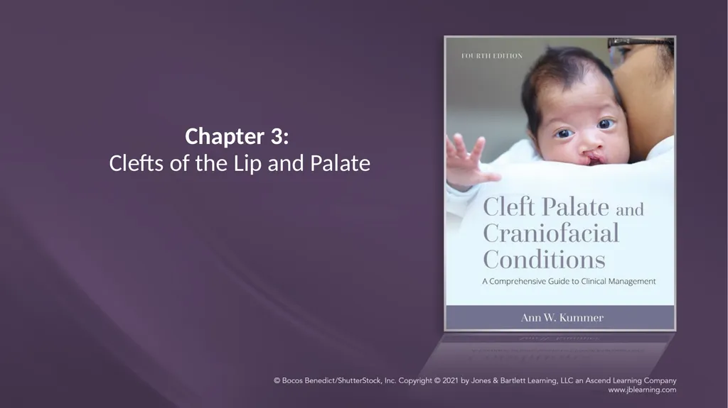
Chapter 3: Clefts of the Lip and Palate Chapter 3:
KL
Published · 64 slides · 0 views

1 / 1









Description
Chapter 3: Clefts of the Lip and Palate Chapter 3: Clefts of the Lip and Palate Part 1: EMBRYOLOGICAL DEVELOPMENT AND CAUSES OF CLEFTS Embryological Development of the Face and Skull Embryological development of the face and skull begins
Related Topics
Share
Embed code
Download this presentation From Below
"Chapter 3: Clefts of the Lip and Palate Chapter 3:" is the property of its rightful owner. Permission is granted to download and print the materials on this website for personal, non-commercial use only, and to display it on your personal computer provided you do not modify the materials and that you retain all copyright notices contained in the materials. By downloading content from our website, you accept the terms of this agreement.
Presentation Transcript
01
Chapter 3:
Clefts of the Lip and Palate<br>
02
Chapter 3: Clefts of the Lip and Palate Part 1:
EMBRYOLOGICAL DEVELOPMENT AND CAUSES OF CLEFTS<br>
03
Embryological Development of the Face and Skull Embryological development of the face and skull begins with the formation of neural crest cells.
Neural crest cells migrate to form various structures, including the lip and palate.<br>
Neural crest cells migrate to form various structures, including the lip and palate.<br>
04
Embryological Development: Lip and Palate Lip begins at 7 weeks gestation
Palate begins at 9 weeks gestation
Development of each is independent<br>
Palate begins at 9 weeks gestation
Development of each is independent<br>
05
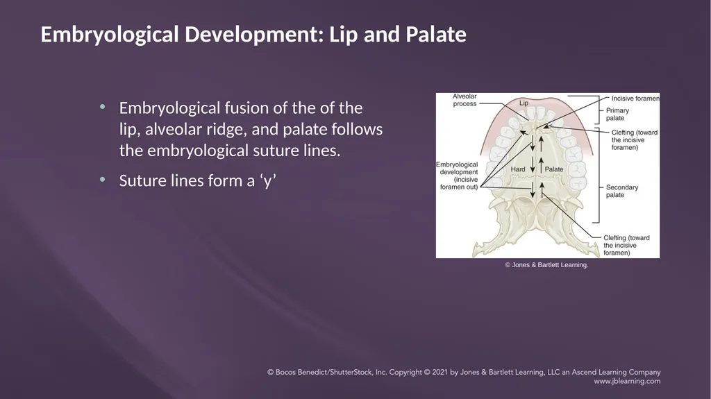
Embryological Development: Lip and Palate Embryological fusion of the of the lip, alveolar ridge, and palate follows the embryological suture lines.
Suture lines form a ‘y’ © Jones & Bartlett Learning.<br>
Suture lines form a ‘y’ © Jones & Bartlett Learning.<br>
06
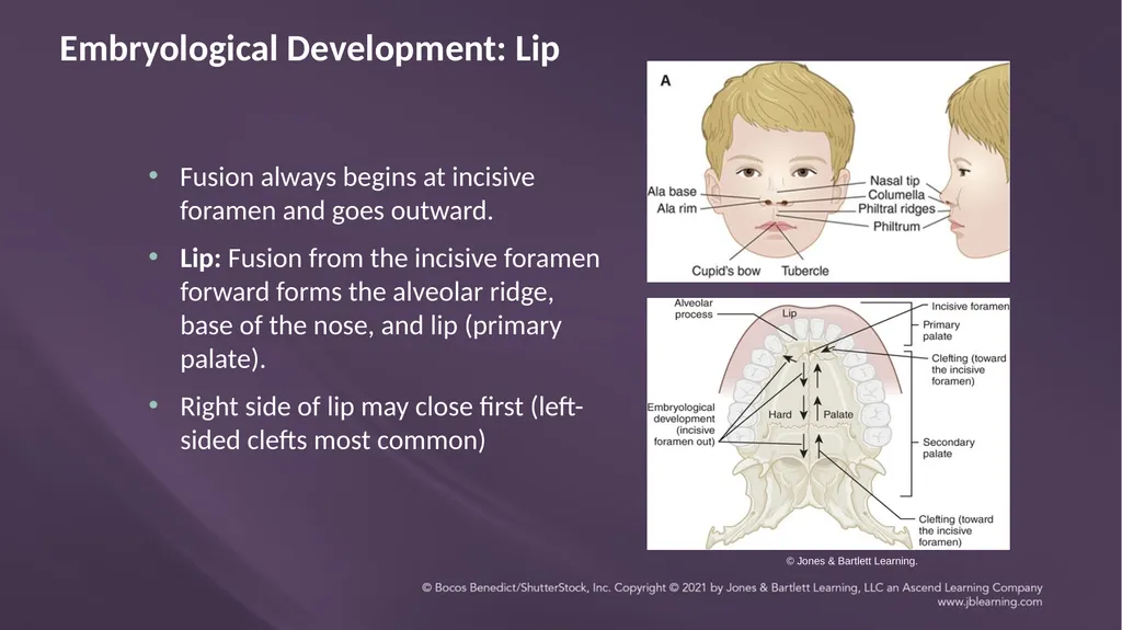
Embryological Development: Lip Fusion always begins at incisive foramen and goes outward.
Lip: Fusion from the incisive foramen forward forms the alveolar ridge, base of the nose, and lip (primary palate).
Right side of lip may close first (left-sided clefts most common) © Jones & Bartlett Learning.<br>
Lip: Fusion from the incisive foramen forward forms the alveolar ridge, base of the nose, and lip (primary palate).
Right side of lip may close first (left-sided clefts most common) © Jones & Bartlett Learning.<br>
07
Embryological Development: Palate Prior to palate formation, the tongue is in a superior and posterior position in the nasopharynx.
Palatal shelves of the maxilla are in a vertical position on each side of the tongue<br>
Palatal shelves of the maxilla are in a vertical position on each side of the tongue<br>
08
Embryological Development: Palate The mandible grows down and forward.
The tongue drops down and forward with the mandible.
The palatal shelves begin to move from vertical to horizontal position, starting with the incisive foramen and moving backward.<br>
The tongue drops down and forward with the mandible.
The palatal shelves begin to move from vertical to horizontal position, starting with the incisive foramen and moving backward.<br>
09
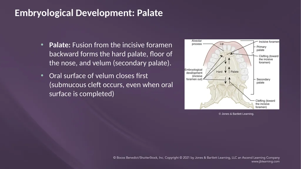
Embryological Development: Palate Palate: Fusion from the incisive foramen backward forms the hard palate, floor of the nose, and velum (secondary palate).
Oral surface of velum closes first (submucous cleft occurs, even when oral surface is completed) © Jones & Bartlett Learning.<br>
Oral surface of velum closes first (submucous cleft occurs, even when oral surface is completed) © Jones & Bartlett Learning.<br>
10
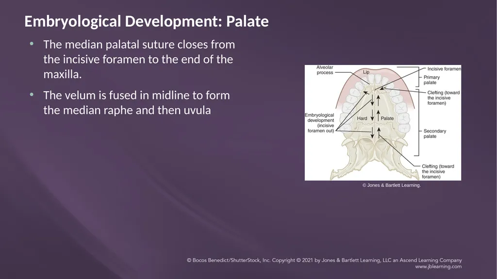
Embryological Development: Palate The median palatal suture closes from the incisive foramen to the end of the maxilla.
The velum is fused in midline to form the median raphe and then uvula © Jones & Bartlett Learning.<br>
The velum is fused in midline to form the median raphe and then uvula © Jones & Bartlett Learning.<br>
11
WHAT IS A CLEFT? Cleft: An abnormal opening or fissure in an anatomical structure
Usually a congenital malformation due to abnormal fusion of anatomical parts during fetal development
Can be acquired due to surgery (e.g., tumor removal)
For cleft lip: Cleft leaves an opening between sections of the lip and alveolar ridge
For cleft palate: Cleft leaves an opening between the sections of the hard palate and velum, resulting an opening between the oral and nasal cavities<br>
12
Variation in Clefts Vary in type: cleft lip and/or cleft palate
Vary in length: complete (all the way to the incisive foramen) or incomplete
Vary in width: very wide to very narrow<br>
Vary in length: complete (all the way to the incisive foramen) or incomplete
Vary in width: very wide to very narrow<br>
13
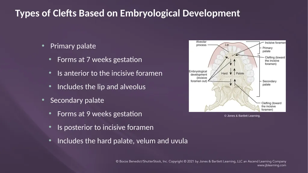
Types of Clefts Based on Embryological Development Primary palate
Forms at 7 weeks gestation
Is anterior to the incisive foramen
Includes the lip and alveolus
Secondary palate
Forms at 9 weeks gestation
Is posterior to incisive foramen
Includes the hard palate, velum and uvula © Jones & Bartlett Learning.<br>
Forms at 7 weeks gestation
Is anterior to the incisive foramen
Includes the lip and alveolus
Secondary palate
Forms at 9 weeks gestation
Is posterior to incisive foramen
Includes the hard palate, velum and uvula © Jones & Bartlett Learning.<br>
14
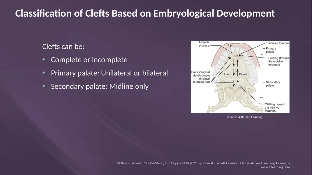
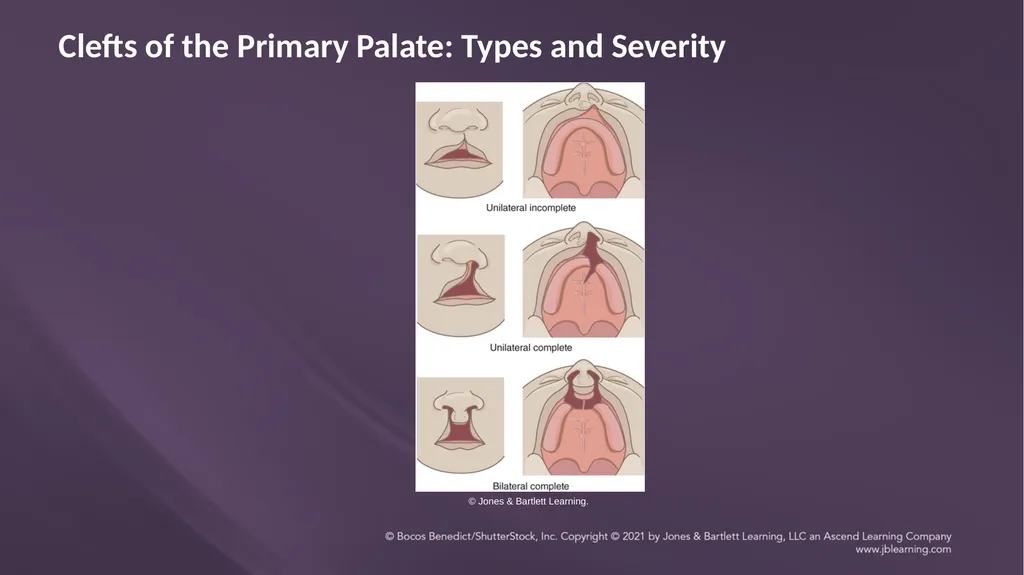
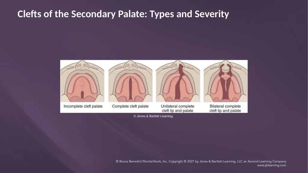
Classification of Clefts Based on Embryological Development Clefts can be:
Complete or incomplete
Primary palate: Unilateral or bilateral
Secondary palate: Midline only © Jones & Bartlett Learning.<br>
Complete or incomplete
Primary palate: Unilateral or bilateral
Secondary palate: Midline only © Jones & Bartlett Learning.<br>
15
Demonstration<br>
16
Causes of Clefts The delay in migration of neural crest cells causes the fetus to miss the normal period of fusion at the sutures.
When sutures don’t fuse, they leave a cleft and dysplasticity (lack of normal development) of surrounding structures.<br>
When sutures don’t fuse, they leave a cleft and dysplasticity (lack of normal development) of surrounding structures.<br>
17
Causes of Clefts: Multifactorial Cause of congenital clefts is usually “multifactorial” due to a combination of the following:
Genetic factors
Environmental factors<br>
Genetic factors
Environmental factors<br>
18
Genetic (Endogenous) Factors Genetic defects are common
Can cause a predisposition for clefting
Recurrence risk: Usually 3-5% in the US<br>
Can cause a predisposition for clefting
Recurrence risk: Usually 3-5% in the US<br>
19
Genetic (Endogenous) Factors Predisposition depends on racial background:
Indigenous American Indians: highest risk
Asians
Caucasians
Africans: lowest risk<br>
Indigenous American Indians: highest risk
Asians
Caucasians
Africans: lowest risk<br>
20
Environmental (Exogenous) Factors Teratogens: Chemical agents that can interfere with embryonic development, causing a malformation
Environment: lead, radiation, pollution
Drugs and ingested substances: phenytoin (Dilantin), valium, corticosteroids, smoking, alcohol
Viruses: rubella, influenza
Maternal nutrition: nutritional deficiencies or obesity with diabetes
Physical interference: Crowding in utero that restricts mandibular growth<br>
21
Chapter 3: Clefts of the Lip and Palate Part 2
CLEFTS OF THE PRIMARY AND SECONDARY PALATE<br>
CLEFTS OF THE PRIMARY AND SECONDARY PALATE<br>
22
Clefts of the Primary Palate<br>
23
Clefts of the Primary Palate: Types and Severity © Jones & Bartlett Learning.<br>
24
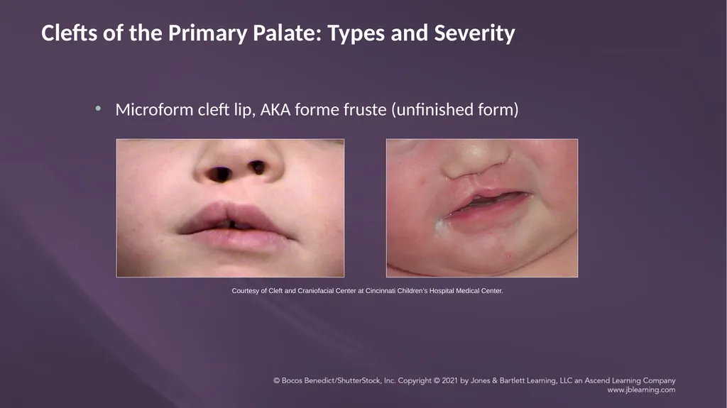
Microform cleft lip, AKA forme fruste (unfinished form) Clefts of the Primary Palate: Types and Severity Courtesy of Cleft and Craniofacial Center at Cincinnati Children’s Hospital Medical Center.<br>
25
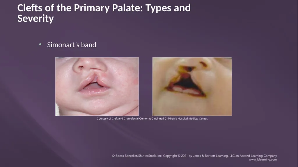
Simonart’s band Clefts of the Primary Palate: Types and Severity Courtesy of Cleft and Craniofacial Center at Cincinnati Children’s Hospital Medical Center.<br>
26
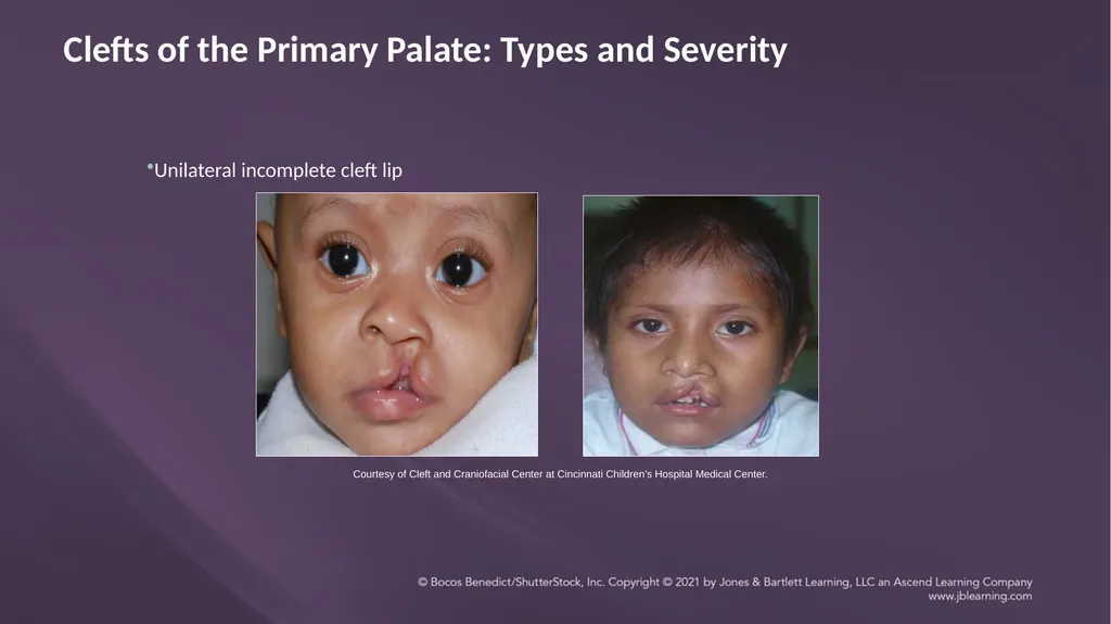
Clefts of the Primary Palate: Types and Severity Courtesy of Cleft and Craniofacial Center at Cincinnati Children’s Hospital Medical Center. Unilateral incomplete cleft lip<br>
27
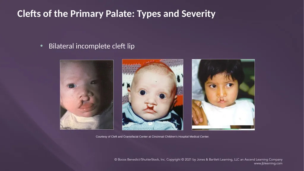
Clefts of the Primary Palate: Types and Severity Bilateral incomplete cleft lip Courtesy of Cleft and Craniofacial Center at Cincinnati Children’s Hospital Medical Center.<br>
28
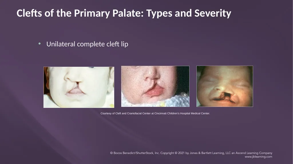
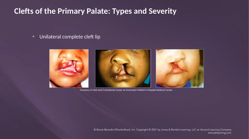
Unilateral complete cleft lip Clefts of the Primary Palate: Types and Severity Courtesy of Cleft and Craniofacial Center at Cincinnati Children’s Hospital Medical Center.<br>
29
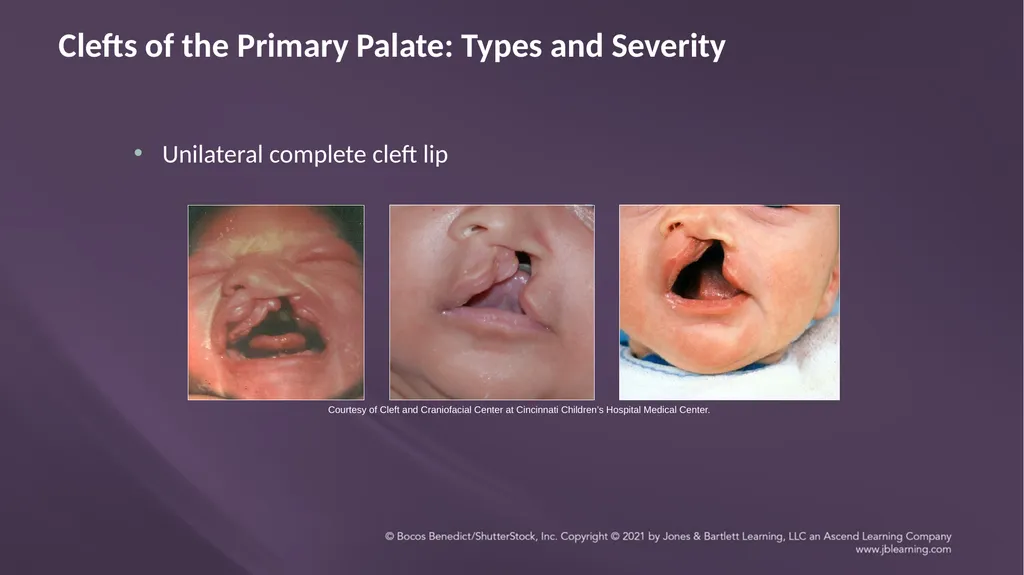
Unilateral complete cleft lip Clefts of the Primary Palate: Types and Severity Courtesy of Cleft and Craniofacial Center at Cincinnati Children’s Hospital Medical Center.<br>
30
Unilateral complete cleft lip Clefts of the Primary Palate: Types and Severity Courtesy of Cleft and Craniofacial Center at Cincinnati Children’s Hospital Medical Center.<br>
31
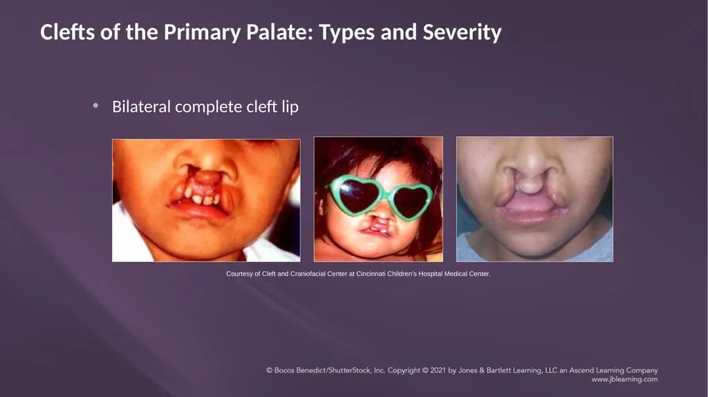
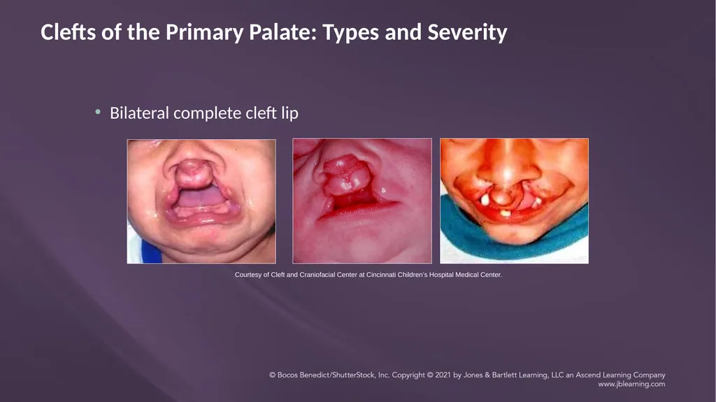
Bilateral complete cleft lip Clefts of the Primary Palate: Types and Severity Courtesy of Cleft and Craniofacial Center at Cincinnati Children’s Hospital Medical Center.<br>
32
Clefts of the Primary Palate: Types and Severity Courtesy of Cleft and Craniofacial Center at Cincinnati Children’s Hospital Medical Center. Bilateral complete cleft lip<br>
33
Clefts of the Primary Palate: Effects on Structure and Function Structure
Cleft lip and orbicularis oris
Wide, flat nose with spreading nasal ala
Short columella
Abnormal dentition
Function
Specific articulation errors
Resonance affected<br>
Cleft lip and orbicularis oris
Wide, flat nose with spreading nasal ala
Short columella
Abnormal dentition
Function
Specific articulation errors
Resonance affected<br>
34
Clefts of the Secondary Palate<br>
35
Clefts of the Secondary Palate: Types and Severity © Jones & Bartlett Learning.<br>
36
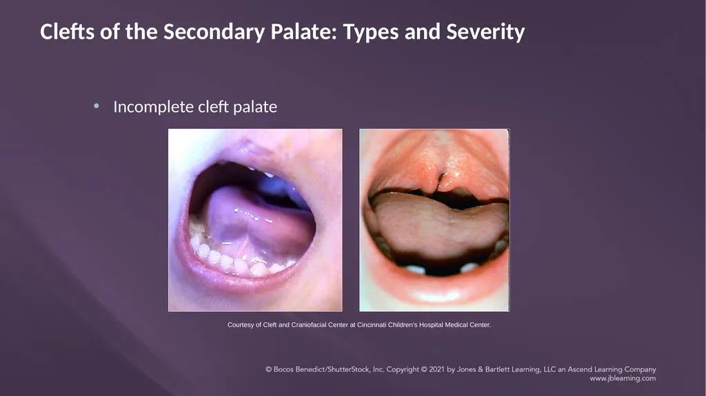
Clefts of the Secondary Palate: Types and Severity Incomplete cleft palate Courtesy of Cleft and Craniofacial Center at Cincinnati Children’s Hospital Medical Center.<br>
37
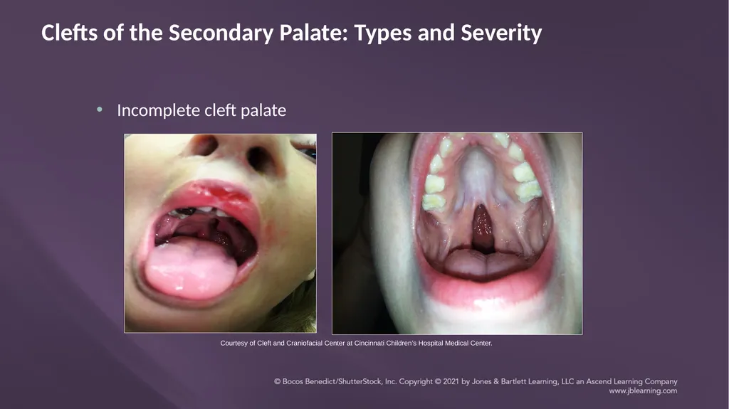
Clefts of the Secondary Palate: Types and Severity Incomplete cleft palate Courtesy of Cleft and Craniofacial Center at Cincinnati Children’s Hospital Medical Center.<br>
38
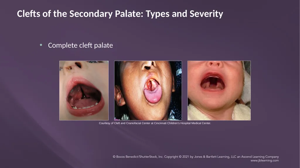
Clefts of the Secondary Palate: Types and Severity Complete cleft palate Courtesy of Cleft and Craniofacial Center at Cincinnati Children’s Hospital Medical Center.<br>
39
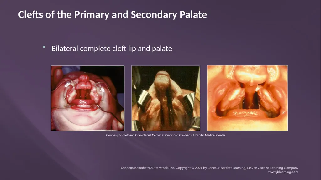
Bilateral complete cleft lip and palate Clefts of the Primary and Secondary Palate Courtesy of Cleft and Craniofacial Center at Cincinnati Children’s Hospital Medical Center.<br>
40
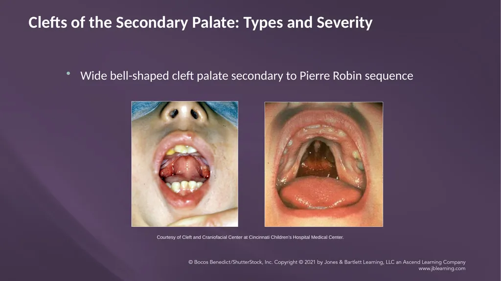
Wide bell-shaped cleft palate secondary to Pierre Robin sequence Clefts of the Secondary Palate: Types and Severity Courtesy of Cleft and Craniofacial Center at Cincinnati Children’s Hospital Medical Center.<br>
41
Clefts of the Secondary Palate: Effects on Structure and Function Structure
Abnormal insertion of the levator velar muscles (called the “cleft muscles of Veau”)
Abnormalities in nasal septum
Function
Velopharyngeal insufficiency (affecting speech and/or resonance)
Feeding problems and nasal regurgitation
Eustachian tube malfunction<br>
Abnormal insertion of the levator velar muscles (called the “cleft muscles of Veau”)
Abnormalities in nasal septum
Function
Velopharyngeal insufficiency (affecting speech and/or resonance)
Feeding problems and nasal regurgitation
Eustachian tube malfunction<br>
42
Submucous Cleft Palate<br>
43
Submucous Cleft: Embryology Fusion of the oral surface of the secondary palate occurs ahead of fusion of the nasal surface
Oral surface of the secondary palate can be completely formed, while the nasal surface (or just the uvula) is incomplete<br>
Oral surface of the secondary palate can be completely formed, while the nasal surface (or just the uvula) is incomplete<br>
44
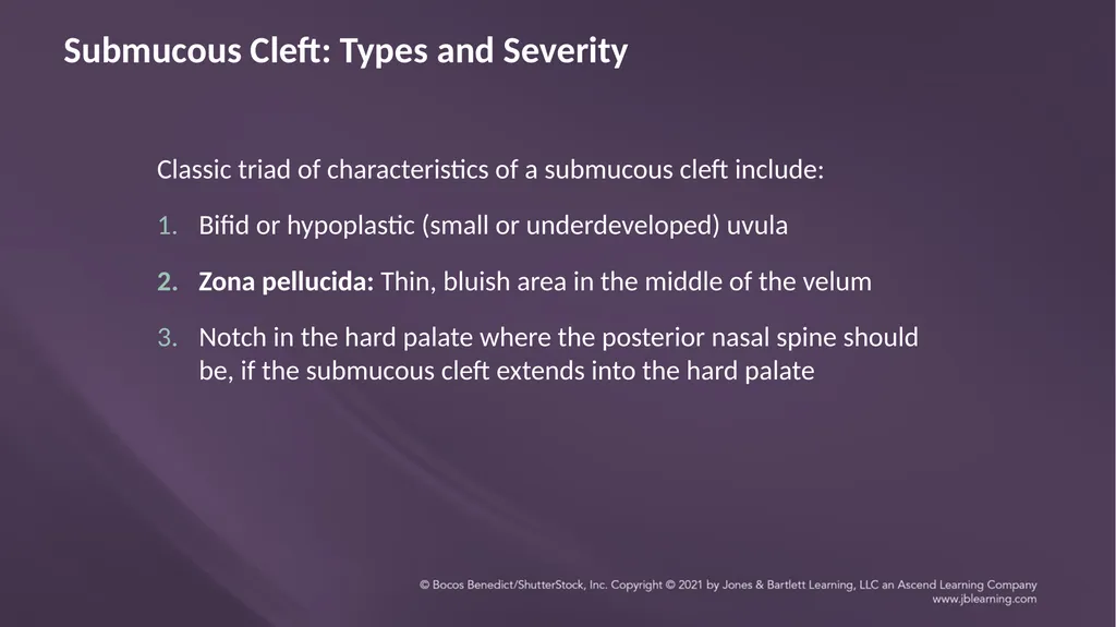
Submucous Cleft: Types and Severity Classic triad of characteristics of a submucous cleft include:
Bifid or hypoplastic (small or underdeveloped) uvula
Zona pellucida: Thin, bluish area in the middle of the velum
Notch in the hard palate where the posterior nasal spine should be, if the submucous cleft extends into the hard palate<br>
Bifid or hypoplastic (small or underdeveloped) uvula
Zona pellucida: Thin, bluish area in the middle of the velum
Notch in the hard palate where the posterior nasal spine should be, if the submucous cleft extends into the hard palate<br>
45
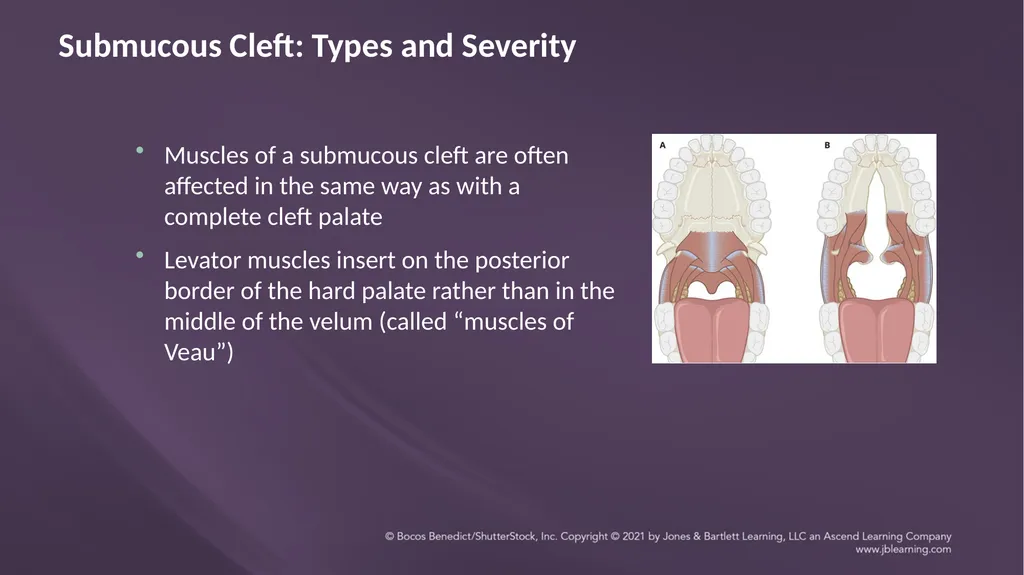
Submucous Cleft: Types and Severity Muscles of a submucous cleft are often affected in the same way as with a complete cleft palate
Levator muscles insert on the posterior border of the hard palate rather than in the middle of the velum (called “muscles of Veau”)<br>
Levator muscles insert on the posterior border of the hard palate rather than in the middle of the velum (called “muscles of Veau”)<br>
46
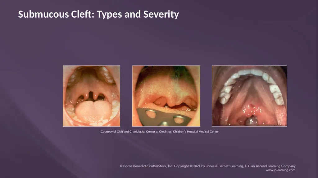
Submucous Cleft: Types and Severity Courtesy of Cleft and Craniofacial Center at Cincinnati Children’s Hospital Medical Center.<br>
47
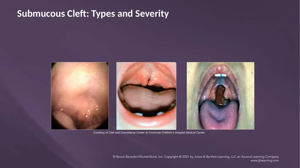
Submucous Cleft: Types and Severity Courtesy of Cleft and Craniofacial Center at Cincinnati Children’s Hospital Medical Center.<br>
48
Submucous Cleft: Types and Severity Courtesy of Cleft and Craniofacial Center
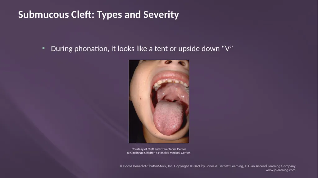
at Cincinnati Children’s Hospital Medical Center. During phonation, it looks like a tent or upside down “V”<br>
at Cincinnati Children’s Hospital Medical Center. During phonation, it looks like a tent or upside down “V”<br>
49
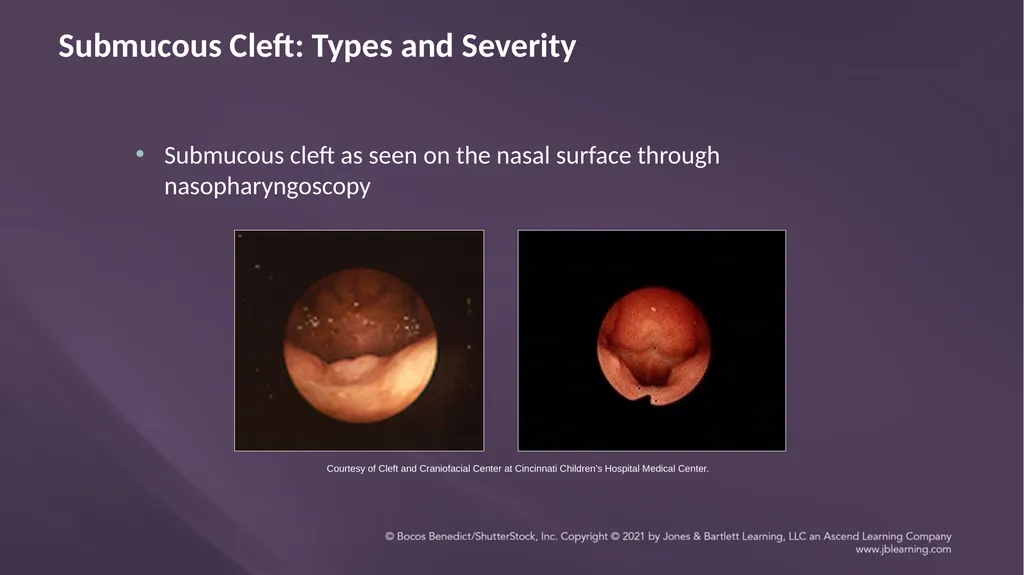
Submucous Cleft: Types and Severity Courtesy of Cleft and Craniofacial Center at Cincinnati Children’s Hospital Medical Center. Submucous cleft as seen on the nasal surface through nasopharyngoscopy<br>
50
Submucous Cleft: Types and Severity Overt (plainly or readily apparent): Visible from the oral surface
Occult (hidden): Normal on the oral surface; abnormal on the nasal surface<br>
Occult (hidden): Normal on the oral surface; abnormal on the nasal surface<br>
51
Submucous Cleft: Effects on Structure and Function Structure
Depends on the extent
May include a minor abnormality of the uvula or a defect of the nasal surface of the velum and hard palate
May include an altered insertion of the levator velar muscles (cleft muscles of Veau)
Function
Velopharyngeal insufficiency (affecting speech and/or resonance)
Feeding problems and nasal regurgitation
Eustachian tube malfunction<br>
52
Facial Clefts<br>
53
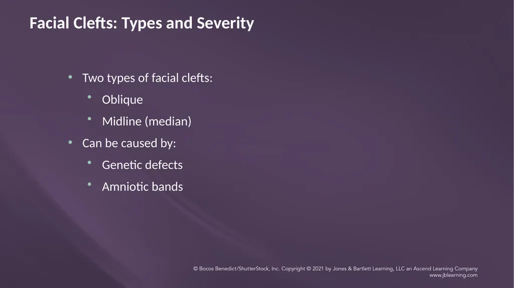
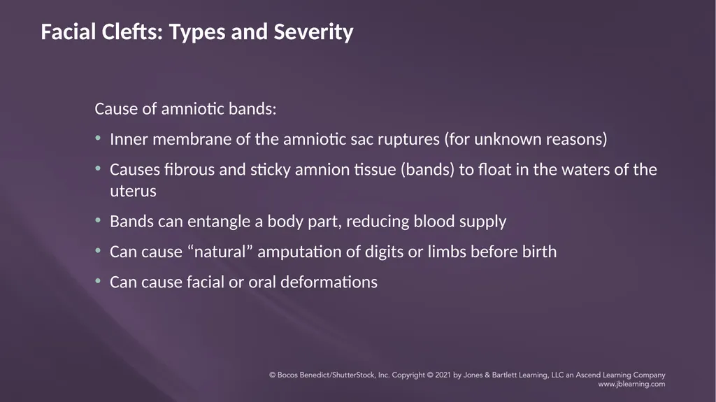
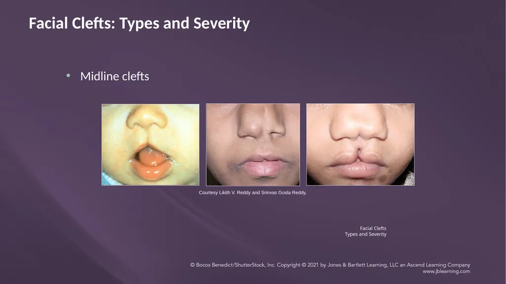
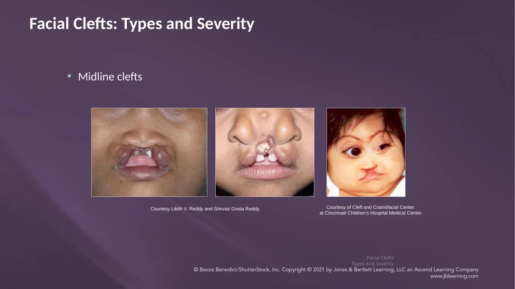
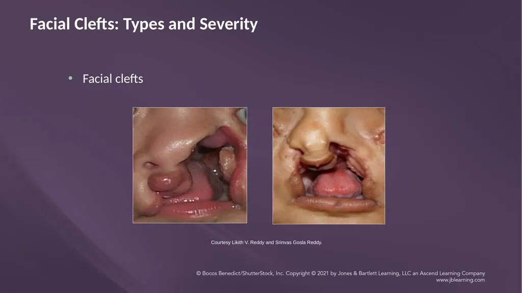
Facial Clefts: Types and Severity Two types of facial clefts:
Oblique
Midline (median)
Can be caused by:
Genetic defects
Amniotic bands<br>
Oblique
Midline (median)
Can be caused by:
Genetic defects
Amniotic bands<br>
54
Facial Clefts: Types and Severity Cause of amniotic bands:
Inner membrane of the amniotic sac ruptures (for unknown reasons)
Causes fibrous and sticky amnion tissue (bands) to float in the waters of the uterus
Bands can entangle a body part, reducing blood supply
Can cause “natural” amputation of digits or limbs before birth
Can cause facial or oral deformations<br>
55
Facial Clefts: Types and Severity Facial Clefts
Types and Severity Midline clefts Courtesy Likith V. Reddy and Srinvas Gosla Reddy.<br>
Types and Severity Midline clefts Courtesy Likith V. Reddy and Srinvas Gosla Reddy.<br>
56
Midline clefts Facial Clefts: Types and Severity Facial Clefts
Types and Severity Courtesy Likith V. Reddy and Srinvas Gosla Reddy. Courtesy of Cleft and Craniofacial Center
at Cincinnati Children’s Hospital Medical Center.<br>
Types and Severity Courtesy Likith V. Reddy and Srinvas Gosla Reddy. Courtesy of Cleft and Craniofacial Center
at Cincinnati Children’s Hospital Medical Center.<br>
57
Facial clefts Facial Clefts: Types and Severity Courtesy Likith V. Reddy and Srinvas Gosla Reddy.<br>
58
Facial Clefts: Effects on Structure and Function Structural
Many structures of the face and skull can be affected
Function
Cognition
Language
Speech
Resonance
Hearing
Feeding/swallowing<br>
Many structures of the face and skull can be affected
Function
Cognition
Language
Speech
Resonance
Hearing
Feeding/swallowing<br>
59
Incidence of Clefts<br>
60
Incidence of Clefts Cleft lip/palate is the fourth most common birth defect and most common congenital defect of the face
Prevalence is about 1/600 in the United States<br>
Prevalence is about 1/600 in the United States<br>
61
Incidence of Clefts Cleft lip (with/without cleft palate) occurs twice as often in males.
Cleft palate occurs twice as often in females.<br>
Cleft palate occurs twice as often in females.<br>
62
Incidence of Clefts Submucous cleft
Essentially unknown because it often does not cause any problems
May become symptomatic after adenoidectomy
Facial cleft
Very rare<br>
Essentially unknown because it often does not cause any problems
May become symptomatic after adenoidectomy
Facial cleft
Very rare<br>
63
SUMMARY<br>
64
Summary Clefts are relatively common.
Clefts of the primary palate include the lip and alveolar ridge.
Clefts of the secondary palate include the uvula, velum, and hard palate.
Submucous clefts can be overt or occult.
A bifid uvula may be indicative of a submucous cleft of the velum.<br>
Clefts of the primary palate include the lip and alveolar ridge.
Clefts of the secondary palate include the uvula, velum, and hard palate.
Submucous clefts can be overt or occult.
A bifid uvula may be indicative of a submucous cleft of the velum.<br>