GLANDERS Anil Kumar Asst. Professor Dept. of VCC UNIT-5 GLANDERS Synonyms: FarcyEquiniaMalleus It is an acute to chronic contagious disease of horse, mules and donkeys characterized by nodules or ulcers in the respiratory tract and on the
"GLANDERS Anil Kumar Asst. Professor Dept. of VCC" is the property of its rightful owner. Permission is granted to
download and print the materials on this website for personal, non-commercial use only, and to display it
on your personal computer provided you do not modify the materials and that you retain all copyright
notices contained in the materials. By downloading content from our website, you accept the terms of this
agreement.
Presentation Transcript
01
GLANDERS Anil Kumar
Asst. Professor
Dept. of VCC UNIT-5<br>
02
GLANDERS
Synonyms: Farcy/Equinia/Malleus
It is an acute to chronic contagious disease of horse, mules and donkeys characterized by nodules or ulcers in the respiratory tract and on the skin.
“Glanders” terms used when the principal lesions are seen in the nostrils, submaxillary glands and lungs & “farcy” when lesions are on the surface of limbs or body.
Etiology:
Pseudomonas mallei
Actinomyces mallei
Burkholderia mallei (Gram –ve bacillus)
Host affected:
It affects the Solipeds (mammals with single hoof like Horses, mules, donkey
In horses chronic form occurs commonly and in mules and donkeys acute forms occurs.
It can also infect Carnivores<br>
03
Cattle & swine are resistant
Human & goat are susceptible
In human it occurs in 4 forms;
Local cutaneous form
Pulmonary form
Septicemic form
Chronic form
In World War-I, the organism was used as Biological agent to infect the Russian horses, mules & human
In World War-II, used it to infect the horses, prisoners of the war & opponent’s horses by Japanese.
Transmission:
Mainly transmitted by direct contact with infected animals
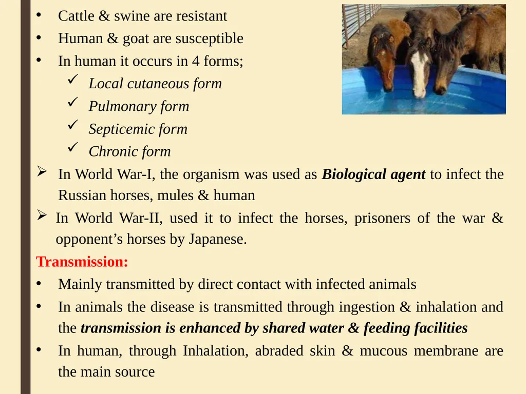
In animals the disease is transmitted through ingestion & inhalation and the transmission is enhanced by shared water & feeding facilities
In human, through Inhalation, abraded skin & mucous membrane are the main source<br>
04
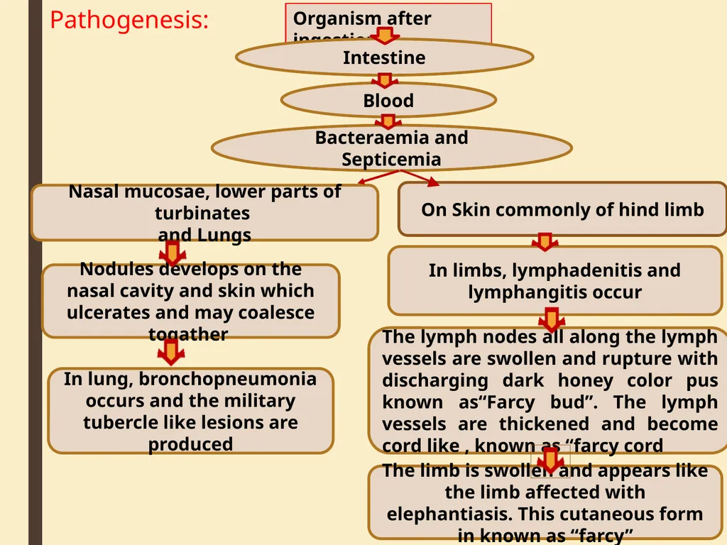
Pathogenesis: Organism after ingestion Nodules develops on the nasal cavity and skin which ulcerates and may coalesce togather In limbs, lymphadenitis and lymphangitis occur The lymph nodes all along the lymph vessels are swollen and rupture with discharging dark honey color pus known as“Farcy bud”. The lymph vessels are thickened and become cord like , known as “farcy cord The limb is swollen and appears like the limb affected with elephantiasis. This cutaneous form in known as “farcy” In lung, bronchopneumonia occurs and the military tubercle like lesions are produced Nasal mucosae, lower parts of turbinates
and Lungs On Skin commonly of hind limb Bacteraemia and Septicemia Blood Intestine<br>
05
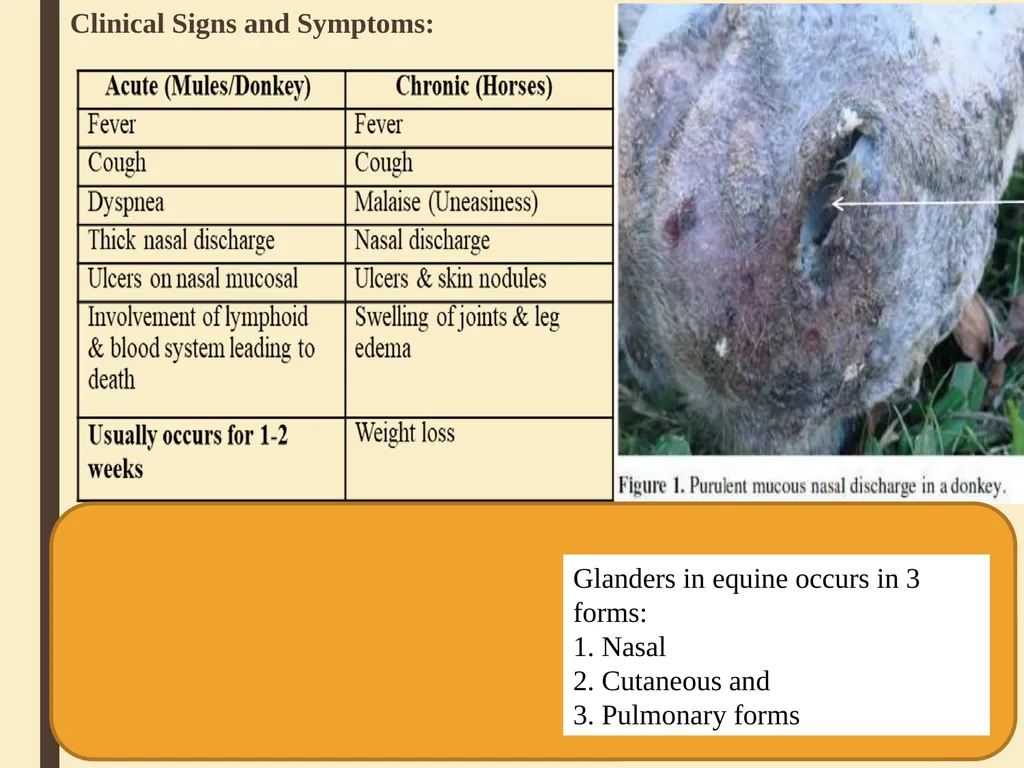
Clinical Signs and Symptoms: Glanders in equine occurs in 3 forms:
1. Nasal
2. Cutaneous and
3. Pulmonary forms<br>
06
Glanders in equine occurs in 3 forms:
1. Nasal
2. Cutaneous and
3. Pulmonary forms
Nasal form:
Characterized by unilateral or bilateral nasal discharge.
The yellowish-green exudate is highly infectious.
The nasal mucosa has nodules and ulcers which may coalesce to form large ulcerated areas.
In some cases the septum may even be perforated.
Nasal lesions are accompanied by enlargement or sometimes rupture and suppuration, of regional lymph nodes
Lesions may also occur in the liver or spleen and, in male animals , glanderous orchitis is a common lesion<br>
07
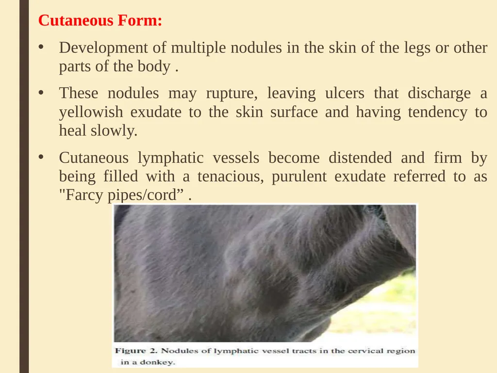
Cutaneous Form:
Development of multiple nodules in the skin of the legs or other parts of the body .
These nodules may rupture, leaving ulcers that discharge a yellowish exudate to the skin surface and having tendency to heal slowly.
Cutaneous lymphatic vessels become distended and firm by being filled with a tenacious, purulent exudate referred to as "Farcy pipes/cord” .<br>
08
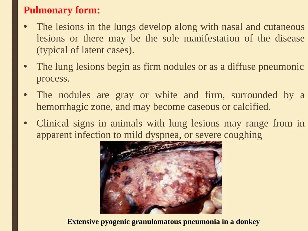
Pulmonary form:
The lesions in the lungs develop along with nasal and cutaneous lesions or there may be the sole manifestation of the disease (typical of latent cases).
The lung lesions begin as firm nodules or as a diffuse pneumonic process.
The nodules are gray or white and firm, surrounded by a hemorrhagic zone, and may become caseous or calcified.
Clinical signs in animals with lung lesions may range from in apparent infection to mild dyspnea, or severe coughing Extensive pyogenic granulomatous pneumonia in a donkey<br>
09
Diagnosis:
Signs & symptoms
Culture & isolation of bacteria
Serological tests :-
Indirect hemagglutination,
Counter-immunoelectrophoresis
Immunofluorescence
Compliment fixation and
ELISA
Compliment fixation and ELISA:
– Most reliable in horses
– Cannot be used in donkey or mule
Mallein test<br>
10
Mallein test:
Mallein is a protein that is not toxic to animals, but animals infected with glanders may become allergic to mallein & exhibit local or systemic hyper- sensitivity reaction after inoculation.
A positive response is indicated by ocular swelling, photosensitivity, lacrimation & suppurative inflammation within 48 hours after injection.
Intradermo-palpebral mallein test used, where 0.1ml of mallein is injected intr-dermally into the lower eye lid by tuberculin syringe and needle and results are recorded after1-2 days later with eyelid swelling, blepharospasms and purulent conjunctivitis.
Treatment:
Cull the affected animals because of zoonotic importance.
If necessary, Antibiotics are used in combination. It is long term treatment up to weeks
Sulphadimidine for 20 days is found highly effective.<br>
11
Prevention & Control
No vaccine is available for animal use.
Quarantine measures (Place and Importation)
Identification and removal of foci of infection through:
Surveillance and monitoring of herds especially in endemic areas
Positive animals should be isolated & slaughtered immediately & carcass should be disposed off by incineration or deep burial
In-contact & exposed animals should be segregated , Re- tested to detect and destroy the positives according to the Glander & Farcy Act ,1899.<br>