HISTOLOGY OF MALE GENITAL SYSTEM Sarangdhar kumar Assistant professor Department of anatomy Rohilkhand medical college Bareilly Male genital system includes Male gonads Testes Duct system Straight tubule Rete testis Efferent ducts
"HISTOLOGY OF MALE GENITAL SYSTEM Sarangdhar kumar" is the property of its rightful owner. Permission is granted to
download and print the materials on this website for personal, non-commercial use only, and to display it
on your personal computer provided you do not modify the materials and that you retain all copyright
notices contained in the materials. By downloading content from our website, you accept the terms of this
agreement.
Presentation Transcript
01
HISTOLOGY OF MALE GENITAL SYSTEM Sarangdhar kumar
Assistant professor
Department of anatomy
Rohilkhand medical college
Bareilly<br>
02
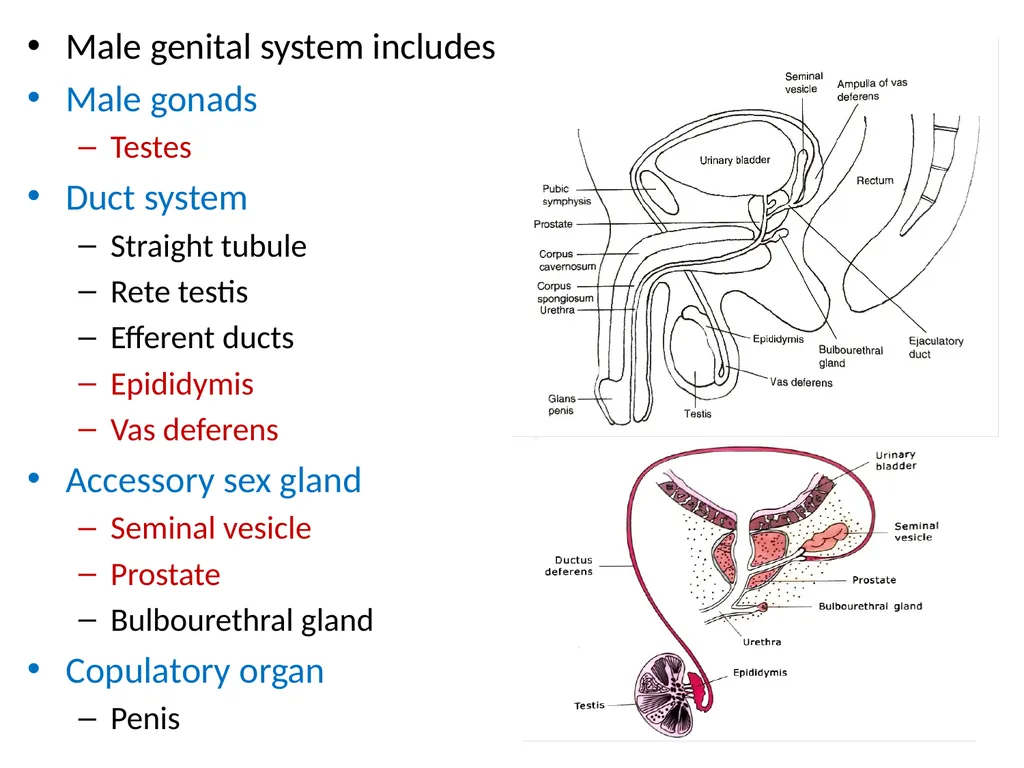
Male genital system includes
Male gonads
Testes
Duct system
Straight tubule
Rete testis
Efferent ducts
Epididymis
Vas deferens
Accessory sex gland
Seminal vesicle
Prostate
Bulbourethral gland
Copulatory organ
Penis<br>
03
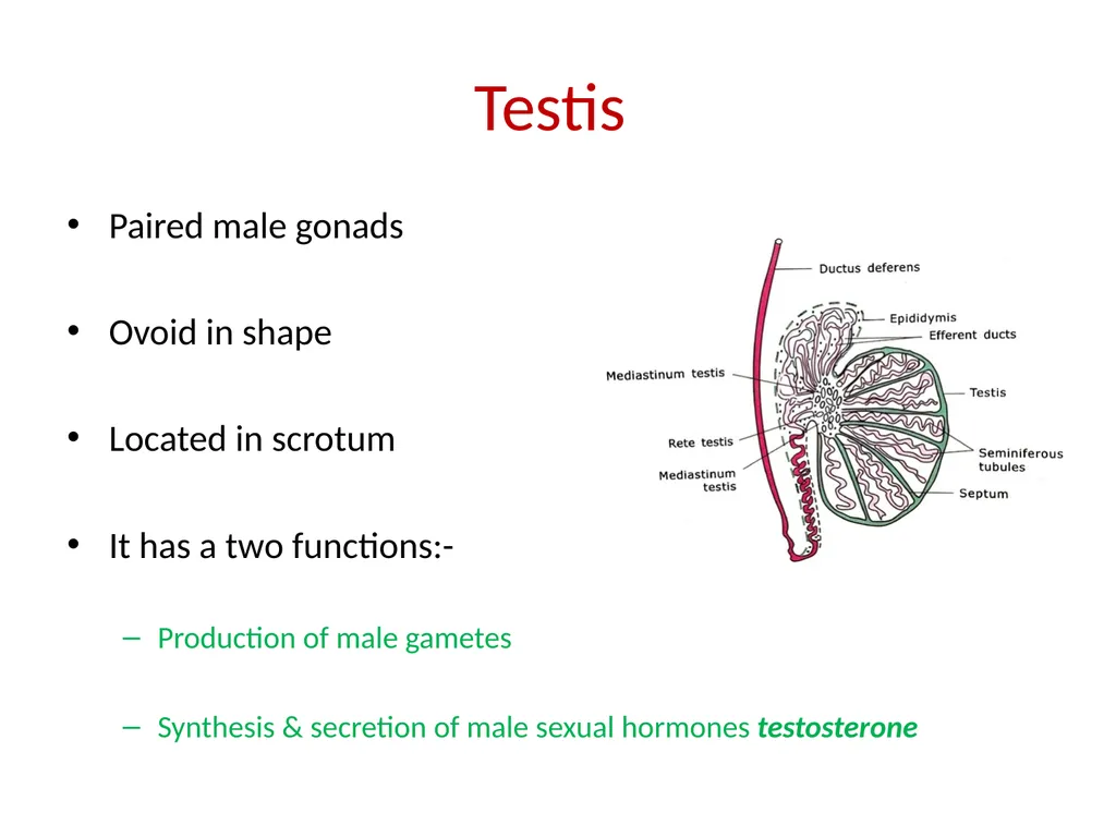
Paired male gonads
Ovoid in shape
Located in scrotum
It has a two functions:-
Production of male gametes
Synthesis & secretion of male sexual hormones testosterone Testis<br>
04
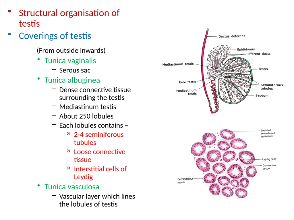
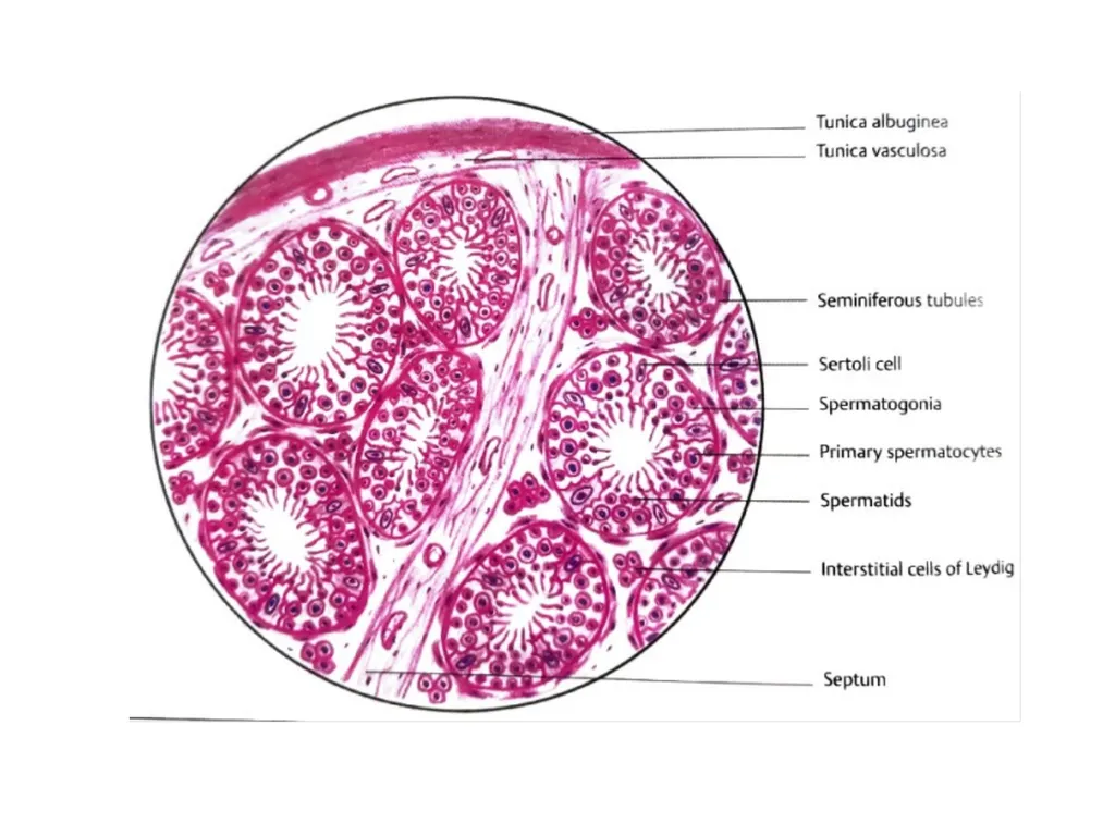
Structural organisation of testis
Coverings of testis
(From outside inwards)
Tunica vaginalis
Serous sac
Tunica albuginea
Dense connective tissue surrounding the testis
Mediastinum testis
About 250 lobules
Each lobules contains –
2-4 seminiferous tubules
Loose connective tissue
Interstitial cells of Leydig
Tunica vasculosa
Vascular layer which lines the lobules of testis<br>
05
Seminiferous tubules
Highly convoluted tubules
Interior of tubules is lined by special type of stratified germinal epithelium(seminiferous epithelium)
Consist of two types of cells
Spermatogenic cells
Differentiate into sperm
Sertoli cells
Supporting cell<br>
06
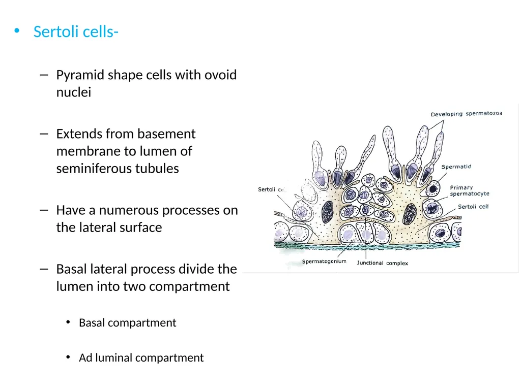
Sertoli cells-
Pyramid shape cells with ovoid nuclei
Extends from basement membrane to lumen of seminiferous tubules
Have a numerous processes on the lateral surface
Basal lateral process divide the lumen into two compartment
Basal compartment
Ad luminal compartment<br>
07
Functions of Sertoli cell
Provide nutrition & mechanical support to spermatogenic cells
Forms blood testis barrier
Phagocytosis of residual bodies
It secretes -
ABP( androgen binding protein)
Inhibin
MIS (mullerian inhibiting substances )
Transferrin (iron transporting molecule)<br>
08
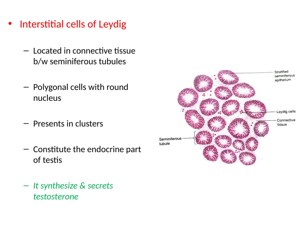
Interstitial cells of Leydig
Located in connective tissue b/w seminiferous tubules
Polygonal cells with round nucleus
Presents in clusters
Constitute the endocrine part of testis
It synthesize & secrets testosterone<br>
09
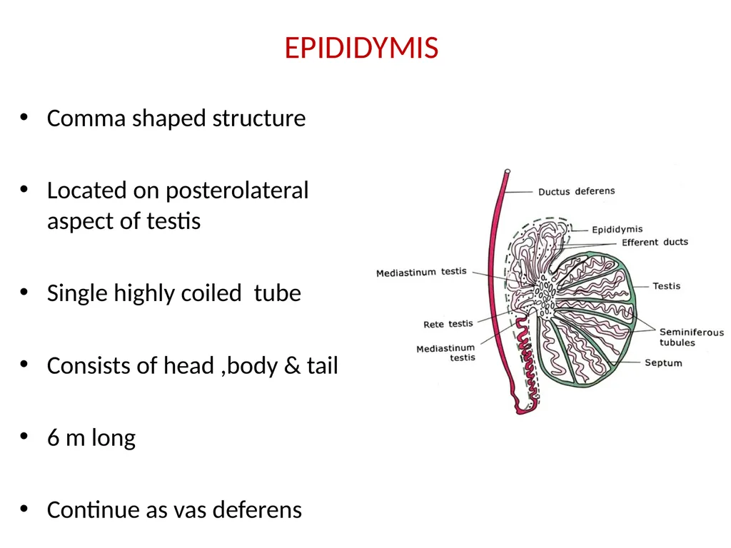
EPIDIDYMIS Comma shaped structure
Located on posterolateral aspect of testis
Single highly coiled tube
Consists of head ,body & tail
6 m long
Continue as vas deferens<br>
10
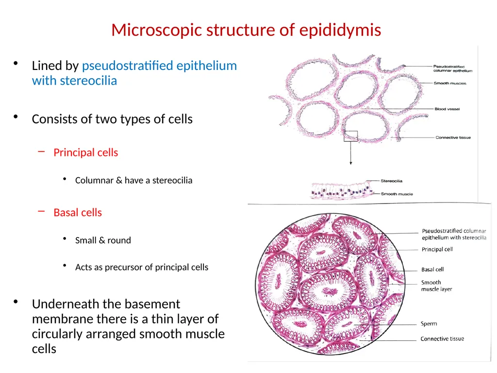
Microscopic structure of epididymis Lined by pseudostratified epithelium with stereocilia
Consists of two types of cells
Principal cells
Columnar & have a stereocilia
Basal cells
Small & round
Acts as precursor of principal cells
Underneath the basement membrane there is a thin layer of circularly arranged smooth muscle cells<br>
11
Functions –
Spermatozoa are stored & make motility
Cells lining the epithelium performs three functions
Absorption
phagocytosis
Secretions
Glycerophosphocholine
Sialic acid
Glycoproteins<br>
12
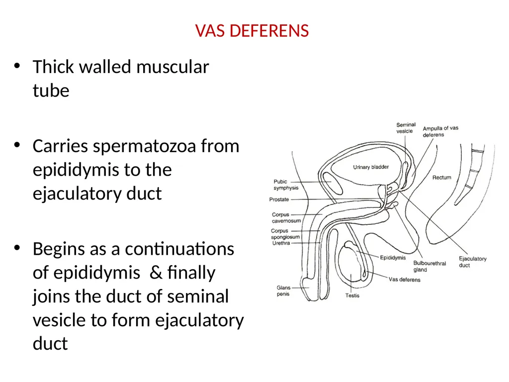
VAS DEFERENS Thick walled muscular tube
Carries spermatozoa from epididymis to the ejaculatory duct
Begins as a continuations of epididymis & finally joins the duct of seminal vesicle to form ejaculatory duct<br>
13
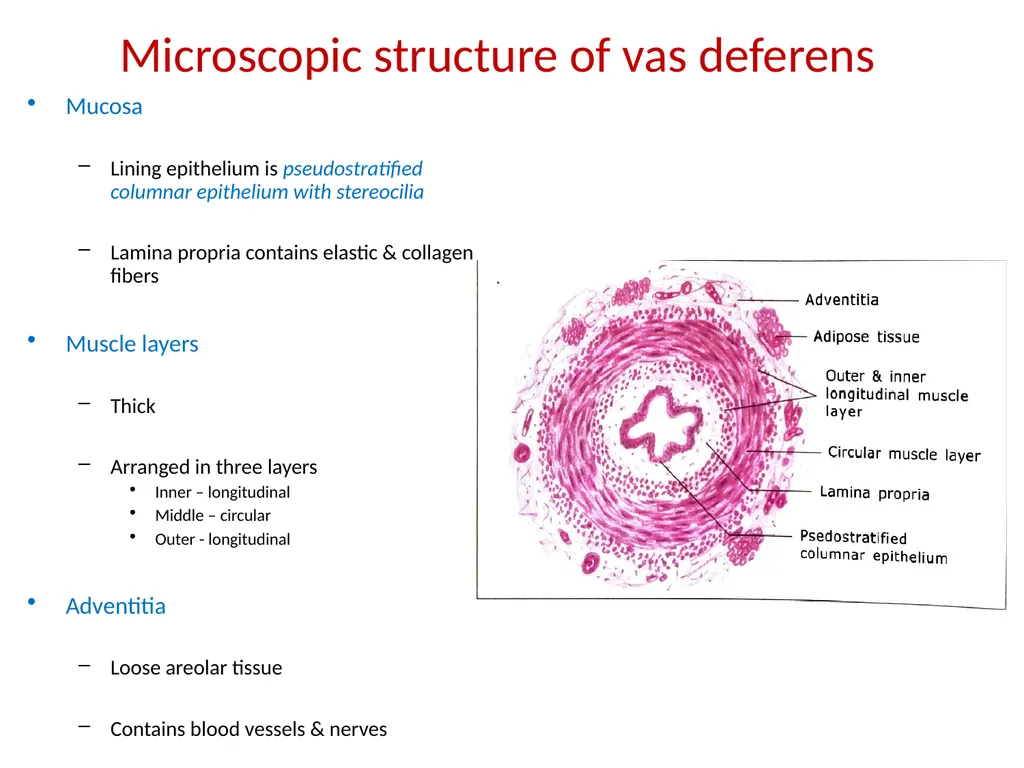
Microscopic structure of vas deferens Mucosa
Lining epithelium is pseudostratified columnar epithelium with stereocilia
Lamina propria contains elastic & collagen fibers
Muscle layers
Thick
Arranged in three layers
Inner – longitudinal
Middle – circular
Outer - longitudinal
Adventitia
Loose areolar tissue
Contains blood vessels & nerves<br>
14
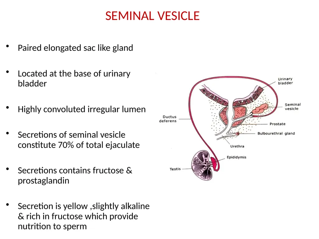
SEMINAL VESICLE Paired elongated sac like gland
Located at the base of urinary bladder
Highly convoluted irregular lumen
Secretions of seminal vesicle constitute 70% of total ejaculate
Secretions contains fructose & prostaglandin
Secretion is yellow ,slightly alkaline & rich in fructose which provide nutrition to sperm<br>
15
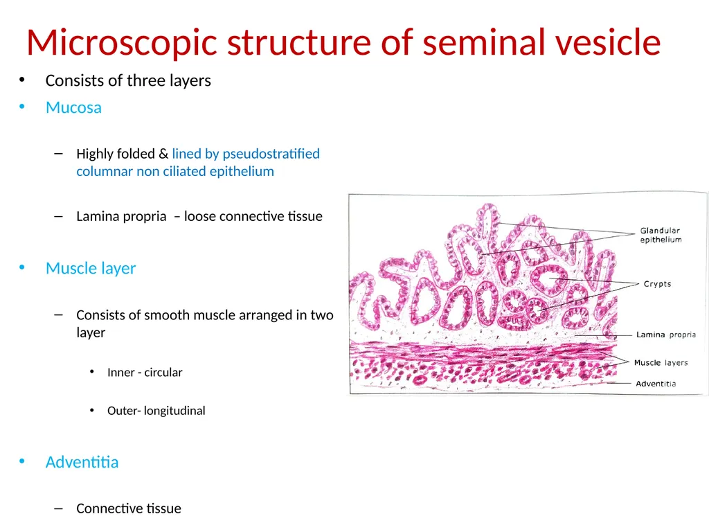
Microscopic structure of seminal vesicle Consists of three layers
Mucosa
Highly folded & lined by pseudostratified columnar non ciliated epithelium
Lamina propria – loose connective tissue
Muscle layer
Consists of smooth muscle arranged in two layer
Inner - circular
Outer- longitudinal
Adventitia
Connective tissue<br>
16
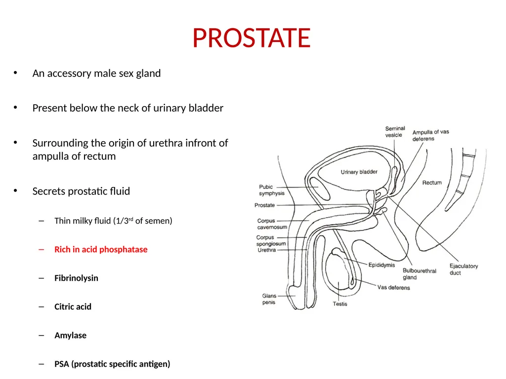
PROSTATE An accessory male sex gland
Present below the neck of urinary bladder
Surrounding the origin of urethra infront of ampulla of rectum
Secrets prostatic fluid
Thin milky fluid (1/3rd of semen)
Rich in acid phosphatase
Fibrinolysin
Citric acid
Amylase
PSA (prostatic specific antigen)<br>
17
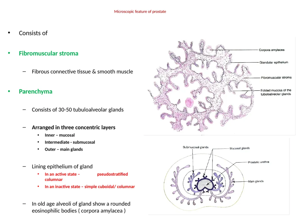
Microscopic feature of prostate Consists of
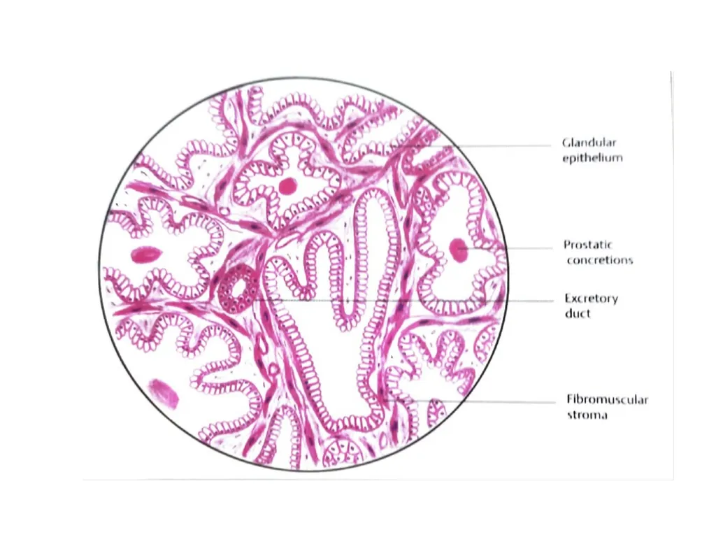
Fibromuscular stroma
Fibrous connective tissue & smooth muscle
Parenchyma
Consists of 30-50 tubuloalveolar glands
Arranged in three concentric layers
Inner – mucosal
Intermediate - submucosal
Outer – main glands
Lining epithelium of gland
In an active state – pseudostratified columnar
In an inactive state – simple cuboidal/ columnar
In old age alveoli of gland show a rounded eosinophilic bodies ( corpora amylacea )<br>
18
Ducts
10-12 in number
Lined by simple columnar epithelium which changes to a transitional epithelium in the terminal part<br>