Histology of Ovary Jaydeep Kumar Nath Asst Professor Department of Zoology Salbari College , Baksa The ovary The ovaries are the female gonads that produces ova. ovaries are present on the right and left sides of the uterus in lower
"Histology of Ovary Jaydeep Kumar Nath Asst" is the property of its rightful owner. Permission is granted to
download and print the materials on this website for personal, non-commercial use only, and to display it
on your personal computer provided you do not modify the materials and that you retain all copyright
notices contained in the materials. By downloading content from our website, you accept the terms of this
agreement.
Presentation Transcript
01
Histology of Ovary Jaydeep Kumar Nath
Asst Professor
Department of Zoology
Salbari College , Baksa<br>
02
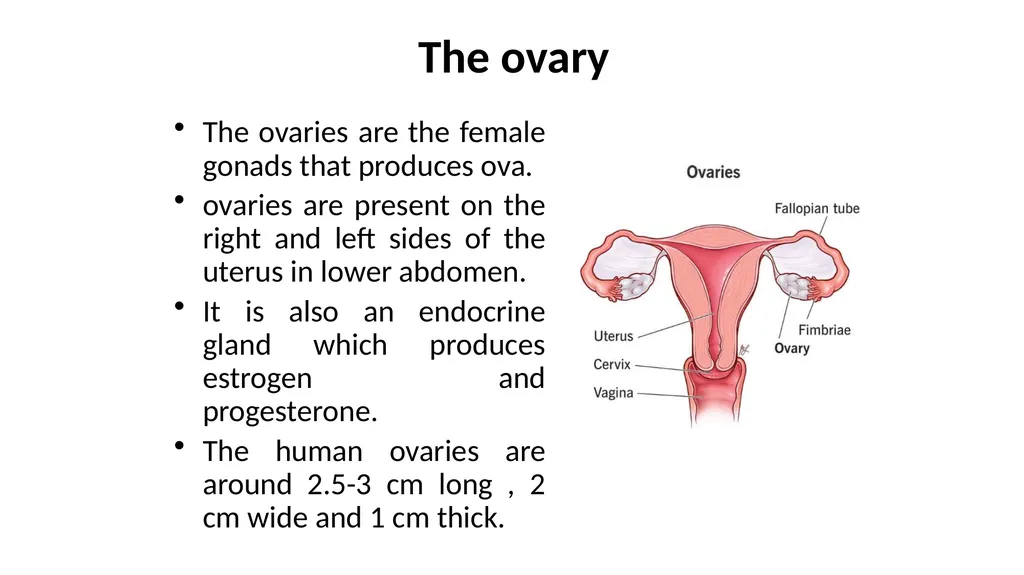
The ovary The ovaries are the female gonads that produces ova.
ovaries are present on the right and left sides of the uterus in lower abdomen.
It is also an endocrine gland which produces estrogen and progesterone.
The human ovaries are around 2.5-3 cm long , 2 cm wide and 1 cm thick.<br>
03
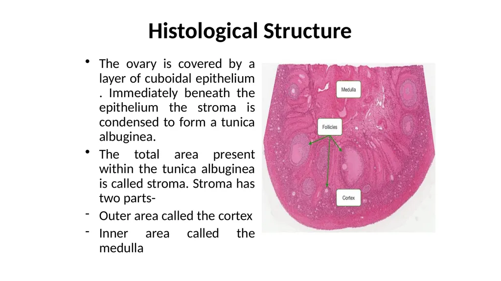
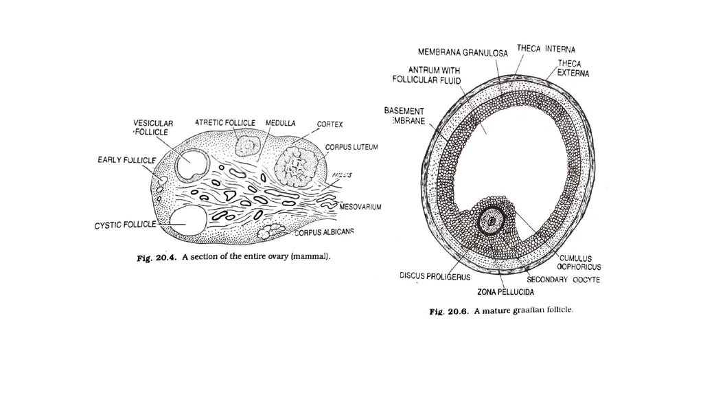
Histological Structure The ovary is covered by a layer of cuboidal epithelium . Immediately beneath the epithelium the stroma is condensed to form a tunica albuginea.
The total area present within the tunica albuginea is called stroma. Stroma has two parts-
Outer area called the cortex
Inner area called the medulla<br>
04
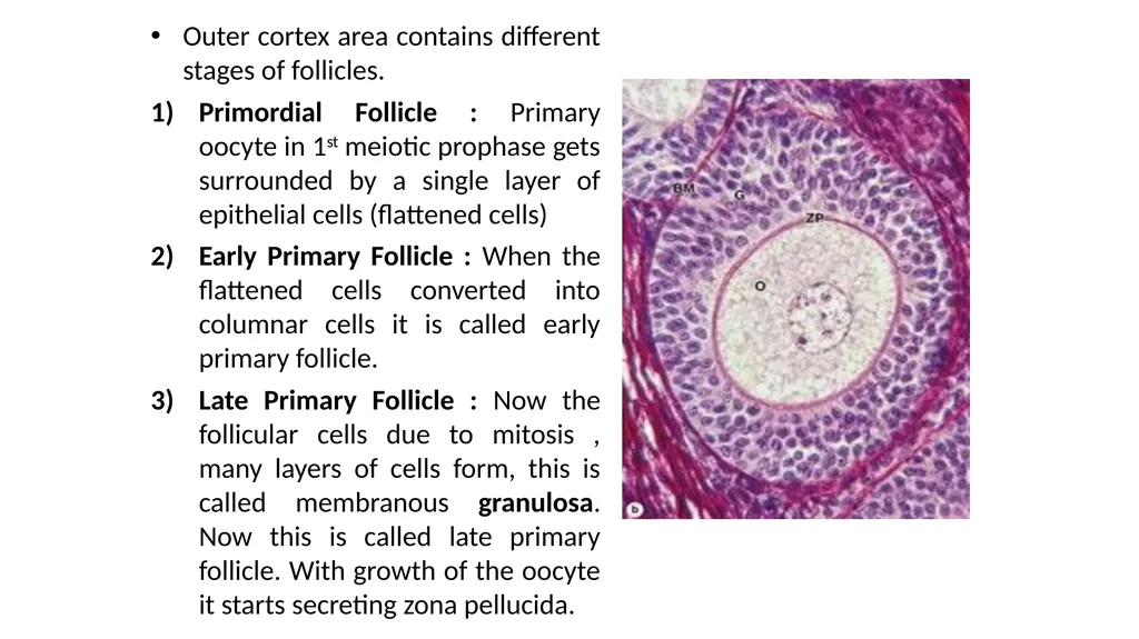
Outer cortex area contains different stages of follicles.
Primordial Follicle : Primary oocyte in 1st meiotic prophase gets surrounded by a single layer of epithelial cells (flattened cells)
Early Primary Follicle : When the flattened cells converted into columnar cells it is called early primary follicle.
Late Primary Follicle : Now the follicular cells due to mitosis , many layers of cells form, this is called membranous granulosa. Now this is called late primary follicle. With growth of the oocyte it starts secreting zona pellucida.<br>
05
4) Secondary Follicle : More layers of follicles are arranged and there is a formation of many small space in granulosa cell layer.
5) Tertiary Follicle : After some time all the space join to form a big space called follicular antrum.
* With further growth , surrounding connective tissue stromal cells specialize forming a sheath theca folliculi
Fibroblasts outside the growing follicles have developed as a steroid secreting theca interna.
Outside the theca interna , another layer of fibrous connective tissue layers form called as theca externa.<br>
06
Mature Graffian follicle
The outermost layer of a mature graaflan follicle is called the theca externa which consists of concentrically-arranged fibres and fusiform cells. Inside the theca externa the cells appear large and ovoid or spindle-shaped and are known as the theca interna. This layer is thought to secrete estrogens and its cells show the typical features associated with cells secreting steroids
Follicles about 0.2 mm in diameter begin to collect pools of fluids. This follicular fluid called liquor folliculi is rich in mucoproteins and is secreted by the follicle cells. It comes to occupy a single cavity, the antrum.
The fluid pushes the ovum and its neighbouring follicle cells, to one side. A peripheral shell consisting of many layers of cuboidal follicle cells surrounds the antrum. It is known as stratum granulosum.
The mass of granulosa cells enclosing the mature ovum projects into the antrum, forming a hillock, the cumulus oophorous.
The ovum is surrounded by the prominent zona pellucida.
The corona radiata is a well-defined, radially arranged layer, of columnar cells immediately surrounding the zona pellucida. The cells of the carona radiata are peeled off from the zona pellucida when the oocyte passes down the oviduct following ovulation.<br>