
Human Reproduction BIOLOGY ,CHAPTER 03,CLASS-XII
KL
Published · 69 slides · 0 views

1 / 1


Description
Human Reproduction BIOLOGY ,CHAPTER 03,CLASS-XII Each sex has pair of gonads, reproductive duct and accessory structures. Sex organs Testis( paired) male , ovaries (paired) Gamete formation, hormones Puberty Gametogenesis - formation of
Related Topics
Share
Embed code
Download this presentation From Below
"Human Reproduction BIOLOGY ,CHAPTER 03,CLASS-XII" is the property of its rightful owner. Permission is granted to download and print the materials on this website for personal, non-commercial use only, and to display it on your personal computer provided you do not modify the materials and that you retain all copyright notices contained in the materials. By downloading content from our website, you accept the terms of this agreement.
Presentation Transcript
01
Human Reproduction BIOLOGY ,CHAPTER 03,CLASS-XII<br>
02
Each sex has pair of gonads, reproductive duct and accessory structures.
Sex organs – Testis( paired) male , ovaries (paired) Gamete formation,
hormones
Puberty
Gametogenesis - formation of gametes (sperms/ ova)
Insemination -- transfer of sperms into the female genital tract
Fertilisation -- fusion of male and female gametes leading to
formation of zygote
Implantation -- development of blastocyst and its attachment to the uterine wall
Gestation -- embryonic development ( from conception to
birth )
Parturition-- delivery of the baby ( child birth ) REPRODUCTIVE EVENTS<br>
03
Male rproductive system<br>
04
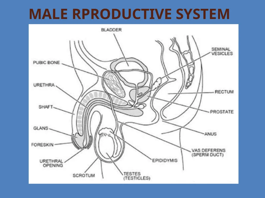
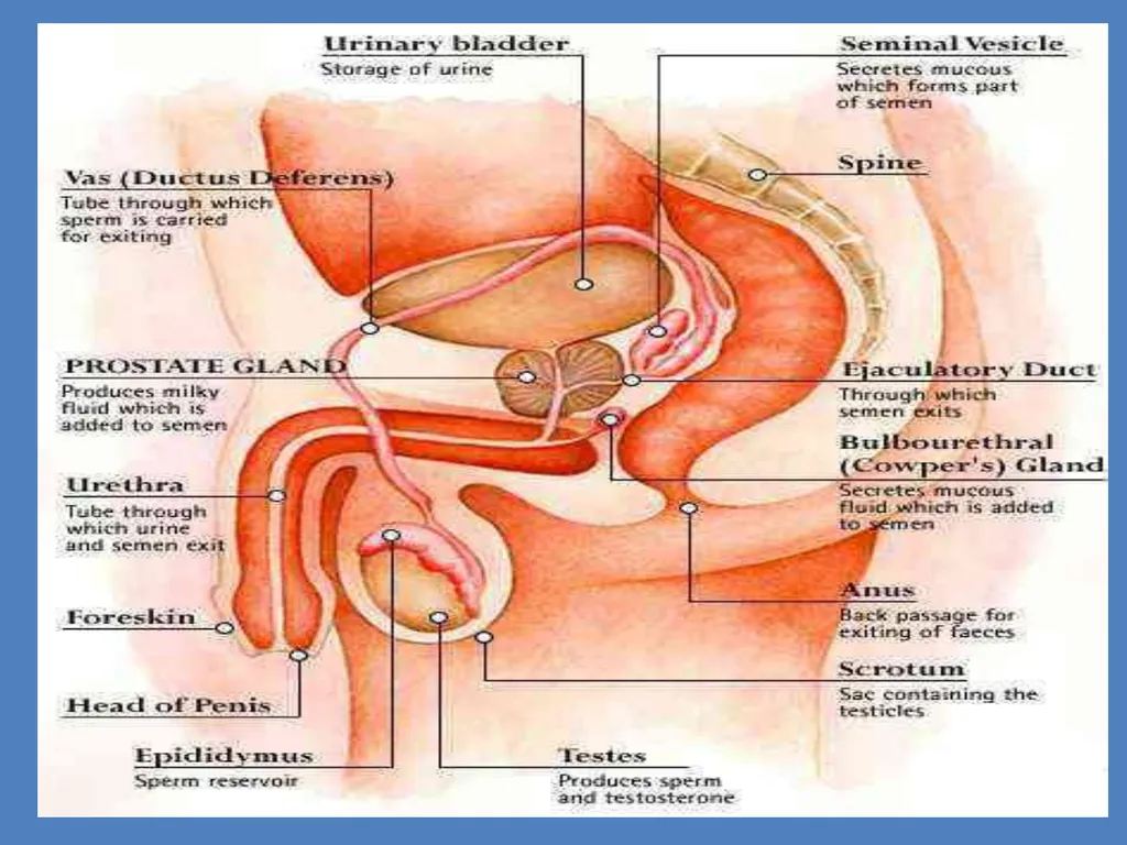
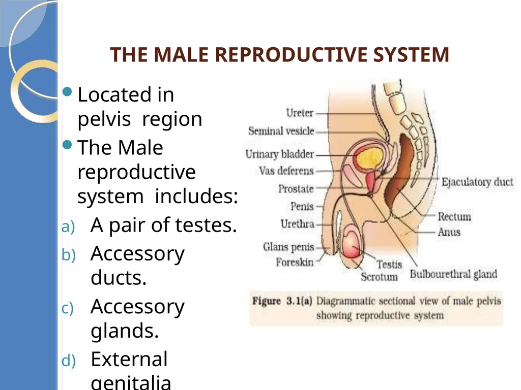
Located in pelvis region
The Male reproductive system includes:
A pair of testes.
Accessory ducts.
Accessory glands.
External genitalia THE MALE REPRODUCTIVE SYSTEM<br>
The Male reproductive system includes:
A pair of testes.
Accessory ducts.
Accessory glands.
External genitalia THE MALE REPRODUCTIVE SYSTEM<br>
05
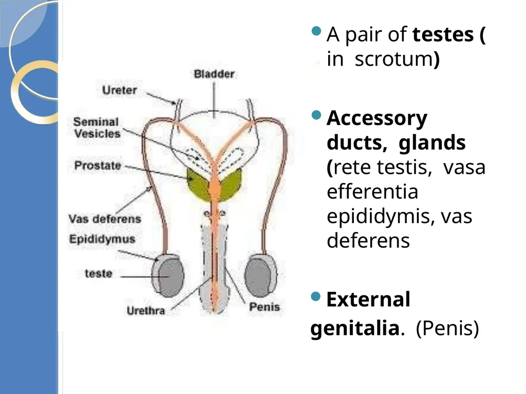
A pair of testes ( in scrotum)
Accessory ducts, glands (rete testis, vasa efferentia epididymis, vas deferens
External genitalia. (Penis)<br>
Accessory ducts, glands (rete testis, vasa efferentia epididymis, vas deferens
External genitalia. (Penis)<br>
06
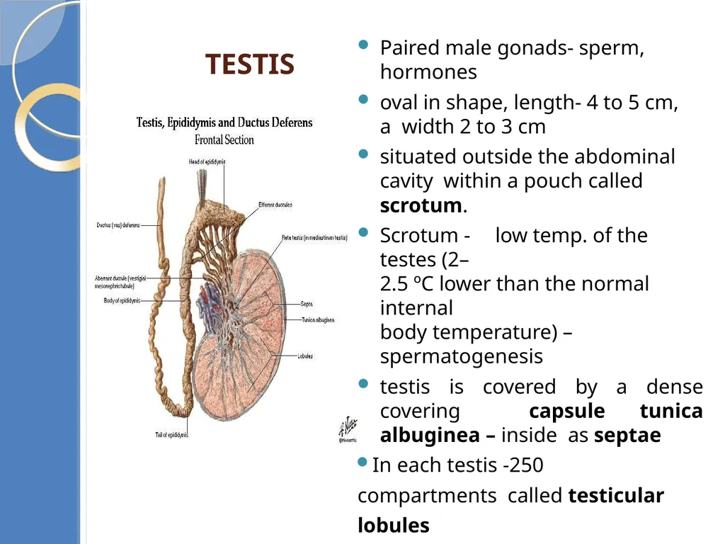
Testis Paired male gonads- sperm, hormones
oval in shape, length- 4 to 5 cm, a width 2 to 3 cm
situated outside the abdominal cavity within a pouch called scrotum.
Scrotum - low temp. of the testes (2–
2.5 ºC lower than the normal internal
body temperature) – spermatogenesis
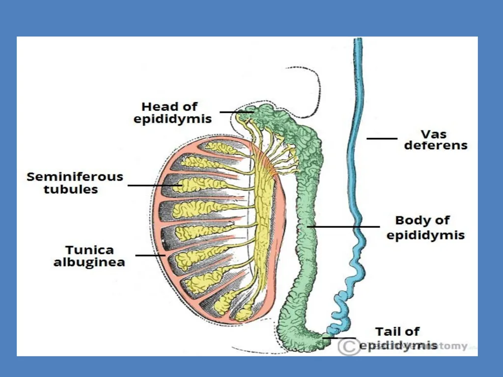
testis is covered by a dense covering capsule tunica albuginea – inside as septae
In each testis -250 compartments called testicular lobules
Each lobule -1-3 convoluted (coiled)
seminiferous tubule<br>
07
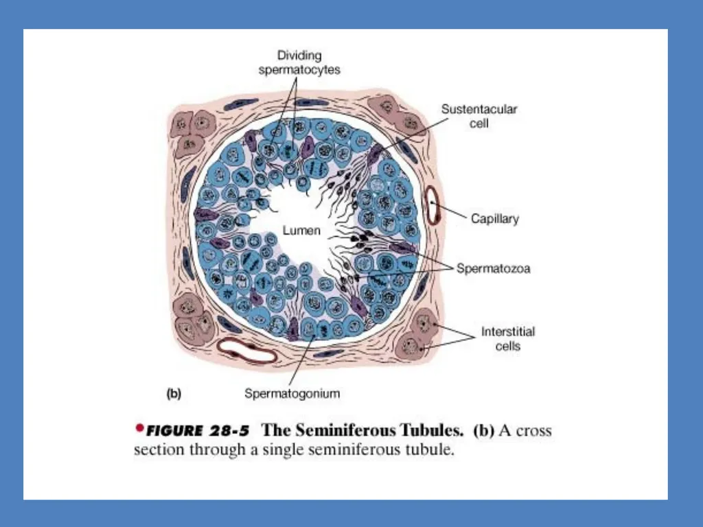
Seminiferous tubule - sperm production Lined on its inside by two types of cells
Male germ cells (spermatogonia) - meiotic divisions - sperm formation
Sertoli cells (supporting cells)
-- provide nutrition to the germ cell
Regions outside the seminiferous tubules called Interstitial spaces, contain small blood vessels & interstitial cells/ Leydig cells
Leydig cells - synthesise & secrete male hormone Androgen ( testosterone)
Other immunologically competent cells are also present<br>
08
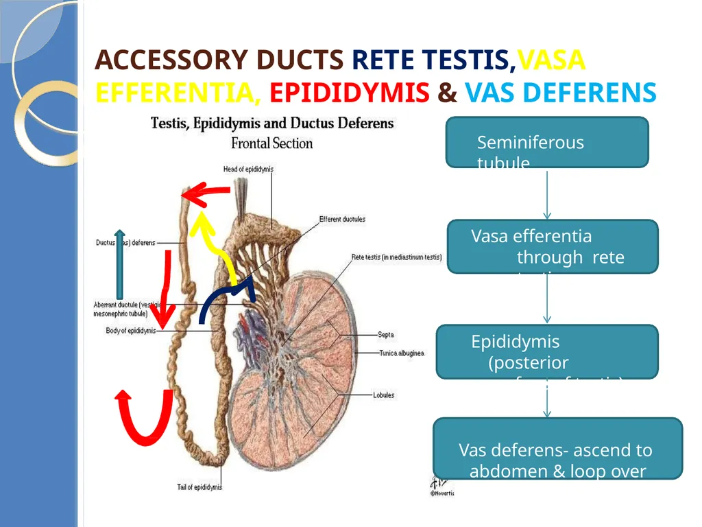
Accessory ducts rete testis,vasa efferentia, epididymis & vas deferens Seminiferous tubule Vasa efferentia through rete testis Epididymis (posterior surface of testis)
Vas deferens- ascend to abdomen & loop over urinary bladder<br>
Vas deferens- ascend to abdomen & loop over urinary bladder<br>
09
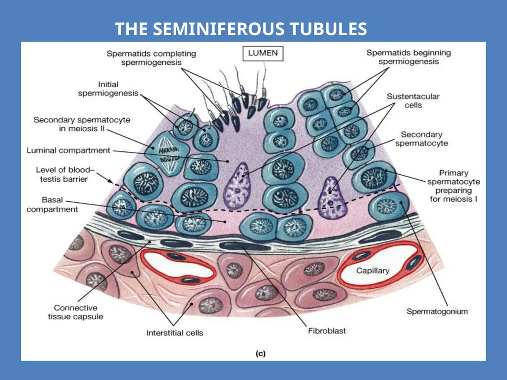
The Seminiferous Tubules<br>
10
Vas deferens receives duct- seminal vesicle
Opens into urethra as
Ejaculatory duct
Function- Stores & transports sperms from testis to urethra
Urethra- originates from urinary bladder, extends through penis to external opening-
urethral meatus<br>
Opens into urethra as
Ejaculatory duct
Function- Stores & transports sperms from testis to urethra
Urethra- originates from urinary bladder, extends through penis to external opening-
urethral meatus<br>
11
External genitalia- Penis External copulatory organ – external genitalia
Made of special tissue- erection to facilitate insemination
Enlarge distal end glans penis covered by loose skin called fore
skin
Accessory Glands
Seminal vesicles (paired)
A Prostate gland
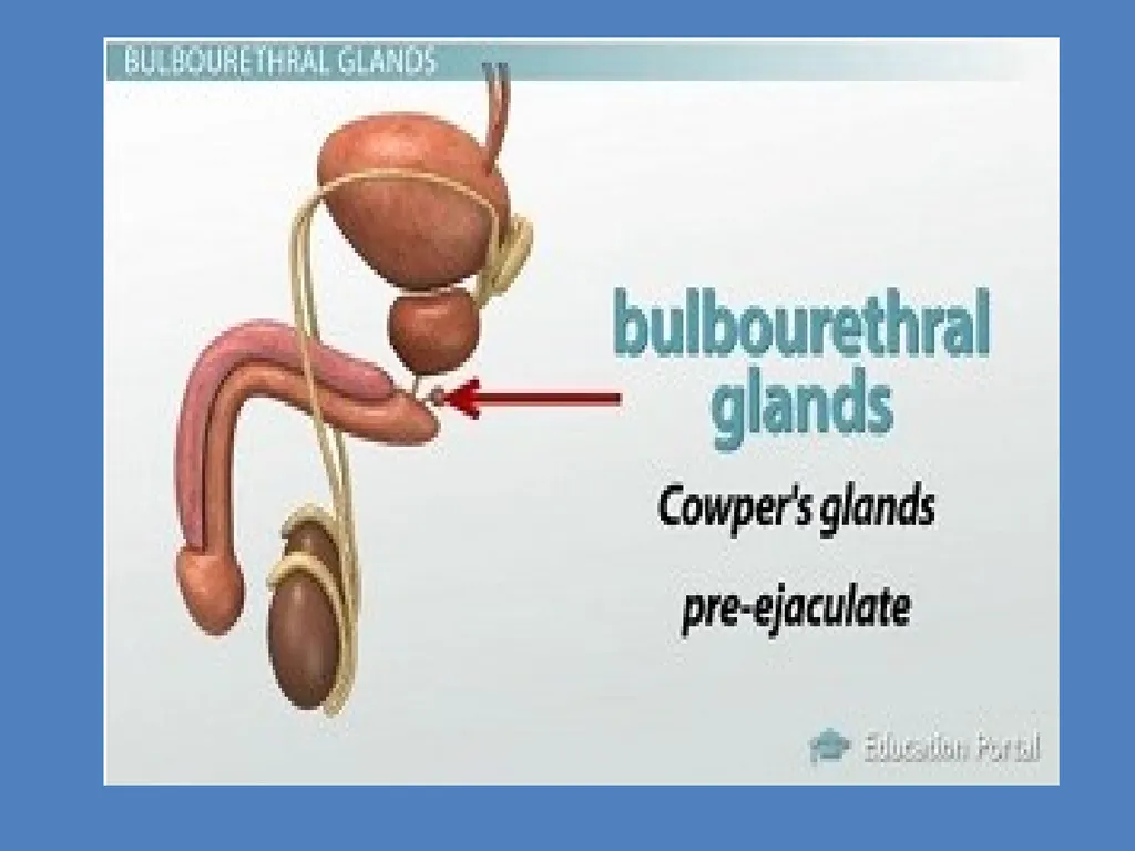
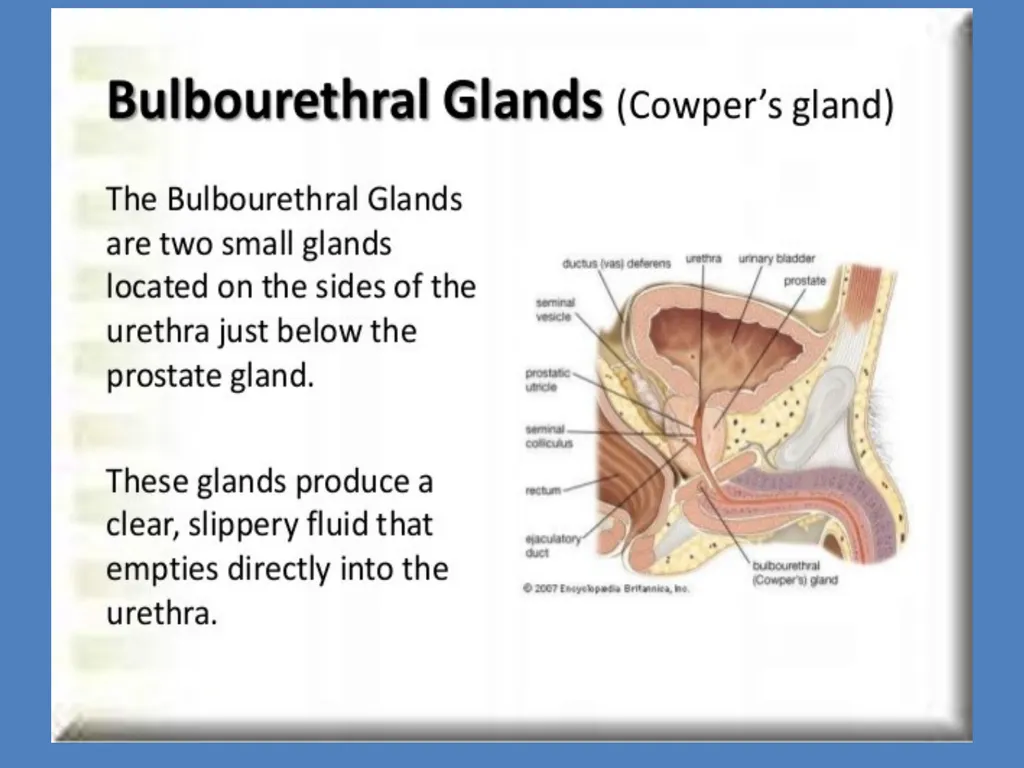
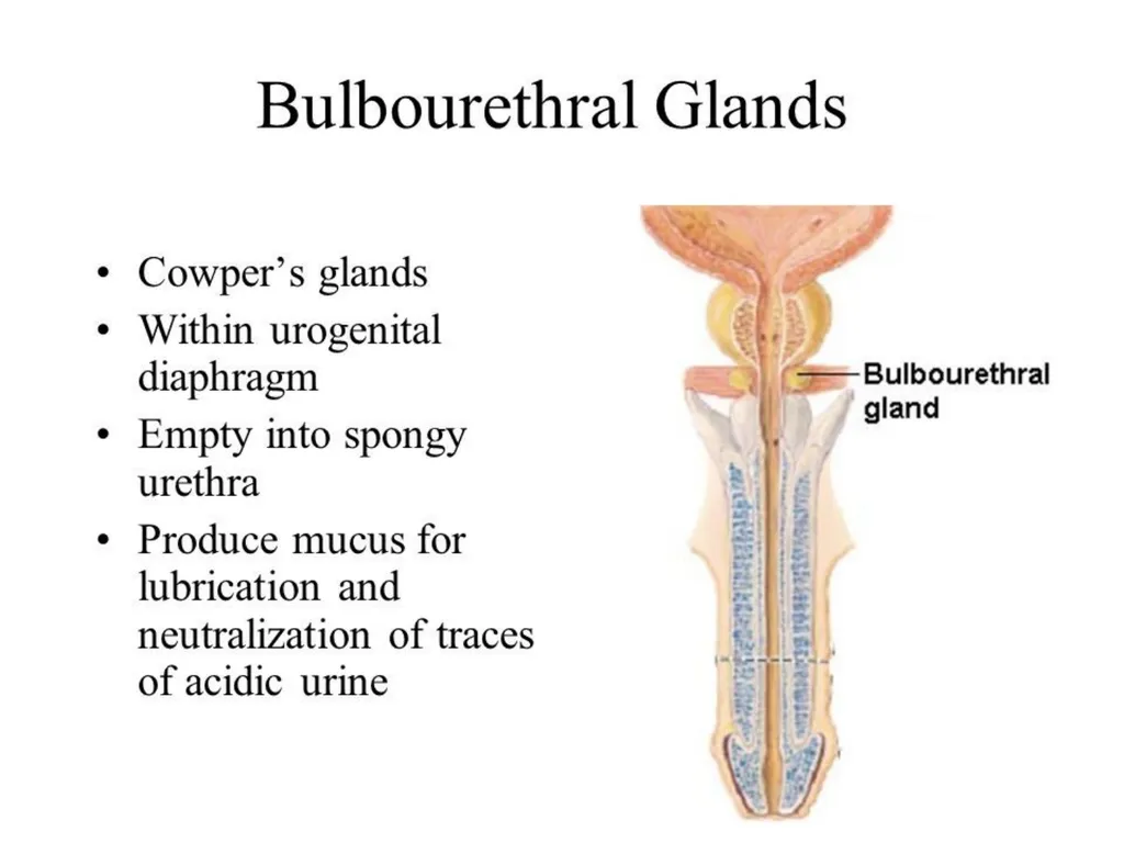
Bulbourethral glands (paired)
Seminal plasma- secretion of all the accessory glands.
Rich in fructose, calcium and certain enzymes.
Helps in lubrication<br>
12
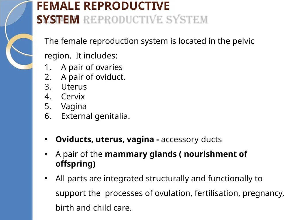
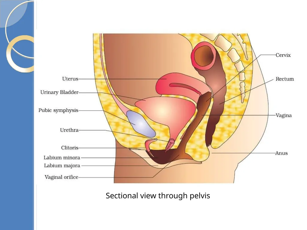
Female reproductive system The female reproduction system is located in the pelvic region. It includes:
A pair of ovaries
A pair of oviduct.
Uterus
Cervix
Vagina
External genitalia.
Oviducts, uterus, vagina - accessory ducts
A pair of the mammary glands ( nourishment of offspring)
All parts are integrated structurally and functionally to support the processes of ovulation, fertilisation, pregnancy, birth and child care.<br>
13
Sectional view through pelvis<br>
14
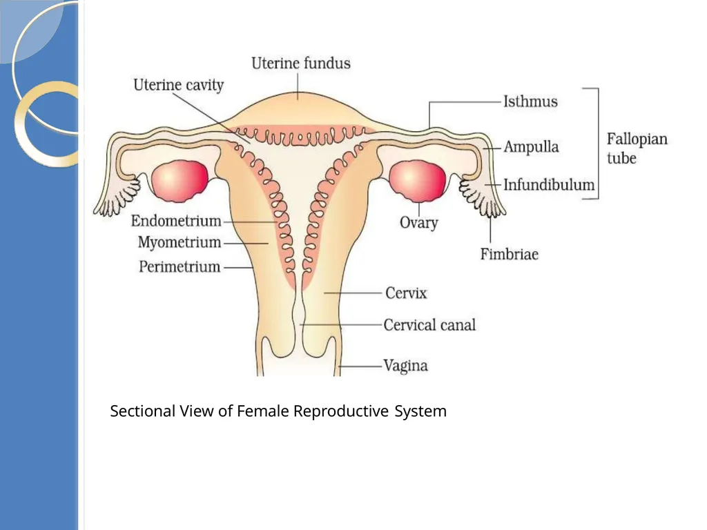
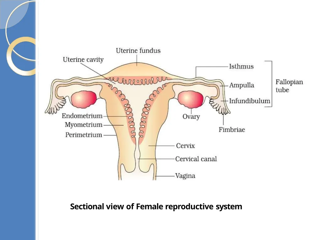
Sectional View of Female Reproductive System<br>
15
Female Gonad: Ovaries Ovaries are the primary female sex organs that produce the female gamete (ovum).
It also produces several female steroid hormones- estrogen &
progesterone
The ovaries located in the lower abdomen.
Each ovary is about 2-4 cm in length.These are Connected to the pelvic wall and uterus by ligaments.
Each ovary is covered by thin epithelium which enclose ovarian stroma
Ovarian Stroma, 2 zones- A peripheral cortex & an inner medulla.
At the peripheral cortex follicles are present & in medulla blood vessels & ovarian ligaments are present<br>
16
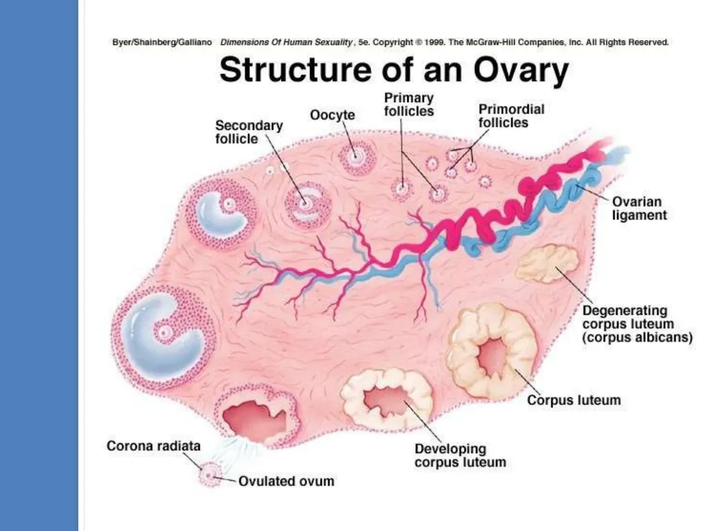
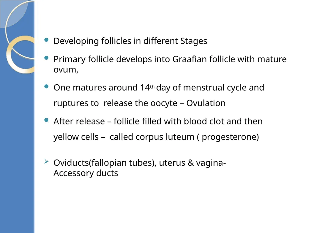
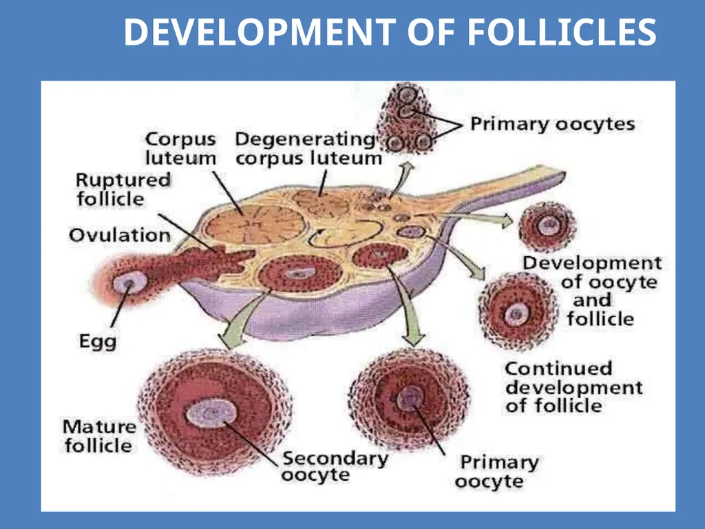
T.S of Ovaries: Developing follicles in different Stages
Primary follicle develops into Graafian follicle with mature ovum,
One matures around 14th day of menstrual cycle and ruptures to release the oocyte – Ovulation
After release – follicle filled with blood clot and then yellow cells – called corpus luteum ( progesterone) Oviducts(fallopian tubes), uterus & vagina- Accessory ducts<br>
17
Sectional view of Female reproductive system<br>
18
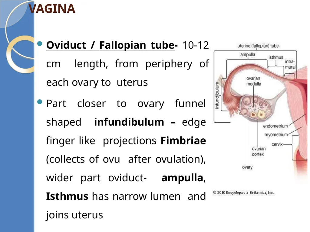
Accessory ducts - Oviduct, Uterus, Vagina Oviduct / Fallopian tube- 10-12 cm length, from periphery of each ovary to uterus
Part closer to ovary funnel shaped infundibulum – edge finger like projections Fimbriae (collects of ovu after ovulation), wider part oviduct- ampulla, Isthmus has narrow lumen and joins uterus<br>
Part closer to ovary funnel shaped infundibulum – edge finger like projections Fimbriae (collects of ovu after ovulation), wider part oviduct- ampulla, Isthmus has narrow lumen and joins uterus<br>
19
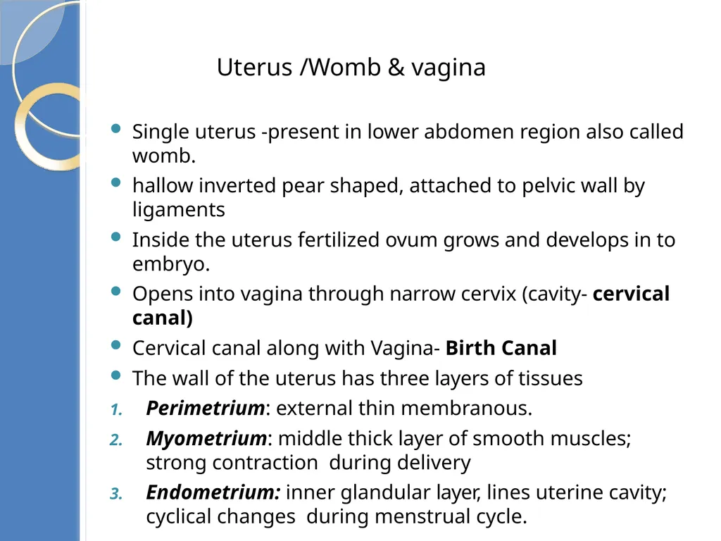
Single uterus -present in lower abdomen region also called womb.
hallow inverted pear shaped, attached to pelvic wall by ligaments
Inside the uterus fertilized ovum grows and develops in to embryo.
Opens into vagina through narrow cervix (cavity- cervical canal)
Cervical canal along with Vagina- Birth Canal
The wall of the uterus has three layers of tissues
Perimetrium: external thin membranous.
Myometrium: middle thick layer of smooth muscles; strong contraction during delivery
Endometrium: inner glandular layer, lines uterine cavity; cyclical changes during menstrual cycle. Uterus /Womb & vagina<br>
20
Mons pubis, labia majora, labia minora, hymen, clitoris
Mons pubis a pad of fatty tissue covered with hair
labia majora - fleshy folds of tissue, surround the vaginal opening.
labia minora – paired folds of tissue under labia majora
Clitoris- tiny finger structure, lies at the upper junction of two labia majora
Hymen - just inside the opening of the vagina, often torn during the first coitus (intercourse)
The presence or absence of hymen is not a reliable indicator of virginity External Genitalia<br>
21
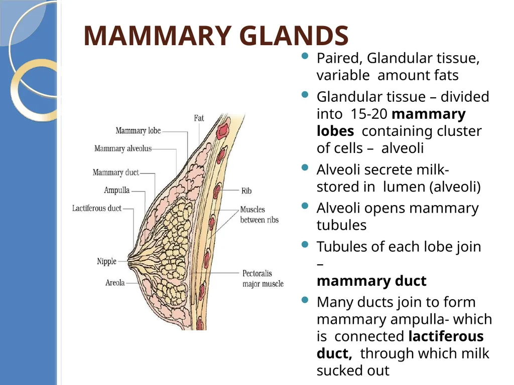
Mammary glands Paired, Glandular tissue, variable amount fats
Glandular tissue – divided into 15-20 mammary lobes containing cluster of cells – alveoli
Alveoli secrete milk- stored in lumen (alveoli)
Alveoli opens mammary tubules
Tubules of each lobe join –
mammary duct
Many ducts join to form mammary ampulla- which is connected lactiferous duct, through which milk sucked out<br>
22
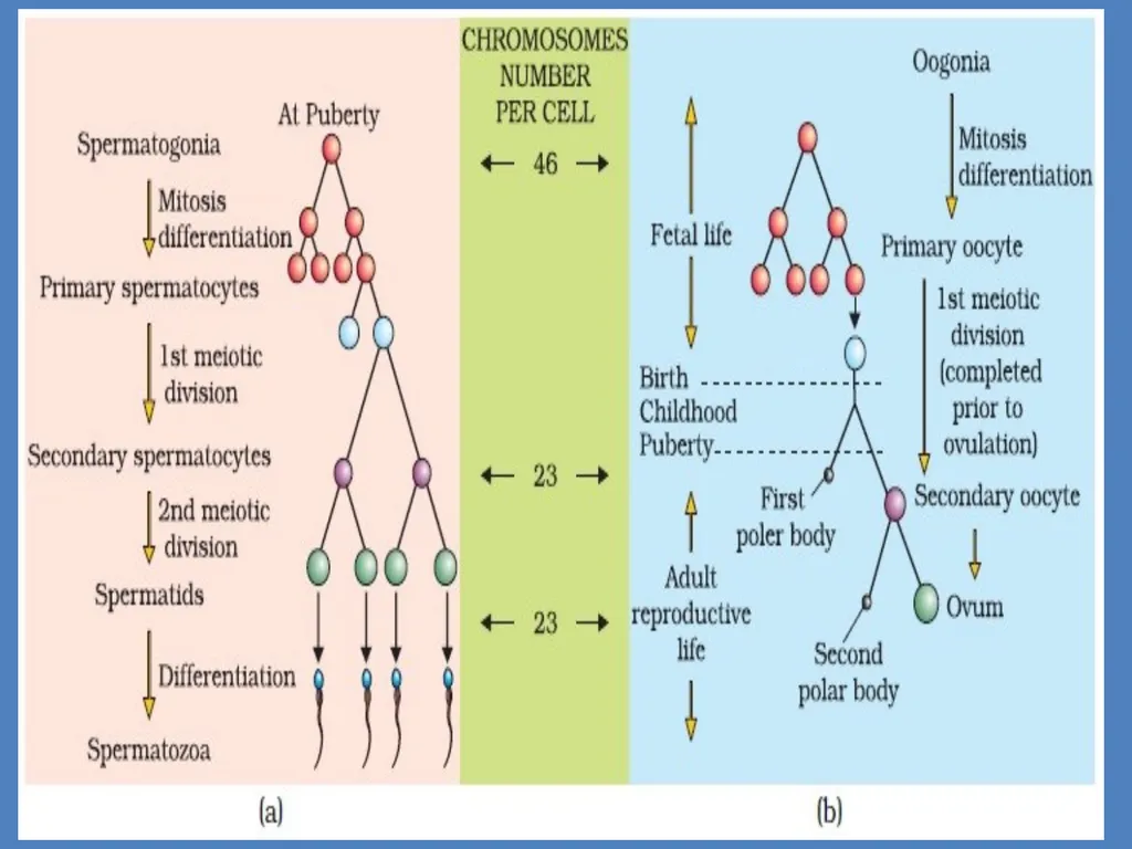
GAMETOGENESIS The process of formation of haploid gametes from diploid germ cells in the gonad is called gametogenesis.
Takes place- primary sex organ (testis & ovary) & produce sperm & ovum
Male – Spermatogenesis (Spermatogonia) & begins at puberty
Female- Oogenesis (Oogonia) & starts at embryonic stage<br>
Takes place- primary sex organ (testis & ovary) & produce sperm & ovum
Male – Spermatogenesis (Spermatogonia) & begins at puberty
Female- Oogenesis (Oogonia) & starts at embryonic stage<br>
23
Spermatogenesis: The process of formation of haploid male gamete sperm in seminiferous tubules of testis is called spermatogenesis.
The inner wall of the seminiferous tubule contains two types of cells as germ cells (spermatogonia cells) and sertoli cells.
germ cells divides and develops into sperms, sertoli cell nourishes the
developing sperms.
The spermatogenesis takes place in two stages as
Spermatidogenesis and Spermiogenesis<br>
24
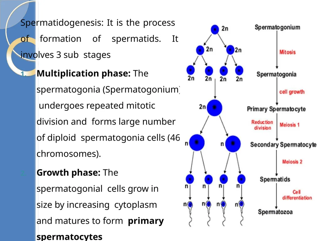
Spermatidogenesis: It is the process of formation of spermatids. It involves 3 sub stages
Multiplication phase: The spermatogonia (Spermatogonium) undergoes repeated mitotic division and forms large number of diploid spermatogonia cells (46 chromosomes).
Growth phase: The spermatogonial cells grow in size by increasing cytoplasm and matures to form primary spermatocytes<br>
Multiplication phase: The spermatogonia (Spermatogonium) undergoes repeated mitotic division and forms large number of diploid spermatogonia cells (46 chromosomes).
Growth phase: The spermatogonial cells grow in size by increasing cytoplasm and matures to form primary spermatocytes<br>
25
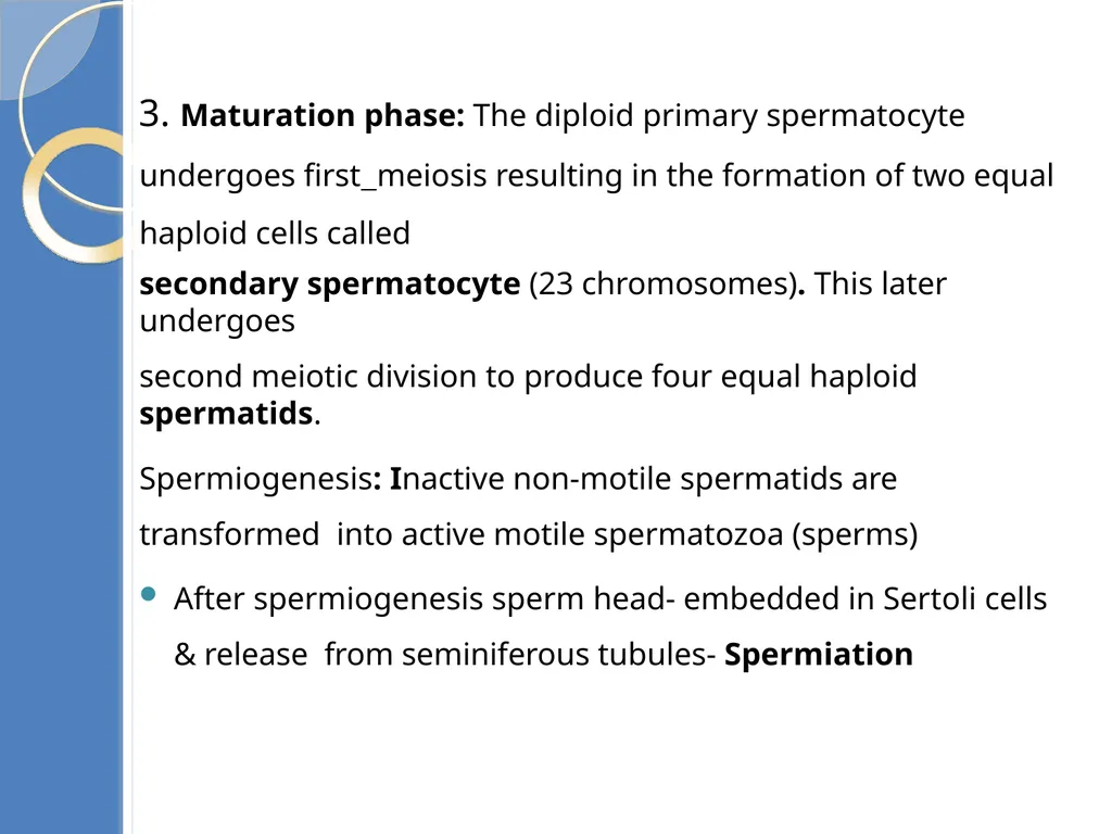
3. Maturation phase: The diploid primary spermatocyte undergoes first meiosis resulting in the formation of two equal haploid cells called
secondary spermatocyte (23 chromosomes). This later undergoes
second meiotic division to produce four equal haploid spermatids.
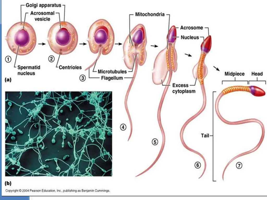
Spermiogenesis: Inactive non-motile spermatids are transformed into active motile spermatozoa (sperms)
After spermiogenesis sperm head- embedded in Sertoli cells & release from seminiferous tubules- Spermiation<br>
26
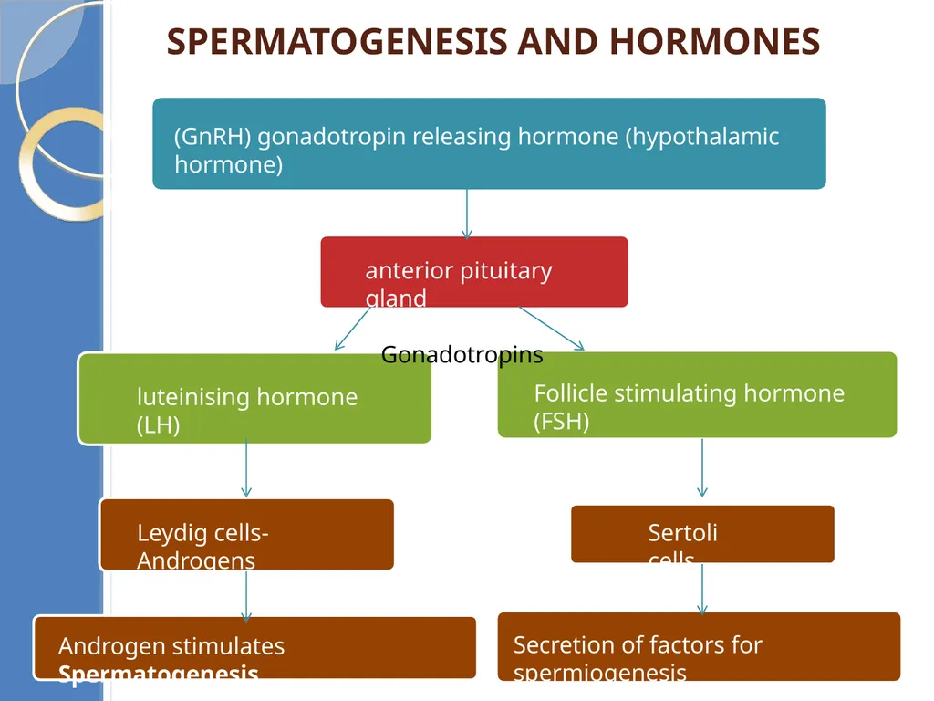
Spermatogenesis and hormones (GnRH) gonadotropin releasing hormone (hypothalamic hormone) luteinising hormone (LH) Leydig cells- Androgens Androgen stimulates Spermatogenesis Follicle stimulating hormone (FSH) Sertoli cells Secretion of factors for spermiogenesis anterior pituitary gland
Gonadotropins<br>
Gonadotropins<br>
27
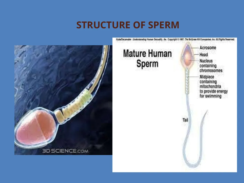
Structure of Sperm<br>
28
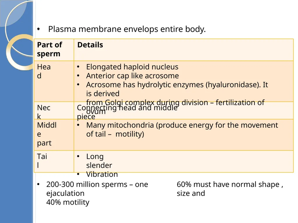
Head Elongated haploid nucleus
Anterior cap like acrosome
Acrosome has hydrolytic enzymes (hyaluronidase). It is derived
from Golgi complex during division – fertilization of ovum Neck Connecting head and middle piece Middle part Many mitochondria (produce energy for the movement of tail – motility) Tail Long slender
Vibration 60% must have normal shape , size and 200-300 million sperms – one ejaculation
40% motility Sperm structure Plasma membrane envelops entire body.
Part of Details
sperm<br>
29
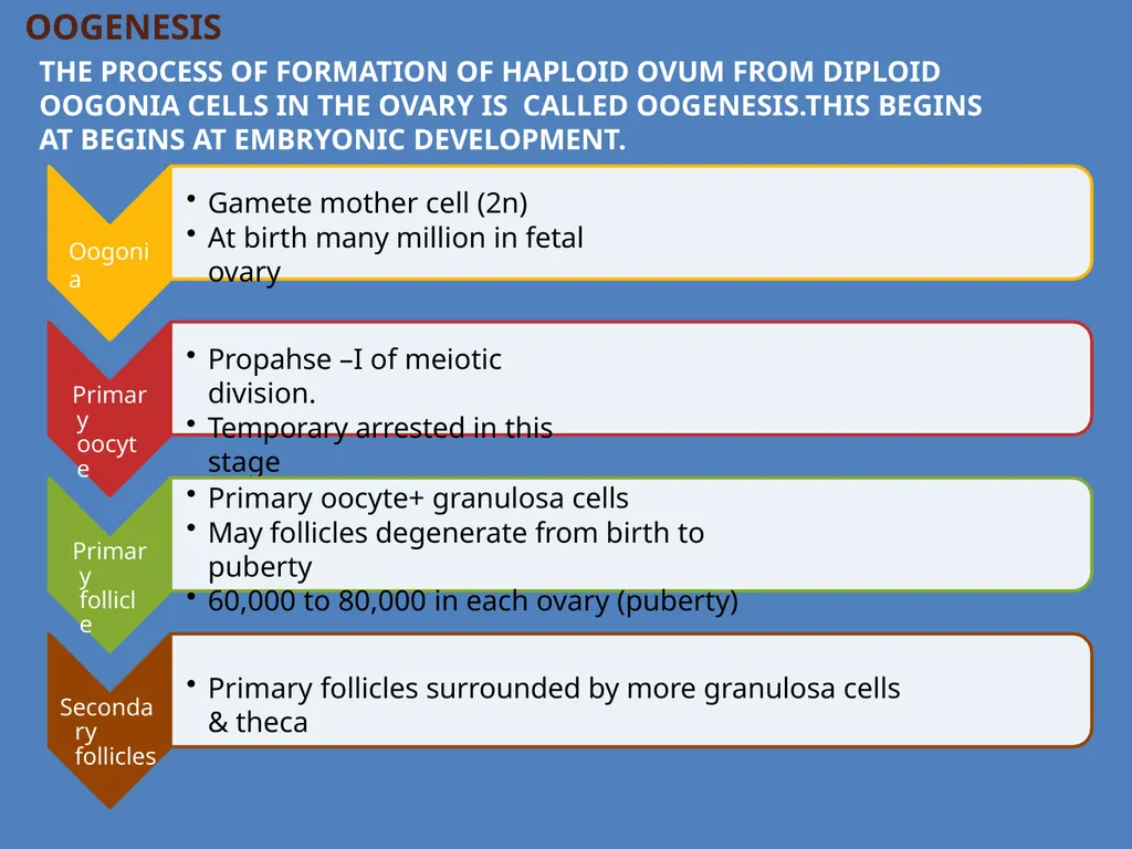
OOGENESIS
The process of formation of haploid ovum from diploid oogonia cells in the ovary is called oogenesis.This begins at begins at embryonic development. Oogonia Gamete mother cell (2n)
At birth many million in fetal ovary Primary oocyte Propahse –I of meiotic division.
Temporary arrested in this stage Primary follicle Primary oocyte+ granulosa cells
May follicles degenerate from birth to puberty
60,000 to 80,000 in each ovary (puberty) Secondary follicles Primary follicles surrounded by more granulosa cells & theca<br>
30
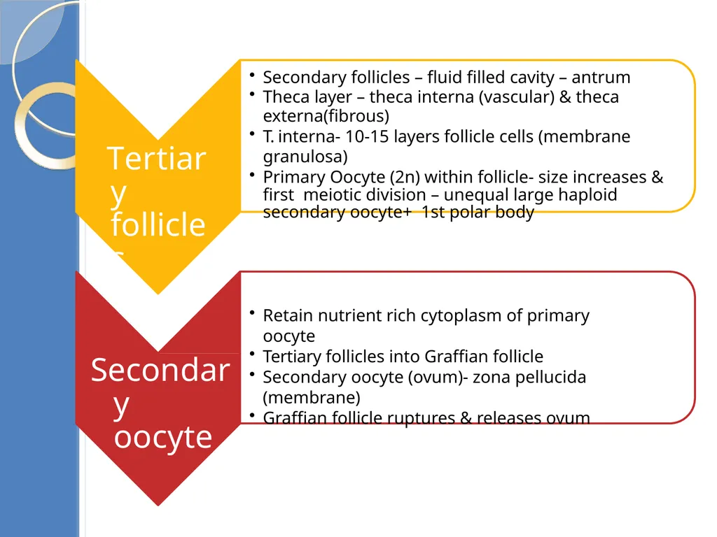
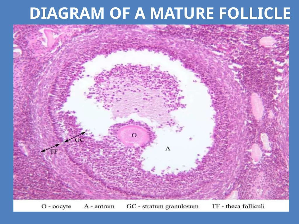
Tertiary follicles Secondary follicles – fluid filled cavity – antrum
Theca layer – theca interna (vascular) & theca
externa(fibrous)
T. interna- 10-15 layers follicle cells (membrane granulosa)
Primary Oocyte (2n) within follicle- size increases & first meiotic division – unequal large haploid secondary oocyte+ 1st polar body Secondary oocyte Retain nutrient rich cytoplasm of primary oocyte
Tertiary follicles into Graffian follicle
Secondary oocyte (ovum)- zona pellucida (membrane)
Graffian follicle ruptures & releases ovum<br>
31
Development of Follicles<br>
32
Diagram of a mature follicle<br>
33
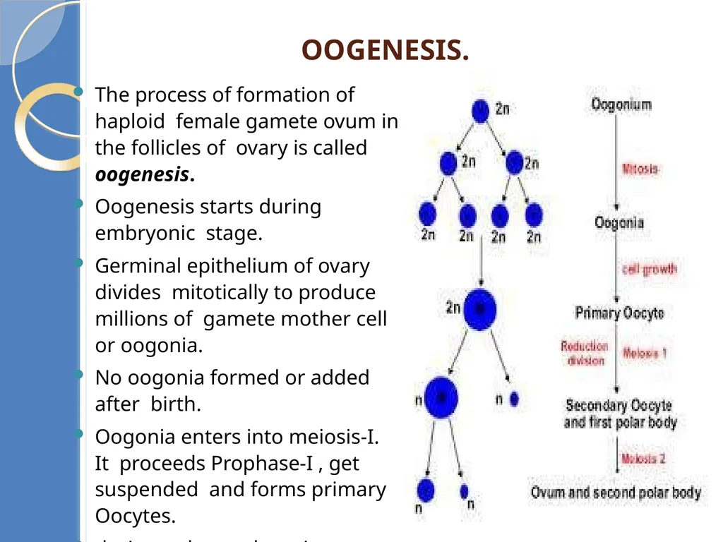
Oogenesis. The process of formation of haploid female gamete ovum in the follicles of ovary is called oogenesis.
Oogenesis starts during embryonic stage.
Germinal epithelium of ovary divides mitotically to produce millions of gamete mother cell or oogonia.
No oogonia formed or added after birth.
Oogonia enters into meiosis-I. It proceeds Prophase-I , get suspended and forms primary Oocytes.
during puberty, the primary oocyte
restarts its first meiotic division.<br>
34
Oogenesis takes place by three stages as follows.
Multiplicative Phase
Growth phase
Maturation phase
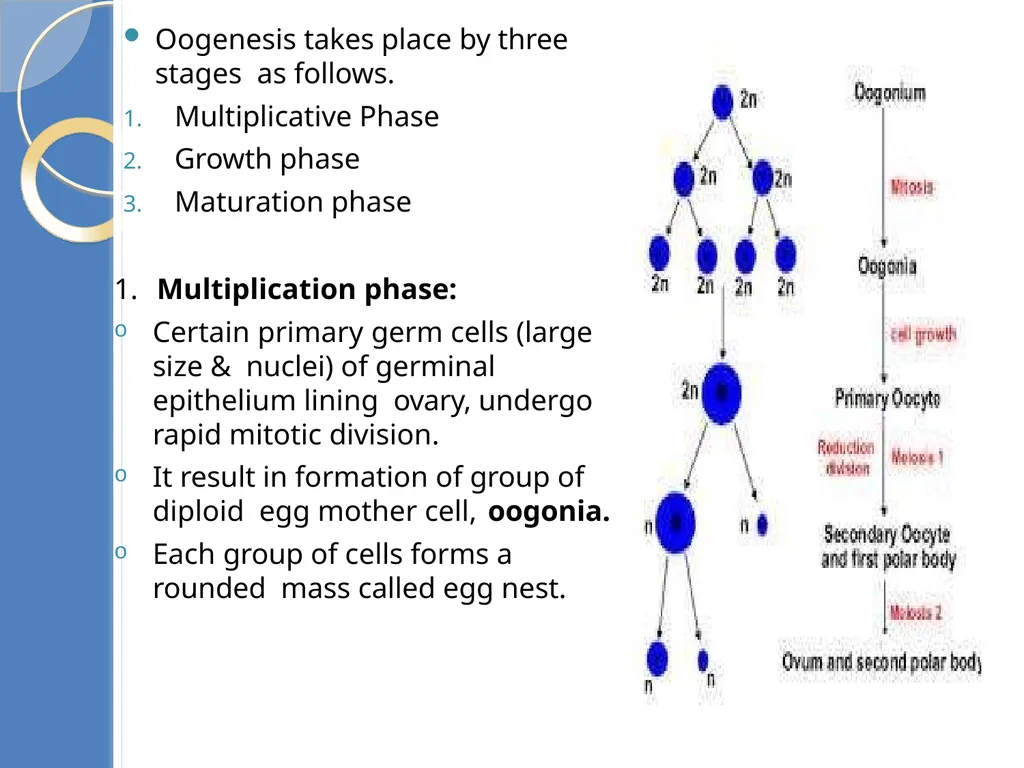
1. Multiplication phase:
Certain primary germ cells (large size & nuclei) of germinal epithelium lining ovary, undergo rapid mitotic division.
It result in formation of group of diploid egg mother cell, oogonia.
Each group of cells forms a rounded mass called egg nest.<br>
35
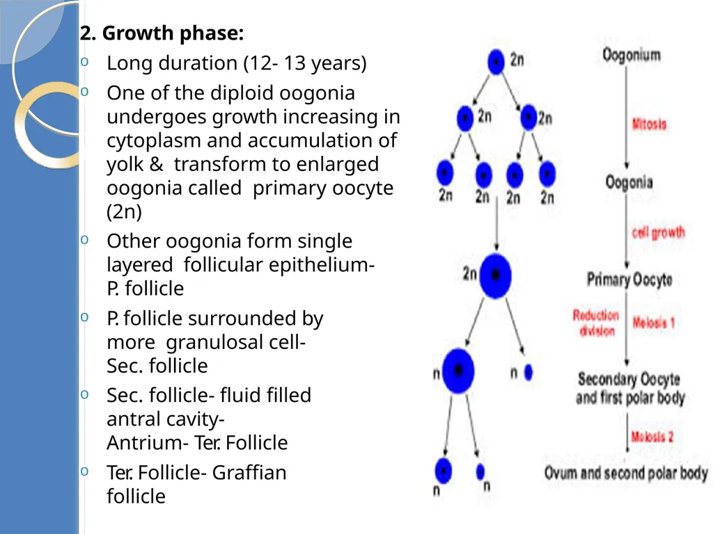
2. Growth phase:
Long duration (12- 13 years)
One of the diploid oogonia undergoes growth increasing in cytoplasm and accumulation of yolk & transform to enlarged oogonia called primary oocyte (2n)
Other oogonia form single layered follicular epithelium- P. follicle
P. follicle surrounded by more granulosal cell- Sec. follicle
Sec. follicle- fluid filled antral cavity-
Antrium- Ter. Follicle
Ter. Follicle- Graffian follicle<br>
36
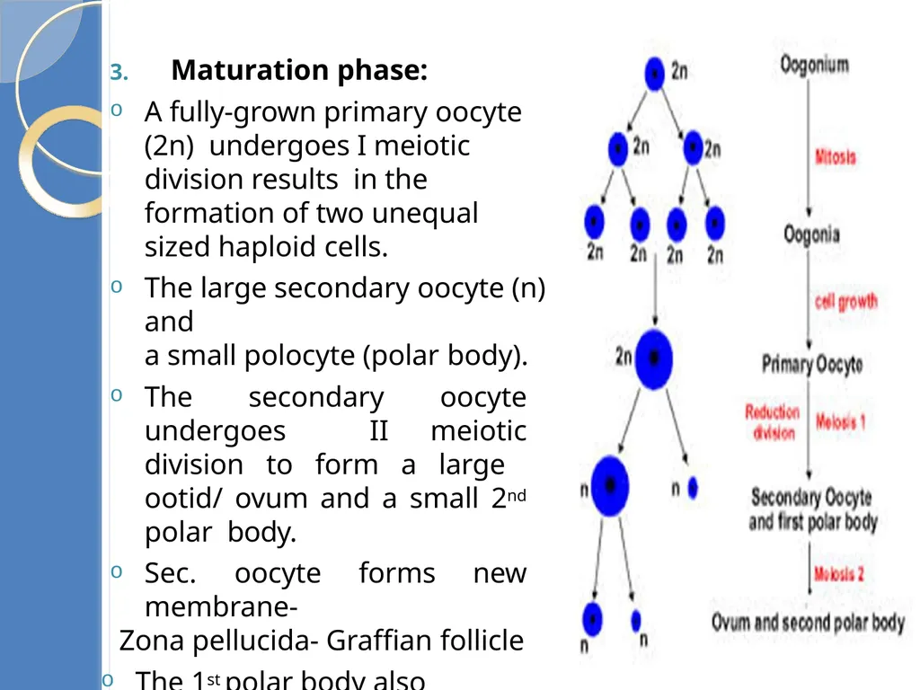
3. Maturation phase:
A fully-grown primary oocyte (2n) undergoes I meiotic division results in the formation of two unequal sized haploid cells.
The large secondary oocyte (n) and
a small polocyte (polar body).
The secondary oocyte undergoes II meiotic division to form a large ootid/ ovum and a small 2nd polar body.
Sec. oocyte forms new membrane-
Zona pellucida- Graffian follicle
The 1st polar body also undergoes
equal division to produce two cells.<br>
37
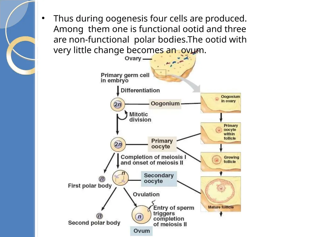
Thus during oogenesis four cells are produced. Among them one is functional ootid and three are non-functional polar bodies.The ootid with very little change becomes an ovum.<br>
38
The three important differences between spermatogeneis and oogenesis.
In oogenesis the unequal cytokinesis during meiosis results in most of the cytoplasm being in one daughter cell which will form the single ovum the other cells (polar bodies) will degenerate. In spermatogeneis all four products of meiosis I and II become mature spermatozoa.
Spermatogenesis is a continuous process throughout the reproductive life of the male as spermatogonia continue to divide by mitosis. At the time of the female’s birth an ovary contains all of the primary oocytes it will ever have.
Spermatogenesis occurs as an uninterrupted sequence; in oogenesis long “resting” periods occur between the formation of the initial steps and final production of the ovum.<br>
39
Menstrual cycle: Reproductive cycle of female primates is called menstrual cycle.
Menstruation is the term given to the periodic discharge of blood, tissue, fluid and mucus from the reproductive organs of sexually mature females. The flow usually lasts from 3 - 6 days each month and is caused by a sudden reduction in the hormones estrogen and progesterone.
The menstrual cycle begins when a female reaches the age of puberty.The first menstruation begins at puberty is called Menarche.
During the menstrual cycle the uterus endometrium prepares itself for implantation of a fertilized egg. If fertilization does not occur the uterus lining is shed from the body.
Menstrual cycle repeated at an average interval of 28 days.
One ovum is released in the middle usually 14th day of each menstrual cycle.<br>
40
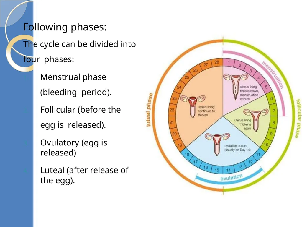
Menstrual cycle has Following phases:
The cycle can be divided into four phases:
Menstrual phase (bleeding period).
Follicular (before the egg is released).
Ovulatory (egg is released)
Luteal (after release of the egg).<br>
The cycle can be divided into four phases:
Menstrual phase (bleeding period).
Follicular (before the egg is released).
Ovulatory (egg is released)
Luteal (after release of the egg).<br>
41
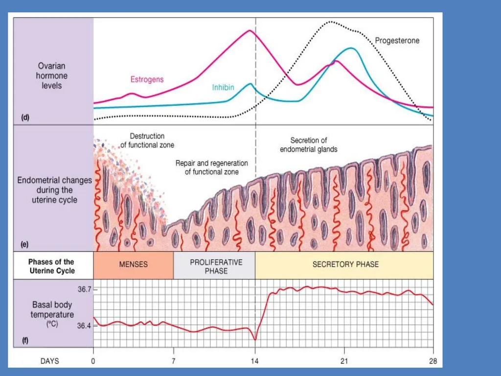
Menstrual cycle<br>
42
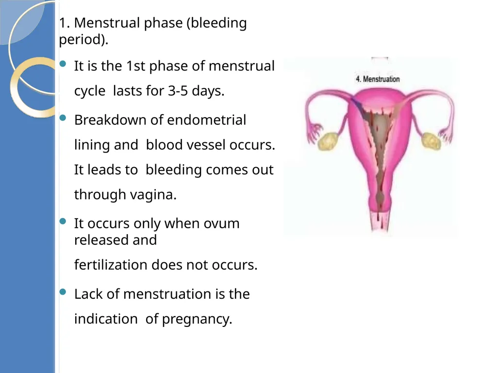
1. Menstrual phase (bleeding period).
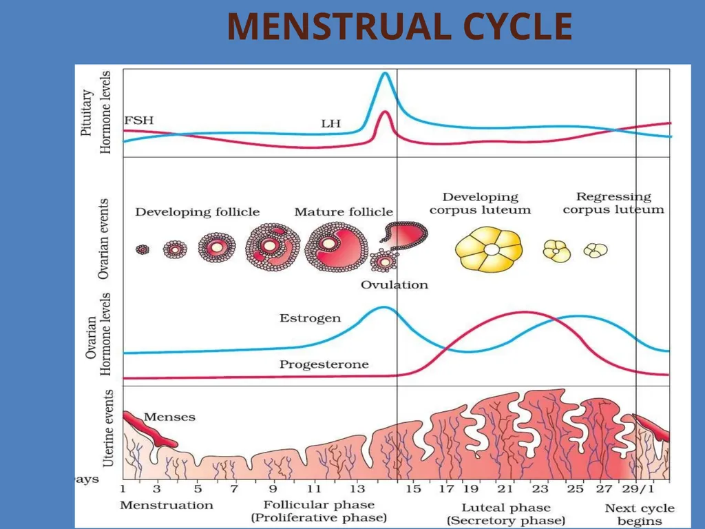
It is the 1st phase of menstrual cycle lasts for 3-5 days.
Breakdown of endometrial lining and blood vessel occurs. It leads to bleeding comes out through vagina.
It occurs only when ovum released and
fertilization does not occurs.
Lack of menstruation is the indication of pregnancy.<br>
It is the 1st phase of menstrual cycle lasts for 3-5 days.
Breakdown of endometrial lining and blood vessel occurs. It leads to bleeding comes out through vagina.
It occurs only when ovum released and
fertilization does not occurs.
Lack of menstruation is the indication of pregnancy.<br>
43
Follicular phase/ Proliferative
phase:
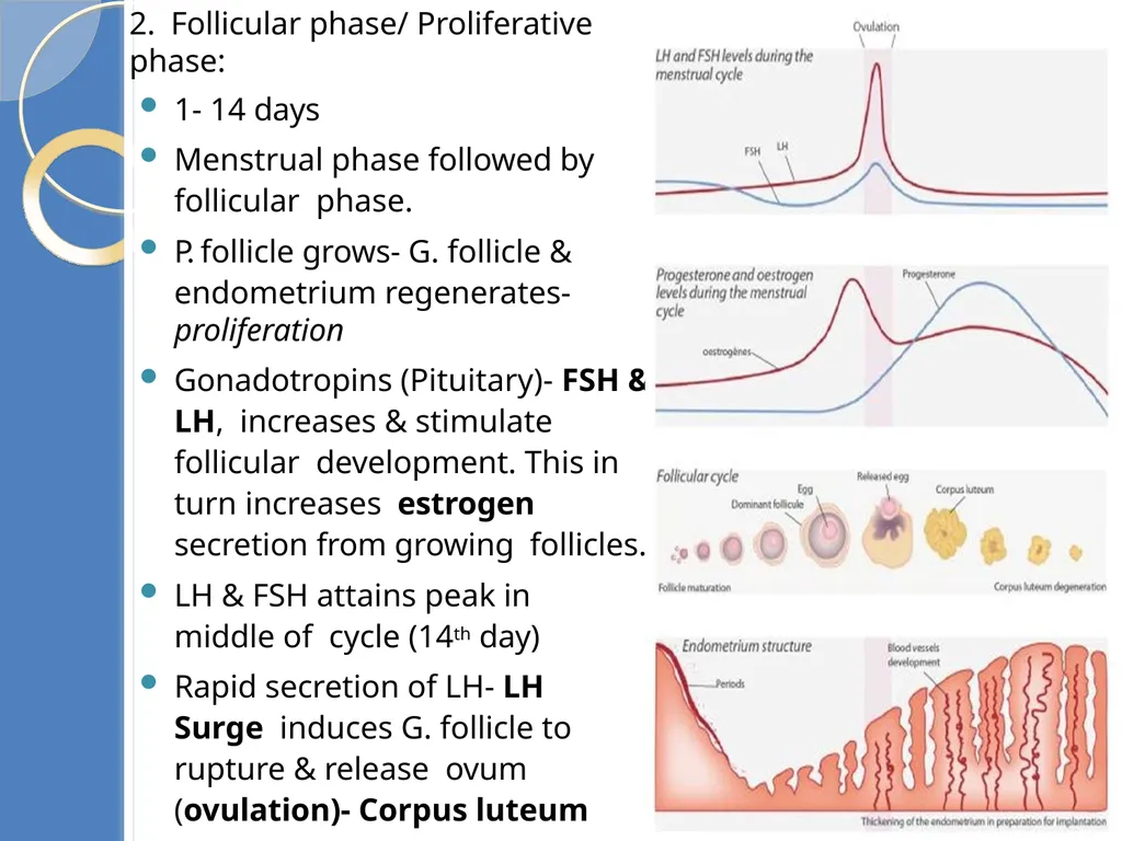
1- 14 days
Menstrual phase followed by follicular phase.
P. follicle grows- G. follicle &
endometrium regenerates- proliferation
Gonadotropins (Pituitary)- FSH & LH, increases & stimulate follicular development. This in turn increases estrogen secretion from growing follicles.
LH & FSH attains peak in middle of cycle (14th day)
Rapid secretion of LH- LH Surge induces G. follicle to rupture & release ovum (ovulation)- Corpus luteum<br>
44
3. Luteal phase/Secretory Phase: This phase begins after ovulation.
Ruptured Graafian follicle transformed into corpus luteum. It produces large
amount of progesterone- essential to maintain & proliferate endometrium
Endometrium- necessary for implantation of fertilized egg/ ovum & does not shed during pregnancy
If fertilization occurs corpus luteum grows further and pregnancy continues. Menstrual cycle stops up.
In absence of fertilization, G. follicle transforms to yellow bodied Corpus luteum
Progesterone level decreases. C. luteum degenerates to Corpus albican
Decrease in Progesterone leads to menstruation
Menstrual cycles ceases at 50 years- Menopause
Cyclic menstruation is indicator of normal reproductive phase & extends between menarche & menopause<br>
45
Fertilization and implantation During copulation (coitus) semen is released by the penis into the vagina is called insemination.
The motile sperms swim rapidly, pass through the cervix, enter into the uterus and finally reach the junction of the isthmus and ampulla (ampullary-isthmic junction) of the fallopian tube
Fertilisation - if the ovum and sperms are transported simultaneously to the ampullary isthmic junction.
The process of fusion of a sperm with an ovum is called
Fertilisation.<br>
46
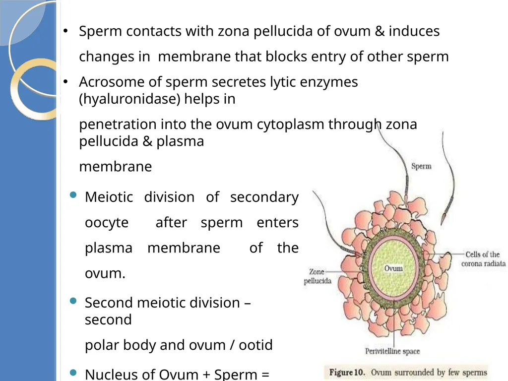
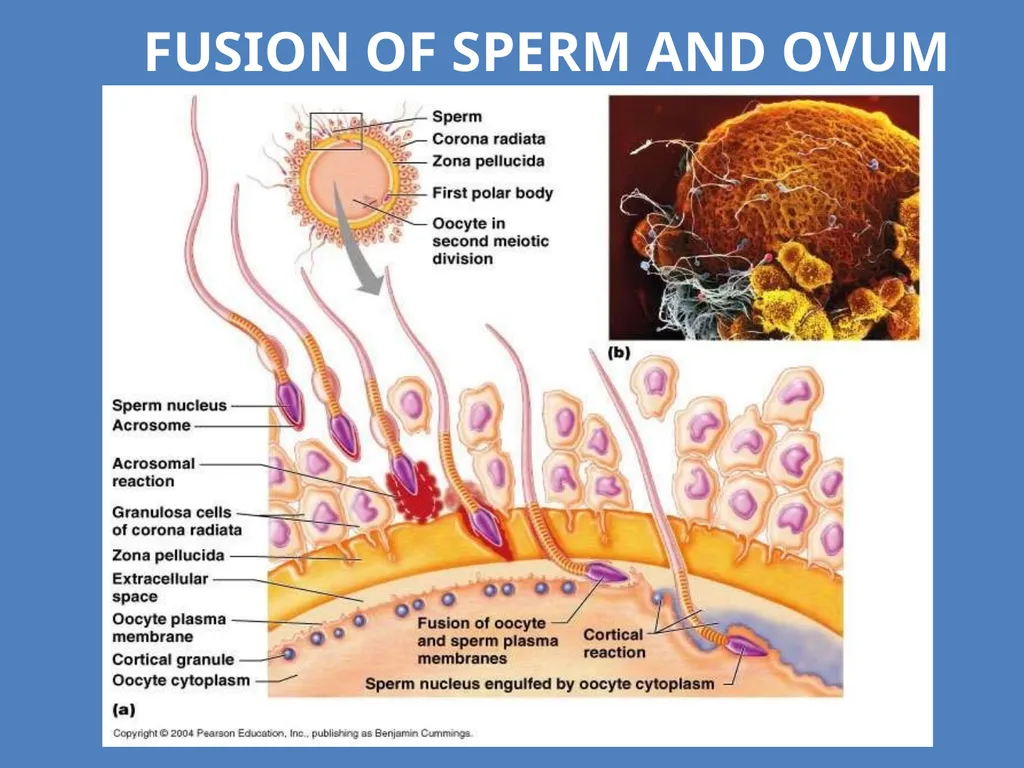
Sperm contacts with zona pellucida of ovum & induces changes in membrane that blocks entry of other sperm
Acrosome of sperm secretes lytic enzymes (hyaluronidase) helps in
penetration into the ovum cytoplasm through zona pellucida & plasma
membrane
Meiotic division of secondary oocyte after sperm enters plasma membrane of the ovum.
Second meiotic division – second
polar body and ovum / ootid
Nucleus of Ovum + Sperm = Zygote
Sex of baby decided this stage<br>
47
Fusion of Sperm and ovum<br>
48
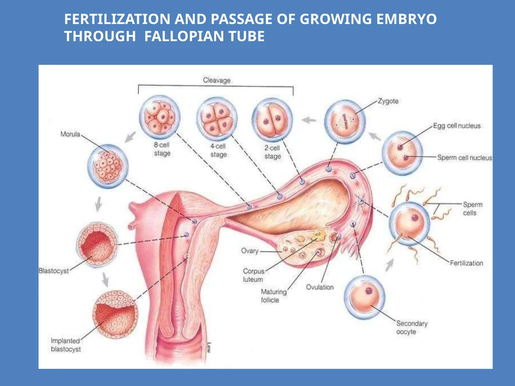
Fertilization and passage of growing embryo through Fallopian tube<br>
49
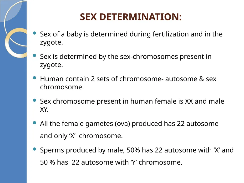
Sex determination: Sex of a baby is determined during fertilization and in the zygote.
Sex is determined by the sex-chromosomes present in zygote.
Human contain 2 sets of chromosome- autosome & sex chromosome.
Sex chromosome present in human female is XX and male XY.
All the female gametes (ova) produced has 22 autosome and only ‘X’ chromosome.
Sperms produced by male, 50% has 22 autosome with ‘X’ and 50 % has 22 autosome with ‘Y’ chromosome.<br>
50
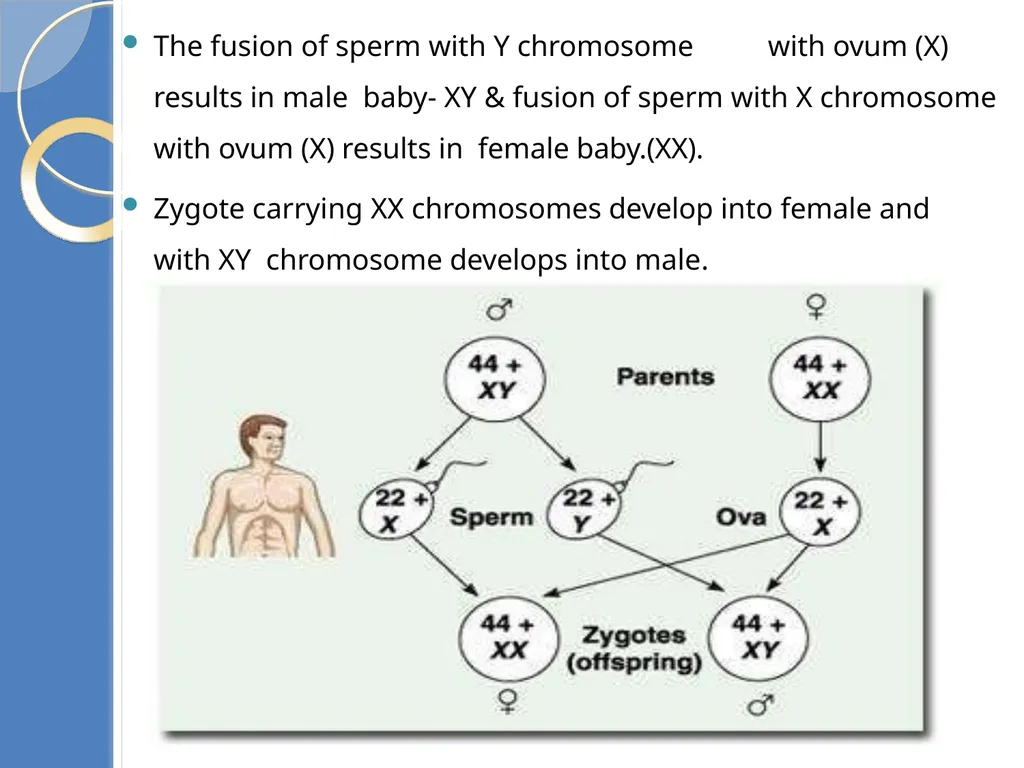
The fusion of sperm with Y chromosome with ovum (X) results in male baby- XY & fusion of sperm with X chromosome with ovum (X) results in female baby.(XX).
Zygote carrying XX chromosomes develop into female and with XY chromosome develops into male.<br>
Zygote carrying XX chromosomes develop into female and with XY chromosome develops into male.<br>
51
Cleavage Zygote from isthmus (oviduct) to uterus– mitotic division, first cleavage in first 36 hrs
2,4,8,16 daughter cells- blastomeres
Embryo with 8 – 16 blastomeres – Morula
Morula – division continues – hollow ball called Blastocyst.
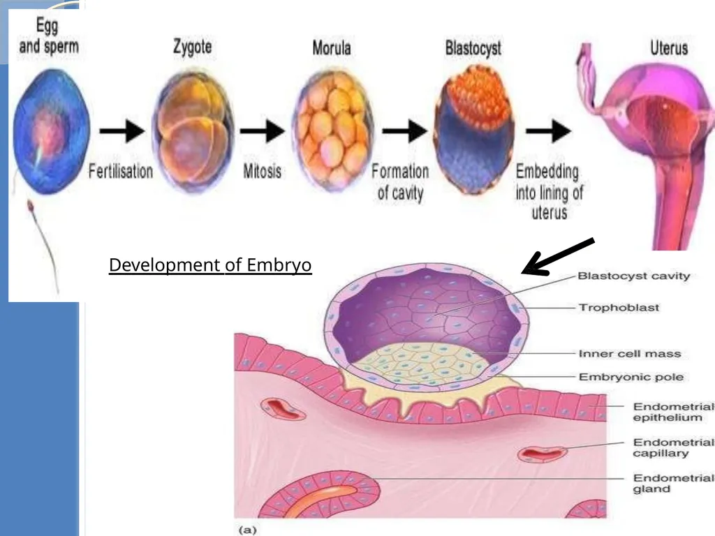
The blastomeres in blastocyst arranged into two layers. An outer layer called trophoblast and an inner cells called inner cell mass.
Trophoblast cells attaches to the endometrium. It helps in implantation and development of placenta.
Inner cell mass gets differentiated into the embryo.
The complete attachment of Blastocyst to the uterine endometrium is called implantation.<br>
52
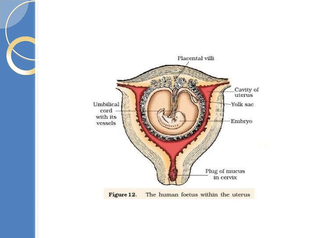
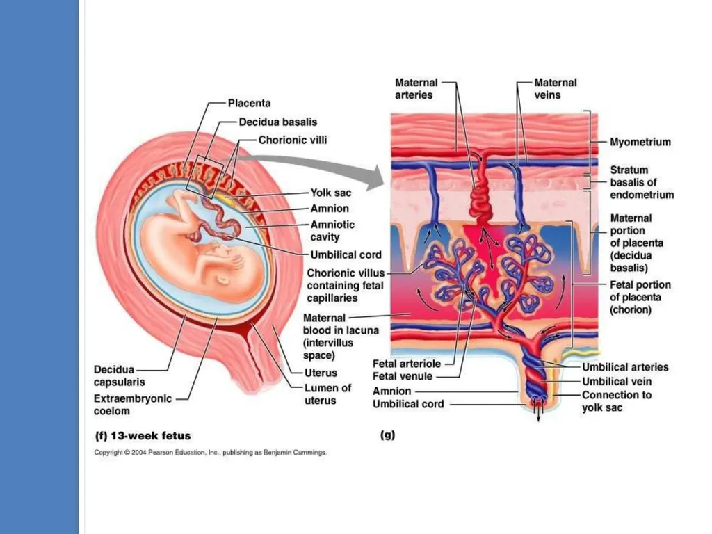
Development of Embryo<br>
53
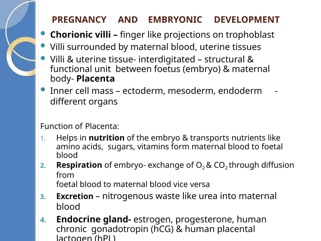
Pregnancy AND Embryonic development Chorionic villi – finger like projections on trophoblast
Villi surrounded by maternal blood, uterine tissues
Villi & uterine tissue- interdigitated – structural & functional unit between foetus (embryo) & maternal body- Placenta
Inner cell mass – ectoderm, mesoderm, endoderm - different organs
Function of Placenta:
Helps in nutrition of the embryo & transports nutrients like amino acids, sugars, vitamins form maternal blood to foetal blood
Respiration of embryo- exchange of O2 & CO2 through diffusion from
foetal blood to maternal blood vice versa
Excretion – nitrogenous waste like urea into maternal blood
Endocrine gland- estrogen, progesterone, human chronic gonadotropin (hCG) & human placental lactogen (hPL)
Antibodies- diphtheria, small pox, measles etc., pass to foetus from
maternal blood
Stores glycogen till liver formation
Effective barrier- toxic chemicals & germs<br>
54
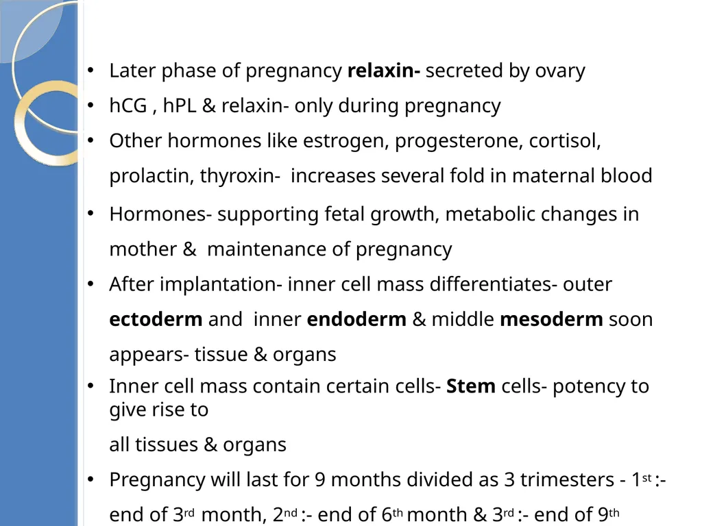
Later phase of pregnancy relaxin- secreted by ovary
hCG , hPL & relaxin- only during pregnancy
Other hormones like estrogen, progesterone, cortisol, prolactin, thyroxin- increases several fold in maternal blood
Hormones- supporting fetal growth, metabolic changes in mother & maintenance of pregnancy
After implantation- inner cell mass differentiates- outer ectoderm and inner endoderm & middle mesoderm soon appears- tissue & organs
Inner cell mass contain certain cells- Stem cells- potency to give rise to
all tissues & organs
Pregnancy will last for 9 months divided as 3 trimesters - 1st :- end of 3rd month, 2nd :- end of 6th month & 3rd :- end of 9th month<br>
55
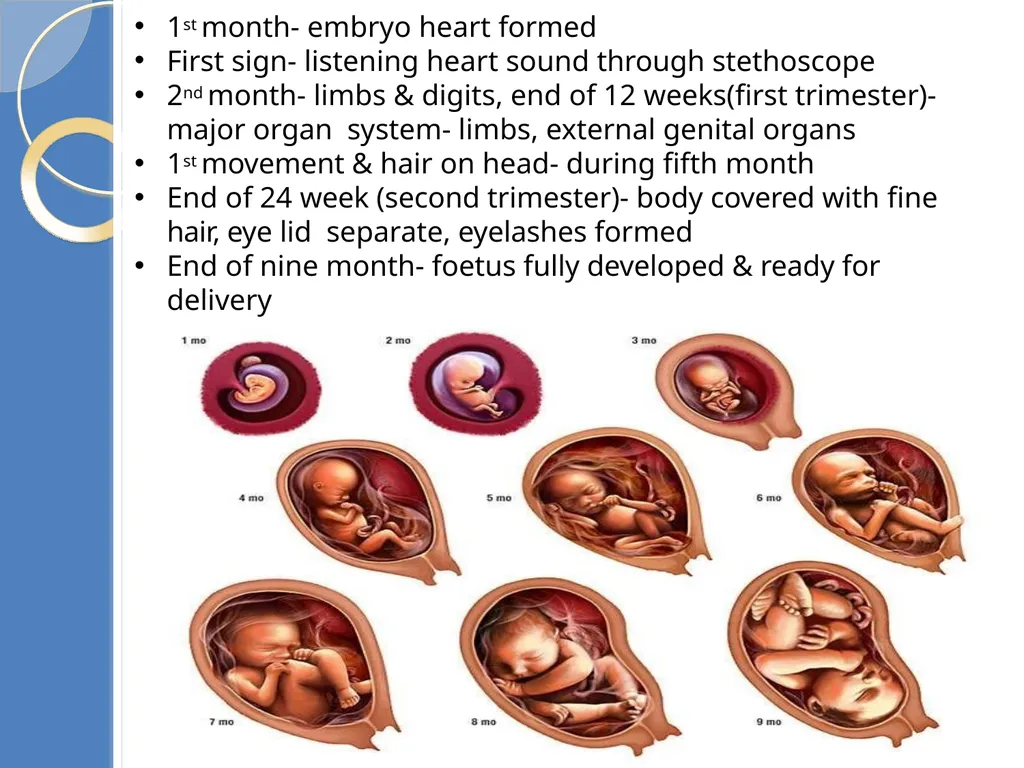
1st month- embryo heart formed
First sign- listening heart sound through stethoscope
2nd month- limbs & digits, end of 12 weeks(first trimester)- major organ system- limbs, external genital organs
1st movement & hair on head- during fifth month
End of 24 week (second trimester)- body covered with fine hair, eye lid separate, eyelashes formed
End of nine month- foetus fully developed & ready for delivery<br>
56
Parturition The period of pregnancy is called gestation period. It is 9 months in human.
The delivery of foetus is called parturition. It occurs by the contraction of uterine Myometrium.
The signal of parturition is originated from the fully developed foetus and the placenta. It induces mild contraction of uterus called fetal ejection reflex.
Hormone (adrenal gland) secreted by foetus diffuses to maternal blood & stimulate oxytocin secretion
Oxytocin causes forceful contraction of myometrium (labour pain) & stimulates further secretion of oxytocin
Stimulatory reflex between uterine contraction & Oxytocin secretion continues inducing stronger contraction & pushes the foetus by dilated cervix (birth canal) facilitated by relaxin- parturition
After delivery the placenta is also expelled out of the uterus.<br>
57
Lactation Mammary gland of female undergo differentiation & produce
milk towards end of pregnancy- Lactation
The mammary gland starts producing milk towards the end of the pregnancy.
Milk produced during initial days of lactation is called colostrum. It contains several antibodies which provide immunity (passive) or resistance to the new born baby.
The milk production is controlled by Lacto trophic or prolactin hormone secreted by pituitary.
Breast feeding during initial period of infant growth is recommended for bringing up a healthy baby<br>