
Transfusion Medicine Rachel S. Bercovitz, MD MS
KL
Published · 81 slides · 0 views

1 / 1
Description
Transfusion Medicine Rachel S. Bercovitz, MD MS Associate Professor of Pediatrics, Northwestern University Feinberg School of Medicine Medical Director, Hemophilia Treatment Center at Ann Robert H. Lurie Childrens Hospital of Chicago
Related Topics
Share
Embed code
Download this presentation From Below
"Transfusion Medicine Rachel S. Bercovitz, MD MS" is the property of its rightful owner. Permission is granted to download and print the materials on this website for personal, non-commercial use only, and to display it on your personal computer provided you do not modify the materials and that you retain all copyright notices contained in the materials. By downloading content from our website, you accept the terms of this agreement.
Presentation Transcript
01
Transfusion Medicine Rachel S. Bercovitz, MD MS
Associate Professor of Pediatrics, Northwestern University Feinberg School of Medicine
Medical Director, Hemophilia Treatment Center at Ann & Robert H. Lurie Children’s Hospital of Chicago<br>
Associate Professor of Pediatrics, Northwestern University Feinberg School of Medicine
Medical Director, Hemophilia Treatment Center at Ann & Robert H. Lurie Children’s Hospital of Chicago<br>
02
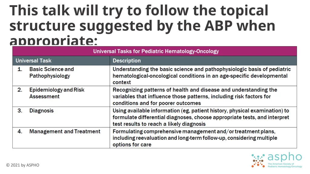
This talk will try to follow the topical structure suggested by the ABP when appropriate:<br>
03
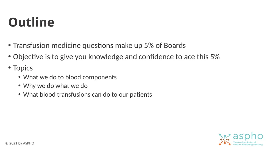
Outline Transfusion medicine questions make up 5% of Boards
Objective is to give you knowledge and confidence to ace this 5%
Topics
What we do to blood components
Why we do what we do
What blood transfusions can do to our patients<br>
Objective is to give you knowledge and confidence to ace this 5%
Topics
What we do to blood components
Why we do what we do
What blood transfusions can do to our patients<br>
04
Component Collection & Storage What we do to blood components<br>
05
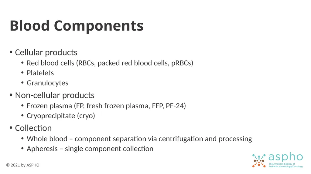
Blood Components Cellular products
Red blood cells (RBCs, packed red blood cells, pRBCs)
Platelets
Granulocytes
Non-cellular products
Frozen plasma (FP, fresh frozen plasma, FFP, PF-24)
Cryoprecipitate (cryo)
Collection
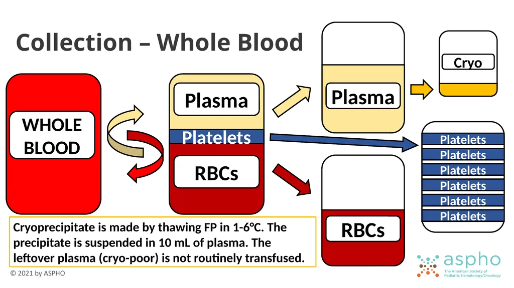
Whole blood – component separation via centrifugation and processing
Apheresis – single component collection<br>
Red blood cells (RBCs, packed red blood cells, pRBCs)
Platelets
Granulocytes
Non-cellular products
Frozen plasma (FP, fresh frozen plasma, FFP, PF-24)
Cryoprecipitate (cryo)
Collection
Whole blood – component separation via centrifugation and processing
Apheresis – single component collection<br>
06
WHOLE BLOOD Collection – Whole Blood Cryoprecipitate is made by thawing FP in 1-6°C. The precipitate is suspended in 10 mL of plasma. The leftover plasma (cryo-poor) is not routinely transfused.<br>
07
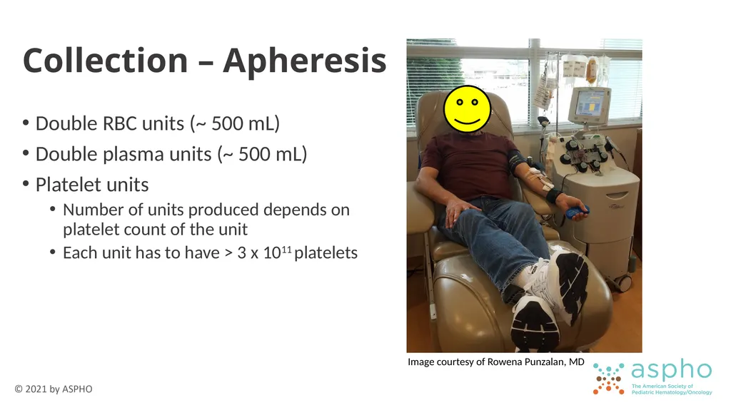
Collection – Apheresis Double RBC units (~ 500 mL)
Double plasma units (~ 500 mL)
Platelet units
Number of units produced depends on platelet count of the unit
Each unit has to have > 3 x 10¹¹ platelets Image courtesy of Rowena Punzalan, MD<br>
Double plasma units (~ 500 mL)
Platelet units
Number of units produced depends on platelet count of the unit
Each unit has to have > 3 x 10¹¹ platelets Image courtesy of Rowena Punzalan, MD<br>
08
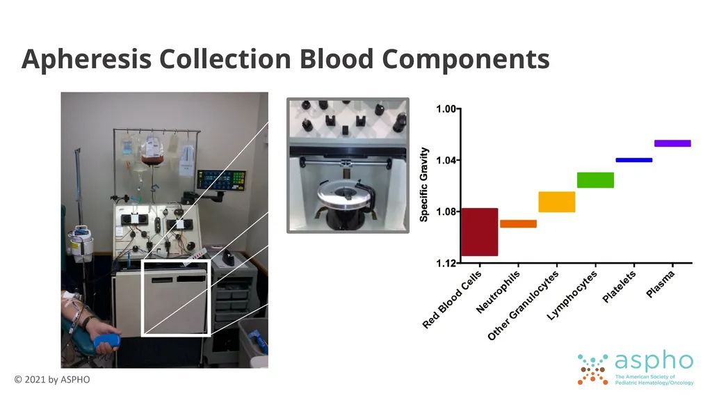
Apheresis Collection Blood Components<br>
09
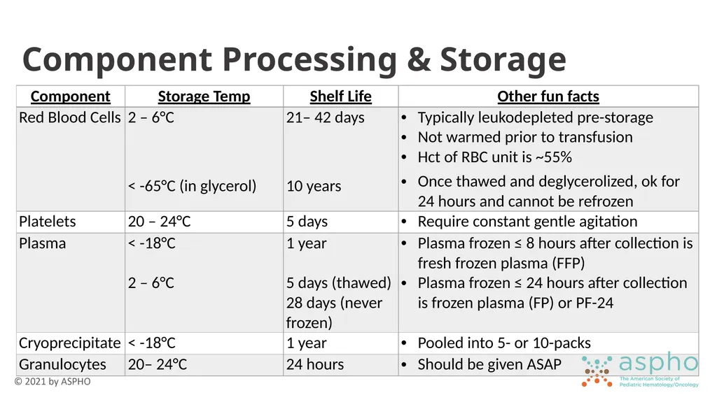
Component Processing & Storage<br>
10
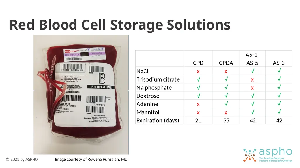
Red Blood Cell Storage Solutions Image courtesy of Rowena Punzalan, MD<br>
11
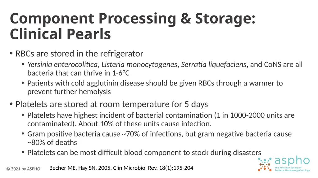
Component Processing & Storage: Clinical Pearls RBCs are stored in the refrigerator
Yersinia enterocolitica, Listeria monocytogenes, Serratia liquefaciens, and CoNS are all bacteria that can thrive in 1-6°C
Patients with cold agglutinin disease should be given RBCs through a warmer to prevent further hemolysis
Platelets are stored at room temperature for 5 days
Platelets have highest incident of bacterial contamination (1 in 1000-2000 units are contaminated). About 10% of these units cause infection.
Gram positive bacteria cause ~70% of infections, but gram negative bacteria cause ~80% of deaths
Platelets can be most difficult blood component to stock during disasters Becher ME, Hay SN. 2005. Clin Microbiol Rev. 18(1):195-204<br>
12
Type & Crossmatch What we do with blood components<br>
13
Terminology Type: Determine ABO and Rh (D) type of blood recipient
Screen: Evaluate for alloantibodies against common and clinically significant RBC antigens
Crossmatch (cross, Xmatch): Electronically or manually confirm compatibility between donor and recipient<br>
Screen: Evaluate for alloantibodies against common and clinically significant RBC antigens
Crossmatch (cross, Xmatch): Electronically or manually confirm compatibility between donor and recipient<br>
14
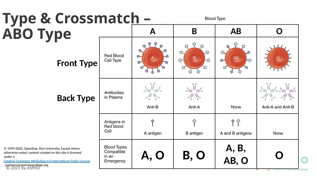
Type & Crossmatch –
ABO Type Front Type Back Type © 1999-2020, OpenStax, Rice University. Except where otherwise noted, content created on this site is licensed under a Creative Commons Attribution 4.0 International Public License; partners@openstaxcollege.org.<br>
15
Type & Crossmatch – ABO Isohemagglutinins (anti-A and anti-B) are IgM antibodies formed by non-A and non-B individuals, respectively, due to environmental exposures
Type O individuals have anti-A and anti-B (IgM) and anti-AB (IgG)
IgM antibodies responsible for acute hemolytic transfusion reaction
Anti-AB IgG is what causes hemolytic disease of the fetus/newborn (HDFN)
Match ABO type
Anti-RBC antibodies should always be avoided
Donor anti-recipient is a minor mismatch (Avoid when possible)
Recipient anti-donor is a major mismatch (Avoid at all costs)<br>
16
Type & Crossmatch – Front & Back Types Different What this looks like (examples)
Patient has A RBCs but no anti-B antibodies
Patient has AB RBCs and also has anti-B antibodies
Causes
Immunosuppressed
Young infants
Patients undergoing ABO-mismatch HSCT
Chimerism
When in doubt, give O RBCs<br>
Patient has A RBCs but no anti-B antibodies
Patient has AB RBCs and also has anti-B antibodies
Causes
Immunosuppressed
Young infants
Patients undergoing ABO-mismatch HSCT
Chimerism
When in doubt, give O RBCs<br>
17
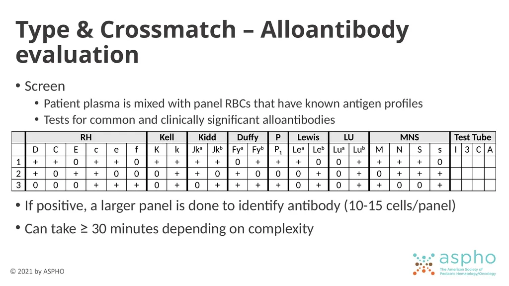
Type & Crossmatch – Alloantibody evaluation Screen
Patient plasma is mixed with panel RBCs that have known antigen profiles
Tests for common and clinically significant alloantibodies
If positive, a larger panel is done to identify antibody (10-15 cells/panel)
Can take ≥ 30 minutes depending on complexity<br>
Patient plasma is mixed with panel RBCs that have known antigen profiles
Tests for common and clinically significant alloantibodies
If positive, a larger panel is done to identify antibody (10-15 cells/panel)
Can take ≥ 30 minutes depending on complexity<br>
18
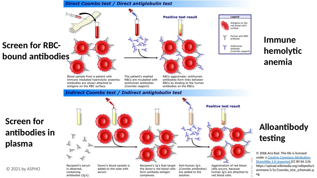
Screen for RBC-bound antibodies Screen for antibodies in plasma Immune hemolytic anemia Alloantibody testing © 2006 Aria Rad. This file is licensed under a Creative Commons Attribution-ShareAlike 3.0 Unported (CC BY-SA 3.0); https://upload.wikimedia.org/wikipedia/commons/1/1c/Coombs_test_schematic.png.<br>
19
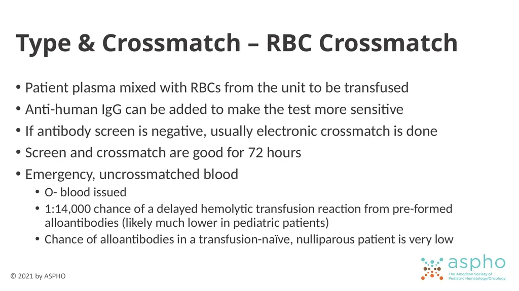
Type & Crossmatch – RBC Crossmatch Patient plasma mixed with RBCs from the unit to be transfused
Anti-human IgG can be added to make the test more sensitive
If antibody screen is negative, usually electronic crossmatch is done
Screen and crossmatch are good for 72 hours
Emergency, uncrossmatched blood
O- blood issued
1:14,000 chance of a delayed hemolytic transfusion reaction from pre-formed alloantibodies (likely much lower in pediatric patients)
Chance of alloantibodies in a transfusion-naïve, nulliparous patient is very low<br>
20
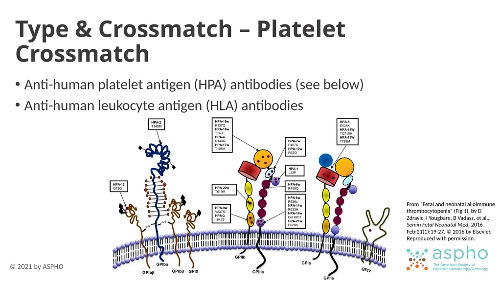
Type & Crossmatch – Platelet Crossmatch Anti-human platelet antigen (HPA) antibodies (see below)
Anti-human leukocyte antigen (HLA) antibodies From “Fetal and neonatal alloimmune thrombocytopenia” (Fig 1), by D Zdravic, I Yougbare, B Vadasz, et al., Semin Fetal Neonatal Med, 2016 Feb;21(1):19-27. © 2016 by Elsevier. Reproduced with permission.<br>
Anti-human leukocyte antigen (HLA) antibodies From “Fetal and neonatal alloimmune thrombocytopenia” (Fig 1), by D Zdravic, I Yougbare, B Vadasz, et al., Semin Fetal Neonatal Med, 2016 Feb;21(1):19-27. © 2016 by Elsevier. Reproduced with permission.<br>
21
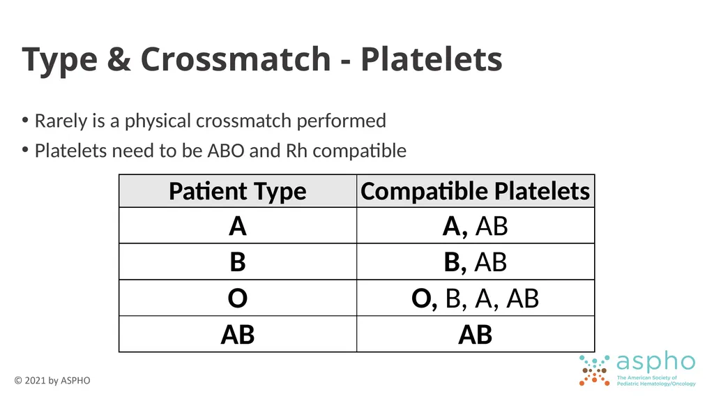
Type & Crossmatch - Platelets Rarely is a physical crossmatch performed
Platelets need to be ABO and Rh compatible<br>
Platelets need to be ABO and Rh compatible<br>
22
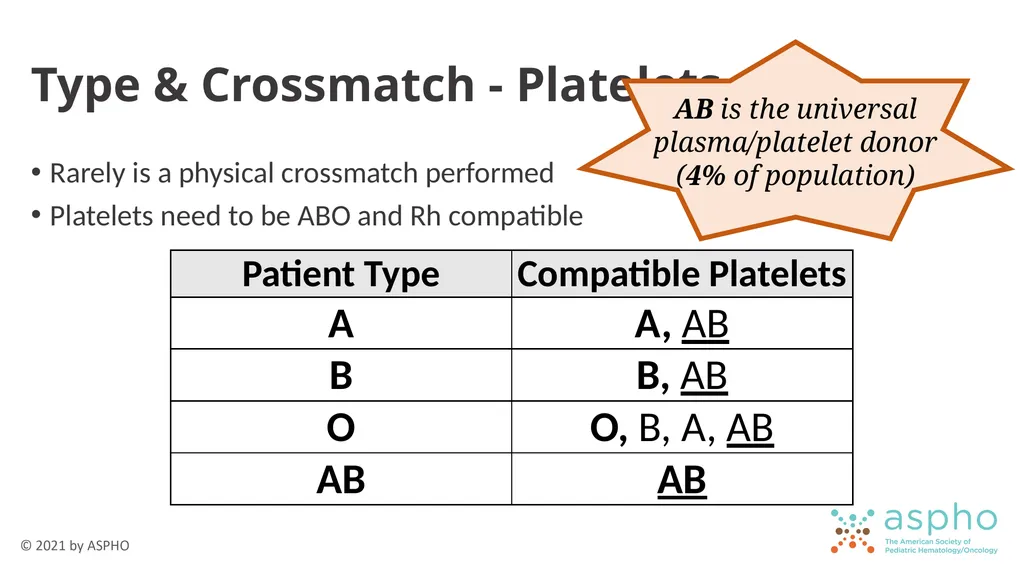
Type & Crossmatch - Platelets Rarely is a physical crossmatch performed
Platelets need to be ABO and Rh compatible<br>
Platelets need to be ABO and Rh compatible<br>
23
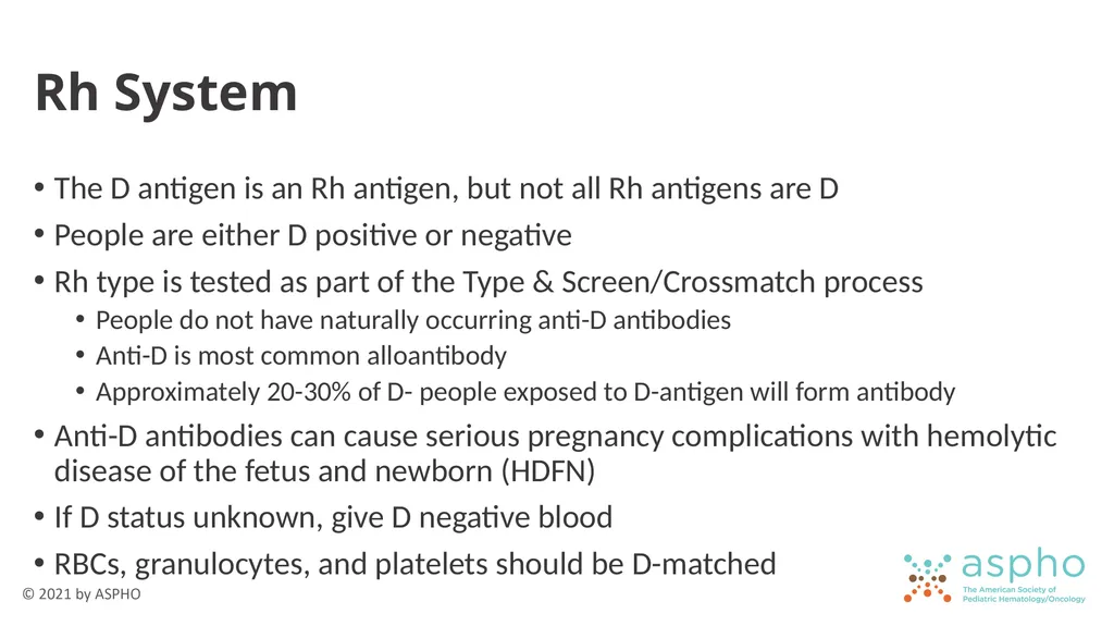
Rh System The D antigen is an Rh antigen, but not all Rh antigens are D
People are either D positive or negative
Rh type is tested as part of the Type & Screen/Crossmatch process
People do not have naturally occurring anti-D antibodies
Anti-D is most common alloantibody
Approximately 20-30% of D- people exposed to D-antigen will form antibody
Anti-D antibodies can cause serious pregnancy complications with hemolytic disease of the fetus and newborn (HDFN)
If D status unknown, give D negative blood
RBCs, granulocytes, and platelets should be D-matched<br>
24
Transfusion Indications Why we do what we do<br>
25
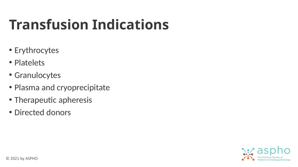
Transfusion Indications Erythrocytes
Platelets
Granulocytes
Plasma and cryoprecipitate
Therapeutic apheresis
Directed donors<br>
Platelets
Granulocytes
Plasma and cryoprecipitate
Therapeutic apheresis
Directed donors<br>
26
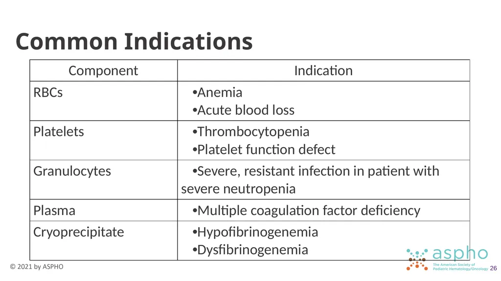
Common Indications 26<br>
27
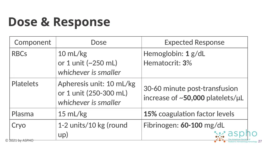
Dose & Response 27<br>
28
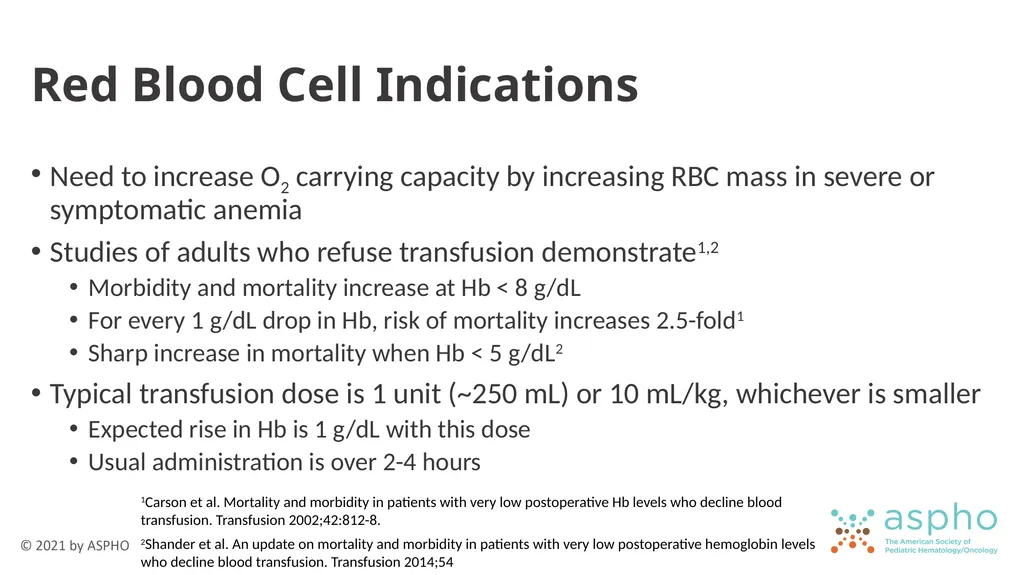
Red Blood Cell Indications Need to increase O2 carrying capacity by increasing RBC mass in severe or symptomatic anemia
Studies of adults who refuse transfusion demonstrate1,2
Morbidity and mortality increase at Hb < 8 g/dL
For every 1 g/dL drop in Hb, risk of mortality increases 2.5-fold1
Sharp increase in mortality when Hb < 5 g/dL2
Typical transfusion dose is 1 unit (~250 mL) or 10 mL/kg, whichever is smaller
Expected rise in Hb is 1 g/dL with this dose
Usual administration is over 2-4 hours 1Carson et al. Mortality and morbidity in patients with very low postoperative Hb levels who decline blood transfusion. Transfusion 2002;42:812-8.
2Shander et al. An update on mortality and morbidity in patients with very low postoperative hemoglobin levels who decline blood transfusion. Transfusion 2014;54<br>
29
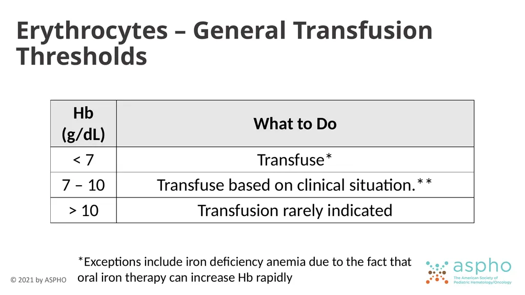
Erythrocytes – General Transfusion Thresholds *Exceptions include iron deficiency anemia due to the fact that oral iron therapy can increase Hb rapidly<br>
30
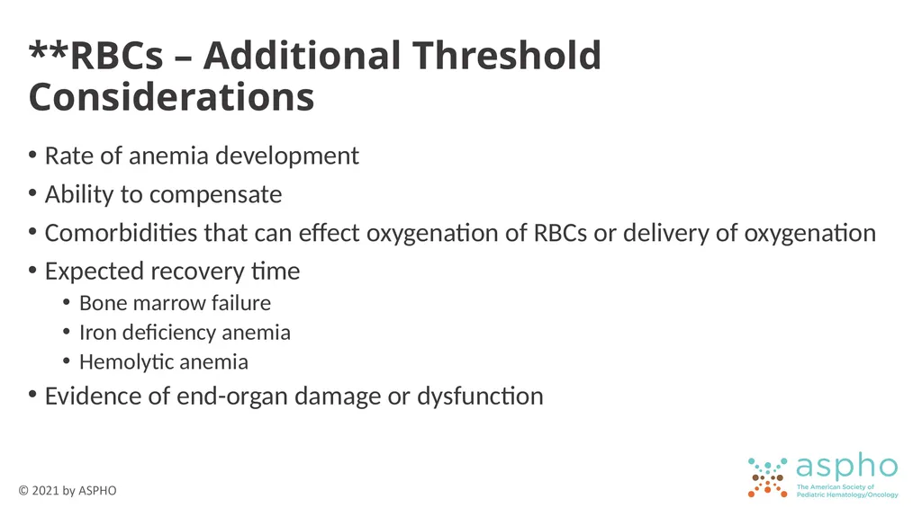
**RBCs – Additional Threshold Considerations Rate of anemia development
Ability to compensate
Comorbidities that can effect oxygenation of RBCs or delivery of oxygenation
Expected recovery time
Bone marrow failure
Iron deficiency anemia
Hemolytic anemia
Evidence of end-organ damage or dysfunction<br>
Ability to compensate
Comorbidities that can effect oxygenation of RBCs or delivery of oxygenation
Expected recovery time
Bone marrow failure
Iron deficiency anemia
Hemolytic anemia
Evidence of end-organ damage or dysfunction<br>
31
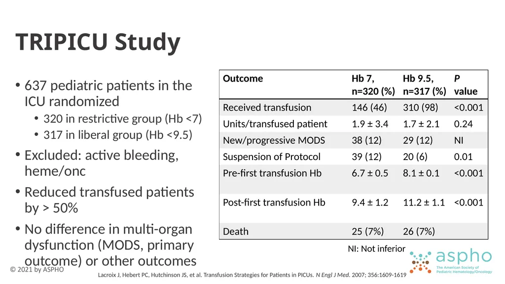
TRIPICU Study 637 pediatric patients in the ICU randomized
320 in restrictive group (Hb <7)
317 in liberal group (Hb <9.5)
Excluded: active bleeding, heme/onc
Reduced transfused patients by > 50%
No difference in multi-organ dysfunction (MODS, primary outcome) or other outcomes NI: Not inferior Lacroix J, Hebert PC, Hutchinson JS, et al. Transfusion Strategies for Patients in PICUs. N Engl J Med. 2007; 356:1609-1619<br>
32
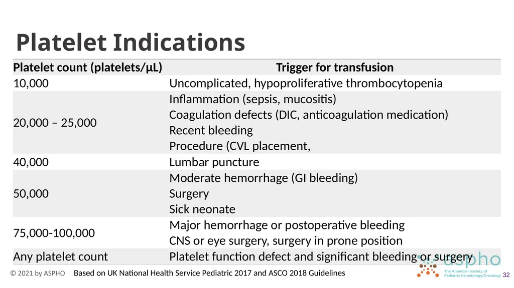
Platelet Indications 32 Based on UK National Health Service Pediatric 2017 and ASCO 2018 Guidelines<br>
33
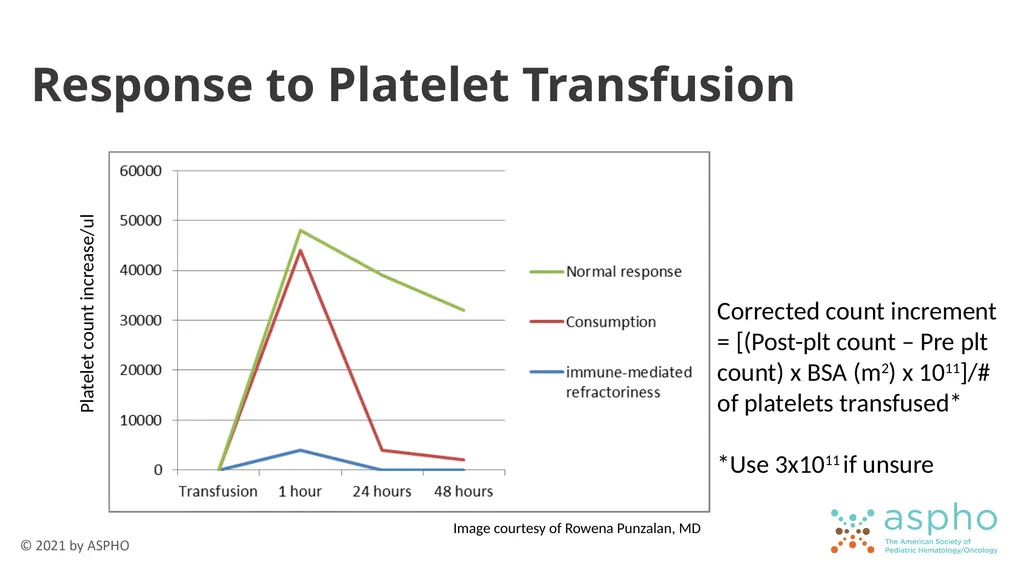
Response to Platelet Transfusion Platelet count increase/ul Corrected count increment = [(Post-plt count – Pre plt count) x BSA (m2) x 1011]/# of platelets transfused*
*Use 3x10¹¹ if unsure Image courtesy of Rowena Punzalan, MD<br>
*Use 3x10¹¹ if unsure Image courtesy of Rowena Punzalan, MD<br>
34
Platelet Refractoriness Consider if patients fail to achieve expected platelet count rise after 2 transfusions
Antigens on platelets
HLA class I
AB antigens on platelets
Platelet glycoproteins
Drug-induced
Typically due to alloantibodies, but patients with autoimmune thrombocytopenia will be refractory to transfused platelets
Anti-HLA class 1 antibodies is most common cause<br>
Antigens on platelets
HLA class I
AB antigens on platelets
Platelet glycoproteins
Drug-induced
Typically due to alloantibodies, but patients with autoimmune thrombocytopenia will be refractory to transfused platelets
Anti-HLA class 1 antibodies is most common cause<br>
35
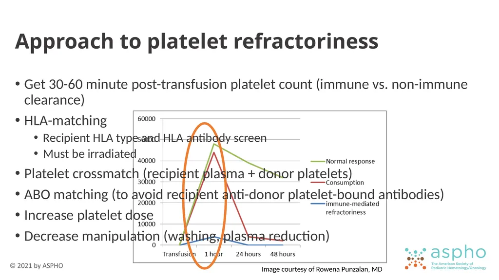
Approach to platelet refractoriness Get 30-60 minute post-transfusion platelet count (immune vs. non-immune clearance)
HLA-matching
Recipient HLA type and HLA antibody screen
Must be irradiated
Platelet crossmatch (recipient plasma + donor platelets)
ABO matching (to avoid recipient anti-donor platelet-bound antibodies)
Increase platelet dose
Decrease manipulation (washing, plasma reduction) Image courtesy of Rowena Punzalan, MD<br>
36
Plasma Indications Support during disseminated intravascular coagulopathy (DIC) or massive transfusion
Factor replacement when factor concentrates are unavailable (Factors V & XI)
Replacement in plasma exchange when indicated
Warfarin reversal
If urgent or no other options available
3- and 4-factor prothrombin complex concentrates not approved in pediatric patients (≤ 16 years old)
Second- or third-line treatment of ACE inhibitor-induced angioedema<br>
37
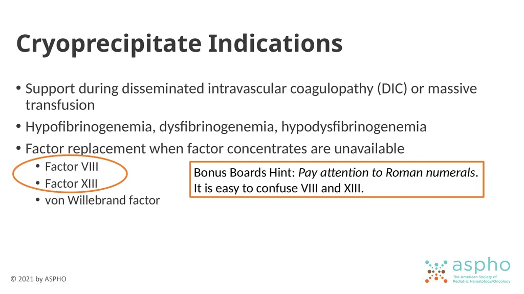
Cryoprecipitate Indications Support during disseminated intravascular coagulopathy (DIC) or massive transfusion
Hypofibrinogenemia, dysfibrinogenemia, hypodysfibrinogenemia
Factor replacement when factor concentrates are unavailable
Factor VIII
Factor XIII
von Willebrand factor Bonus Boards Hint: Pay attention to Roman numerals. It is easy to confuse VIII and XIII.<br>
Hypofibrinogenemia, dysfibrinogenemia, hypodysfibrinogenemia
Factor replacement when factor concentrates are unavailable
Factor VIII
Factor XIII
von Willebrand factor Bonus Boards Hint: Pay attention to Roman numerals. It is easy to confuse VIII and XIII.<br>
38
Granulocyte Indications There are no data that demonstrate benefit of granulocyte transfusion compared to standard antimicrobial therapy
No definitive guidelines
Absolute neutrophil count < 500/µL
Clinical evidence of bacterial or fungal infection
Poor response to antimicrobial therapy (typically 3-5 days)
Consideration of co-morbidities and anticipated neutrophil recovery
Question of efficacy of granulocyte transfusion unlikely to be settled after most recent randomized controlled trial (2008-2014) enrolled < 50% of needed subjects<br>
39
Transfusion Indications – Directed Donor Directed donors – family members (less commonly friends) donate blood to a specific patient rather than to the general blood supply
DIRECTED DONATION IS NOT RECOMMENDED
Less safe – family and friends may lie re: qualifications to avoid embarrassment of not donating or because they feel they are exceptional
Creates inventory issues and potential scenario where directed-donor unit is transfused to another patient
Directed-donor units are not given to other patients if patient does not need it
Risk of transfusion-associated graft-vs-host disease (TA-GVHD)
Exception: Patient has a rare RBC or platelet phenotype and family member is only one able to provide compatible blood<br>
40
Therapeutic Apheresis What we do to blood components and what they do to our patients<br>
41
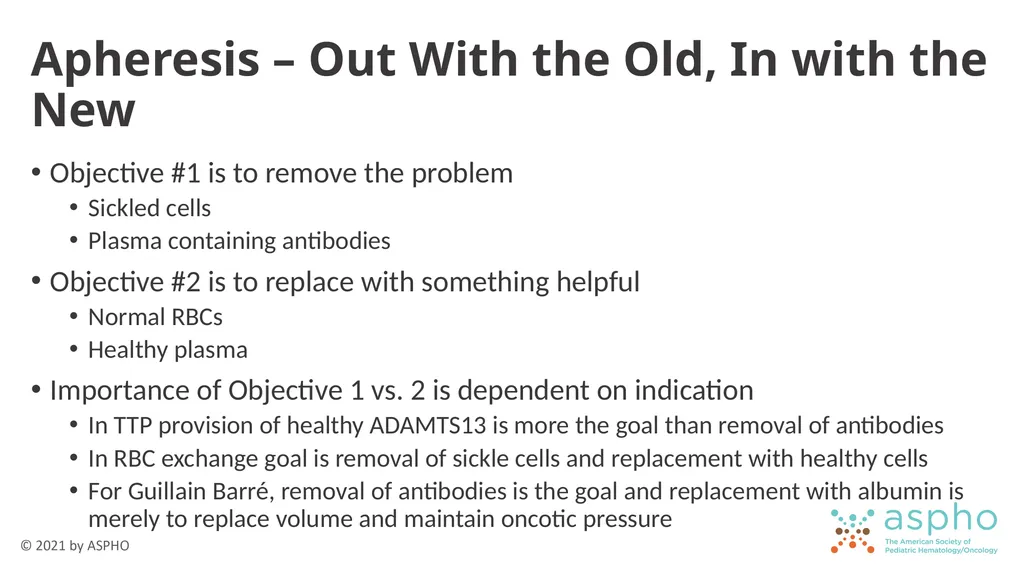
Apheresis – Out With the Old, In with the New Objective #1 is to remove the problem
Sickled cells
Plasma containing antibodies
Objective #2 is to replace with something helpful
Normal RBCs
Healthy plasma
Importance of Objective 1 vs. 2 is dependent on indication
In TTP provision of healthy ADAMTS13 is more the goal than removal of antibodies
In RBC exchange goal is removal of sickle cells and replacement with healthy cells
For Guillain Barré, removal of antibodies is the goal and replacement with albumin is merely to replace volume and maintain oncotic pressure<br>
42
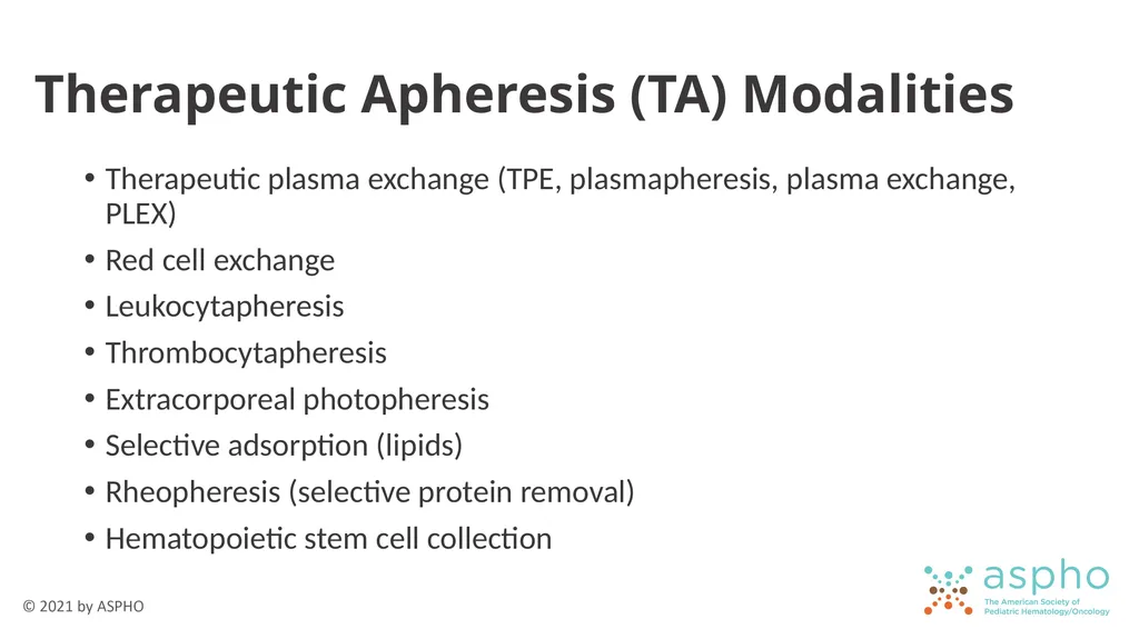
Therapeutic Apheresis (TA) Modalities Therapeutic plasma exchange (TPE, plasmapheresis, plasma exchange, PLEX)
Red cell exchange
Leukocytapheresis
Thrombocytapheresis
Extracorporeal photopheresis
Selective adsorption (lipids)
Rheopheresis (selective protein removal)
Hematopoietic stem cell collection<br>
Red cell exchange
Leukocytapheresis
Thrombocytapheresis
Extracorporeal photopheresis
Selective adsorption (lipids)
Rheopheresis (selective protein removal)
Hematopoietic stem cell collection<br>
43
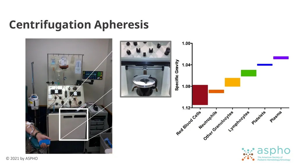
Centrifugation Apheresis<br>
44
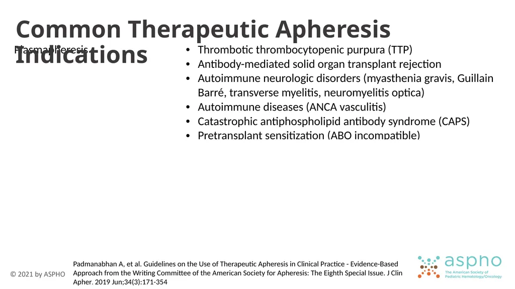
Common Therapeutic Apheresis Indications Padmanabhan A, et al. Guidelines on the Use of Therapeutic Apheresis in Clinical Practice - Evidence-Based Approach from the Writing Committee of the American Society for Apheresis: The Eighth Special Issue. J Clin Apher. 2019 Jun;34(3):171-354<br>
45
Considerations for Therapeutic Apheresis Refer to the ASFA Apheresis Guidelines
Indication
Replacement fluid (plasma versus albumin for TPE)
Frequency and duration
Vascular access: how many procedures
Peripheral access preferred
Central venous catheter able to accommodate negative pressure of withdrawal
Miscellaneous items: Patient age/size re: blood prime or sedation for procedure, medications that can be removed through apheresis Padmanabhan A, et al. Guidelines on the Use of Therapeutic Apheresis in Clinical Practice - Evidence-Based Approach from the Writing Committee of the American Society for Apheresis: The Eighth Special Issue. J Clin Apher. 2019 Jun;34(3):171-354<br>
46
Risks of Therapeutic Apheresis Direct risks of procedure
Hypotension due to fluid shifts and extracorporeal fluid volume
If citrate is used, hypocalcemia ± hypomagnesemia
Fatigue
Reduced coagulation factors (esp. fibrinogen) if using albumin as replacement fluid. Risk is correlated with frequency of procedure.
Indirect risks
Transfusion reaction – if plasma used for TPE or from RBC exchange
Risks of central venous catheter – infection, thrombus<br>
47
Other Apheresis Procedures Collection of hematopoietic stem cells for allogeneic or autologous transplant
Collection of cells for cellular therapy
CAR T-Cell Therapy
Anti-cancer vaccines
Genetic modification of stem cells to treat hemoglobinopathy
NK cells<br>
Collection of cells for cellular therapy
CAR T-Cell Therapy
Anti-cancer vaccines
Genetic modification of stem cells to treat hemoglobinopathy
NK cells<br>
48
Uncommon Apheresis Procedures Leukocytapheresis
Used for symptomatic hyperleukocytosis in leukemia
Acute myeloid leukemia (AML): WBC > 100 x 109/L
Acute lymphoblastic leukemia (ALL): WBC > 400 x 109/L
Can reduce early mortality (≤ 3 weeks after diagnosis), but no affect on long-term event-free or overall survival
Procedure can reduce WBCs by 30-60%
Chemotherapy should be given ASAP to prevent reaccumulation
Thrombocytapheresis
Very rare in pediatrics
Symptomatic thrombocytosis (bleeding or clotting) with platelet count > 1000-1500/dL<br>
49
Component Modification What we do to blood components<br>
50
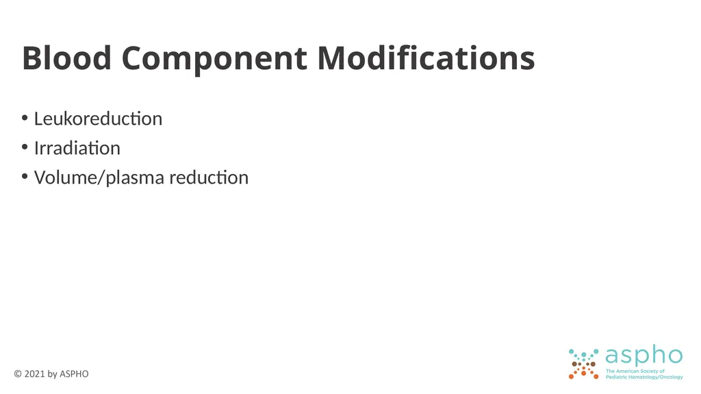
Blood Component Modifications Leukoreduction
Irradiation
Volume/plasma reduction<br>
Irradiation
Volume/plasma reduction<br>
51
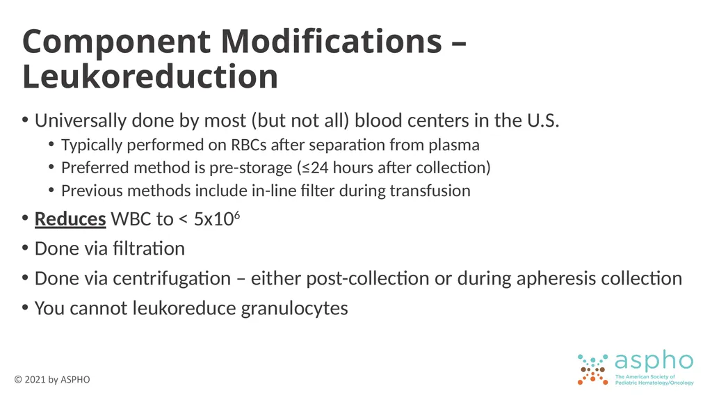
Component Modifications – Leukoreduction Universally done by most (but not all) blood centers in the U.S.
Typically performed on RBCs after separation from plasma
Preferred method is pre-storage (≤24 hours after collection)
Previous methods include in-line filter during transfusion
Reduces WBC to < 5x10⁶
Done via filtration
Done via centrifugation – either post-collection or during apheresis collection
You cannot leukoreduce granulocytes<br>
52
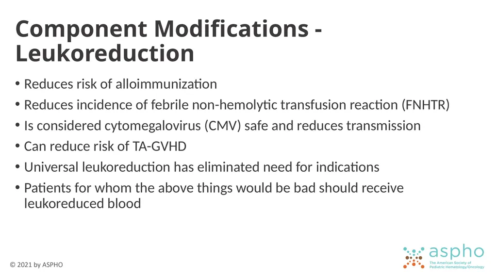
Component Modifications - Leukoreduction Reduces risk of alloimmunization
Reduces incidence of febrile non-hemolytic transfusion reaction (FNHTR)
Is considered cytomegalovirus (CMV) safe and reduces transmission
Can reduce risk of TA-GVHD
Universal leukoreduction has eliminated need for indications
Patients for whom the above things would be bad should receive leukoreduced blood<br>
53
Component Modifications – Irradiation Blood products are exposed to radiation
Devices using x-rays
Devices using gamma rays from cesium-137 or cobalt-60
Irradiation prevents proliferation of donor T lymphocytes to prevent TA-GVHD
Done to cellular products (RBCs, platelets, granulocytes)
Do not irradiate hematopoietic stem cells
The goal is to have these cells engraft
Irradiation would defeat the purpose of a stem cell transplant<br>
54
Component Modification – Irradiation Does not change out-date of platelets or granulocytes
Shortens RBC shelf life to 28 days
From day of collection
From day of irradiation
Whichever is shorter
Best to irradiate RBCs at issue
Reduces free potassium in the unit
Otherwise, if >24-48 hours, wash or volume reduce the product<br>
Shortens RBC shelf life to 28 days
From day of collection
From day of irradiation
Whichever is shorter
Best to irradiate RBCs at issue
Reduces free potassium in the unit
Otherwise, if >24-48 hours, wash or volume reduce the product<br>
55
Irradiation Indications Fetal and neonatal recipients of intrauterine transfusions
Selected immunocompromised recipients
Recipients of cellular components known to be from a blood relative
Recipients who have undergone marrow or peripheral blood progenitor cell transplantation
Recipients of cellular components whose donor is selected for HLA compatibility<br>
Selected immunocompromised recipients
Recipients of cellular components known to be from a blood relative
Recipients who have undergone marrow or peripheral blood progenitor cell transplantation
Recipients of cellular components whose donor is selected for HLA compatibility<br>
56
Selected Immunocompromised Recipients Pre-term neonates
Young children who may not yet have presented with an immunodeficiency (typically < 1 year, up to < 6 years)
Congenital immune deficiencies
Aplastic anemia
Hematologic malignancy
Patients receiving chemotherapy (including autologous stem cell transplant)
Allogeneic stem cell transplant recipients and donors
Receipt of purine analogue chemotherapy<br>
57
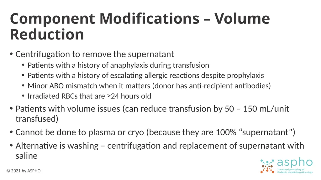
Component Modifications – Volume Reduction Centrifugation to remove the supernatant
Patients with a history of anaphylaxis during transfusion
Patients with a history of escalating allergic reactions despite prophylaxis
Minor ABO mismatch when it matters (donor has anti-recipient antibodies)
Irradiated RBCs that are ≥24 hours old
Patients with volume issues (can reduce transfusion by 50 – 150 mL/unit transfused)
Cannot be done to plasma or cryo (because they are 100% “supernatant”)
Alternative is washing – centrifugation and replacement of supernatant with saline<br>
58
Transfusion Reactions What blood transfusions can do to our patients<br>
59
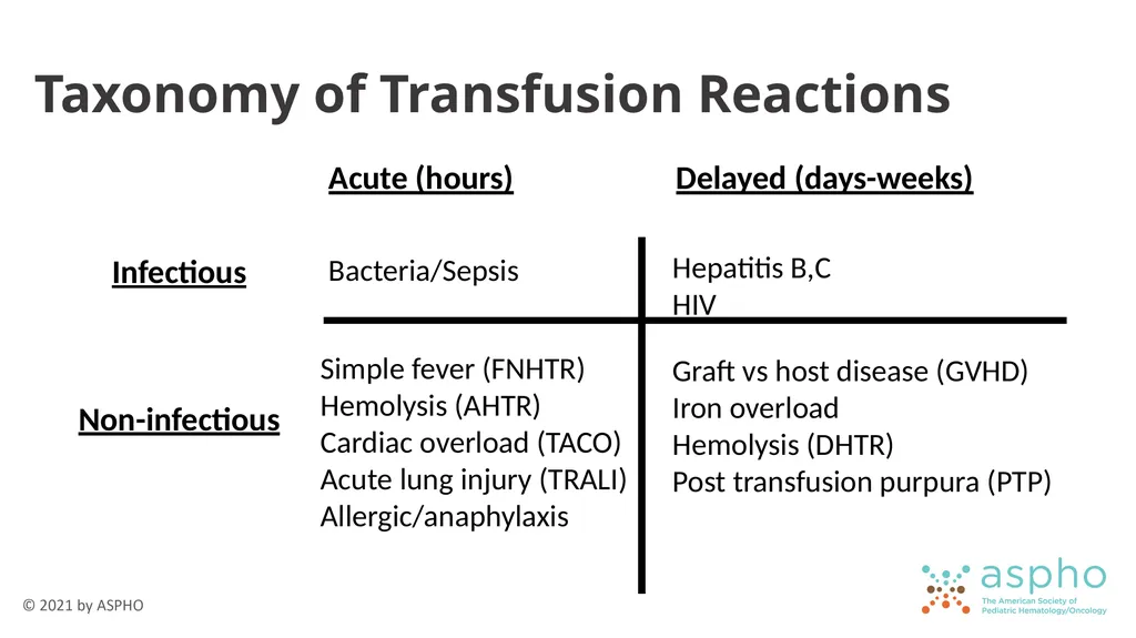
Taxonomy of Transfusion Reactions Acute (hours) Delayed (days-weeks) Infectious Non-infectious Hepatitis B,C
HIV Bacteria/Sepsis Simple fever (FNHTR)
Hemolysis (AHTR)
Cardiac overload (TACO)
Acute lung injury (TRALI)
Allergic/anaphylaxis Graft vs host disease (GVHD)
Iron overload
Hemolysis (DHTR)
Post transfusion purpura (PTP)<br>
HIV Bacteria/Sepsis Simple fever (FNHTR)
Hemolysis (AHTR)
Cardiac overload (TACO)
Acute lung injury (TRALI)
Allergic/anaphylaxis Graft vs host disease (GVHD)
Iron overload
Hemolysis (DHTR)
Post transfusion purpura (PTP)<br>
60
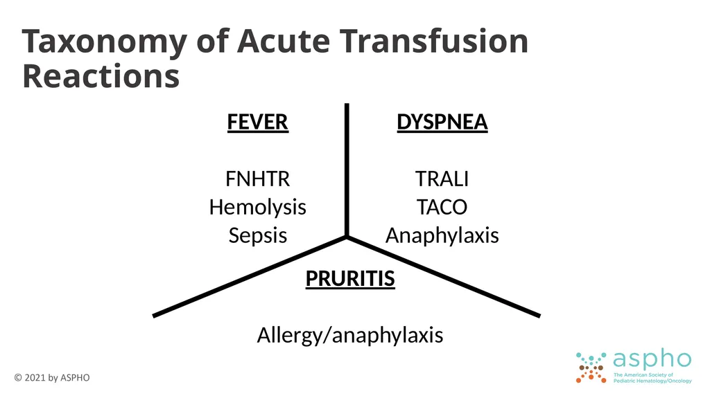
Taxonomy of Acute Transfusion Reactions FEVER
FNHTR
Hemolysis
Sepsis DYSPNEA
TRALI
TACO
Anaphylaxis PRURITIS
Allergy/anaphylaxis<br>
FNHTR
Hemolysis
Sepsis DYSPNEA
TRALI
TACO
Anaphylaxis PRURITIS
Allergy/anaphylaxis<br>
61
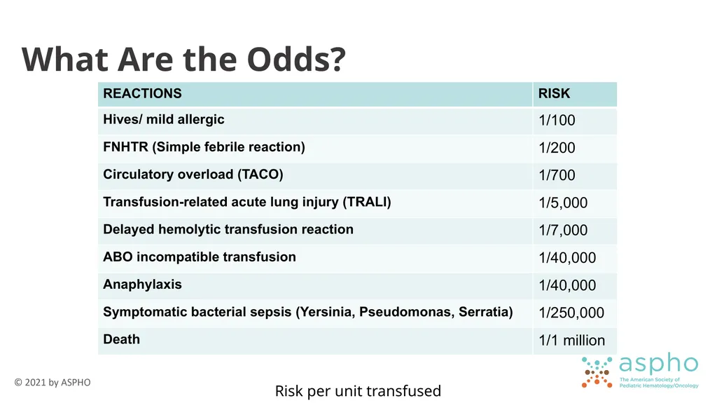
What Are the Odds? Risk per unit transfused<br>
62
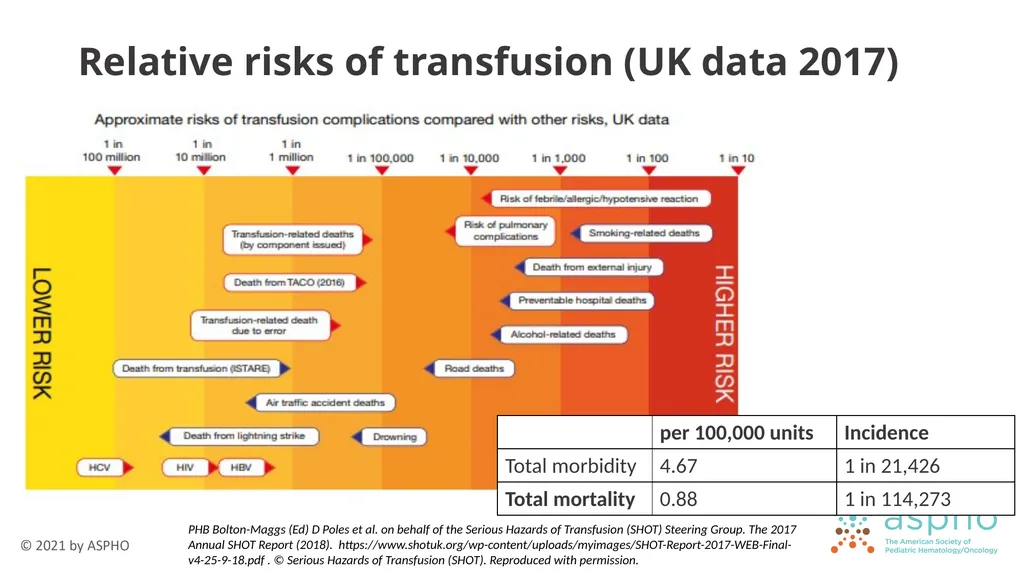
Relative risks of transfusion (UK data 2017) PHB Bolton-Maggs (Ed) D Poles et al. on behalf of the Serious Hazards of Transfusion (SHOT) Steering Group. The 2017
Annual SHOT Report (2018). https://www.shotuk.org/wp-content/uploads/myimages/SHOT-Report-2017-WEB-Final-v4-25-9-18.pdf . © Serious Hazards of Transfusion (SHOT). Reproduced with permission.<br>
63
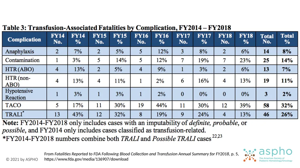
From Fatalities Reported to FDA Following Blood Collection and Transfusion Annual Summary for FY2018, p. 5,
https://www.fda.gov/media/136907/download<br>
64
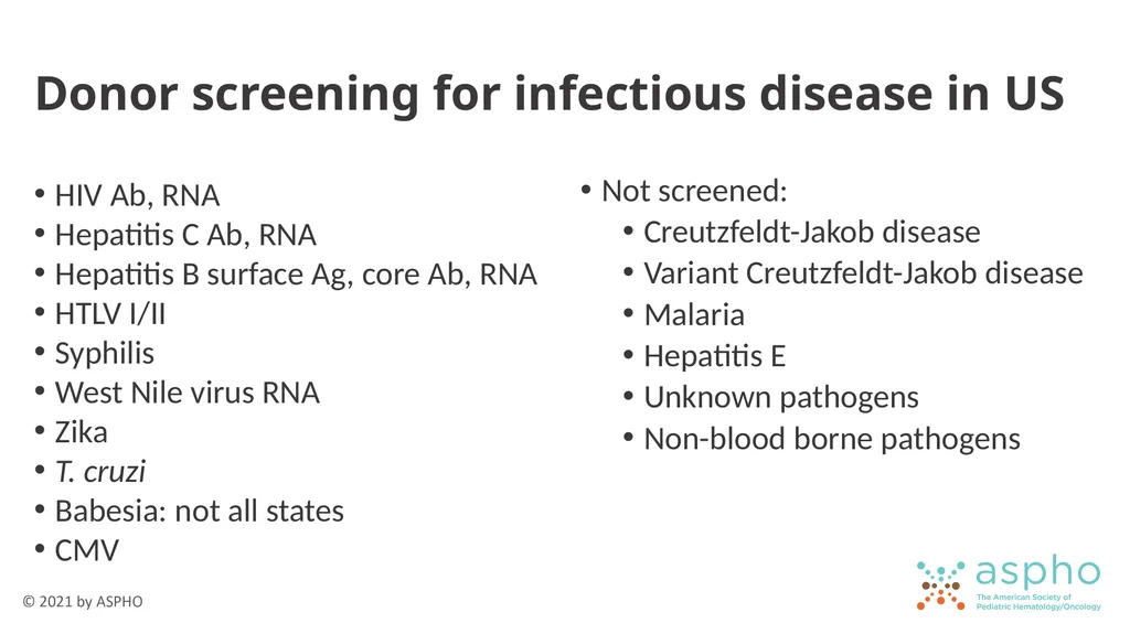
Donor screening for infectious disease in US HIV Ab, RNA
Hepatitis C Ab, RNA
Hepatitis B surface Ag, core Ab, RNA
HTLV I/II
Syphilis
West Nile virus RNA
Zika
T. cruzi
Babesia: not all states
CMV Not screened:
Creutzfeldt-Jakob disease
Variant Creutzfeldt-Jakob disease
Malaria
Hepatitis E
Unknown pathogens
Non-blood borne pathogens<br>
Hepatitis C Ab, RNA
Hepatitis B surface Ag, core Ab, RNA
HTLV I/II
Syphilis
West Nile virus RNA
Zika
T. cruzi
Babesia: not all states
CMV Not screened:
Creutzfeldt-Jakob disease
Variant Creutzfeldt-Jakob disease
Malaria
Hepatitis E
Unknown pathogens
Non-blood borne pathogens<br>
65
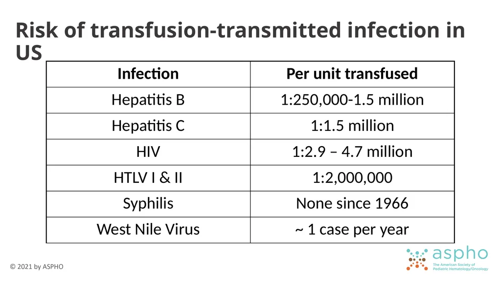
Risk of transfusion-transmitted infection in US<br>
66
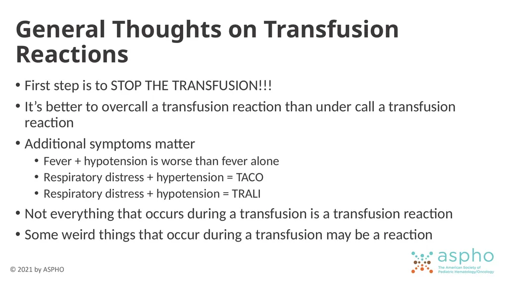
General Thoughts on Transfusion Reactions First step is to STOP THE TRANSFUSION!!!
It’s better to overcall a transfusion reaction than under call a transfusion reaction
Additional symptoms matter
Fever + hypotension is worse than fever alone
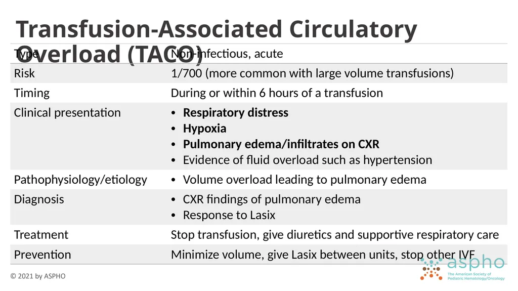
Respiratory distress + hypertension = TACO
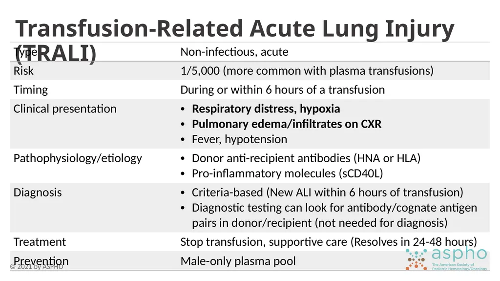
Respiratory distress + hypotension = TRALI
Not everything that occurs during a transfusion is a transfusion reaction
Some weird things that occur during a transfusion may be a reaction<br>
67
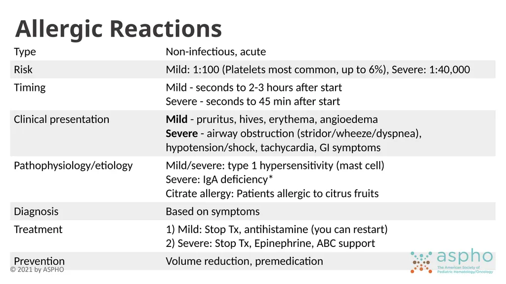
Allergic Reactions<br>
68
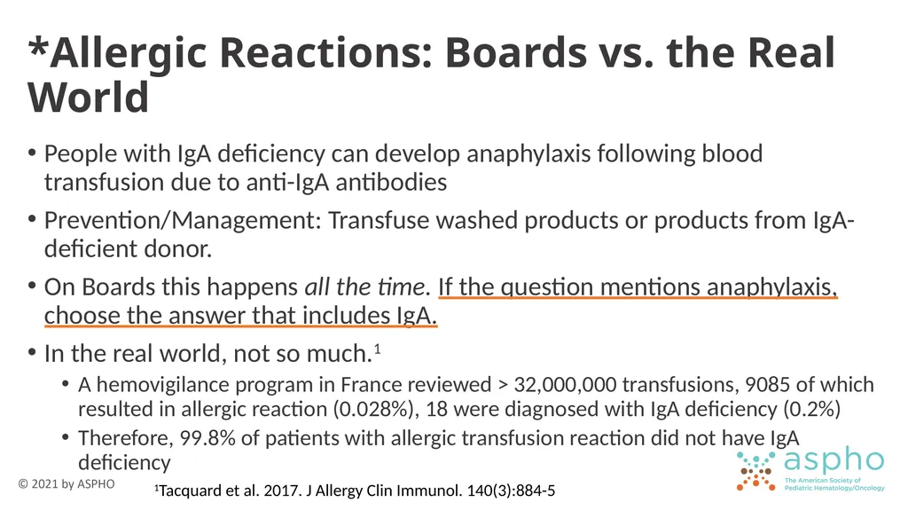
*Allergic Reactions: Boards vs. the Real World People with IgA deficiency can develop anaphylaxis following blood transfusion due to anti-IgA antibodies
Prevention/Management: Transfuse washed products or products from IgA-deficient donor.
On Boards this happens all the time. If the question mentions anaphylaxis, choose the answer that includes IgA.
In the real world, not so much.1
A hemovigilance program in France reviewed > 32,000,000 transfusions, 9085 of which resulted in allergic reaction (0.028%), 18 were diagnosed with IgA deficiency (0.2%)
Therefore, 99.8% of patients with allergic transfusion reaction did not have IgA deficiency 1Tacquard et al. 2017. J Allergy Clin Immunol. 140(3):884-5<br>
69
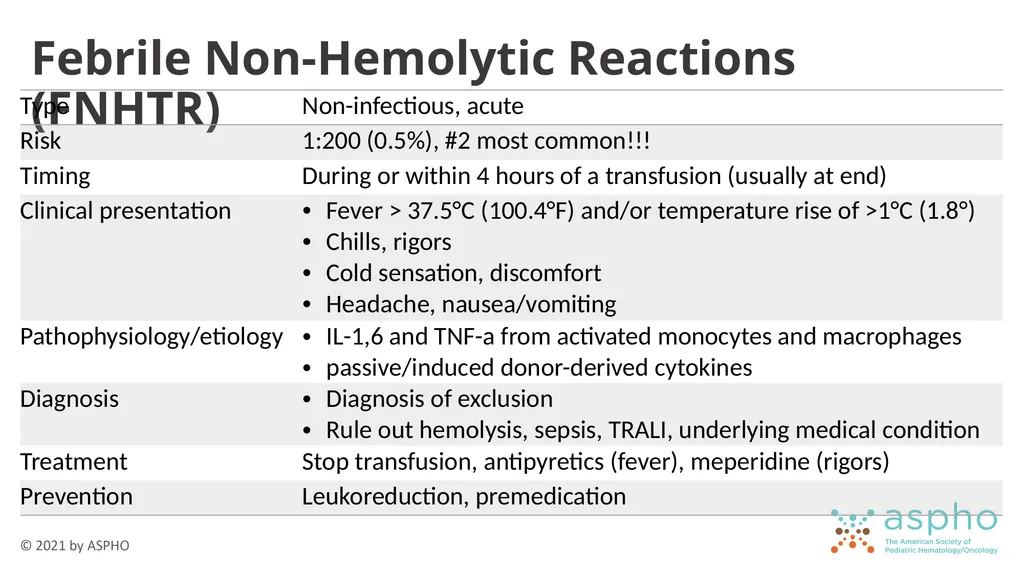
Febrile Non-Hemolytic Reactions (FNHTR)<br>
70
Transfusion-Related Acute Lung Injury (TRALI)<br>
71
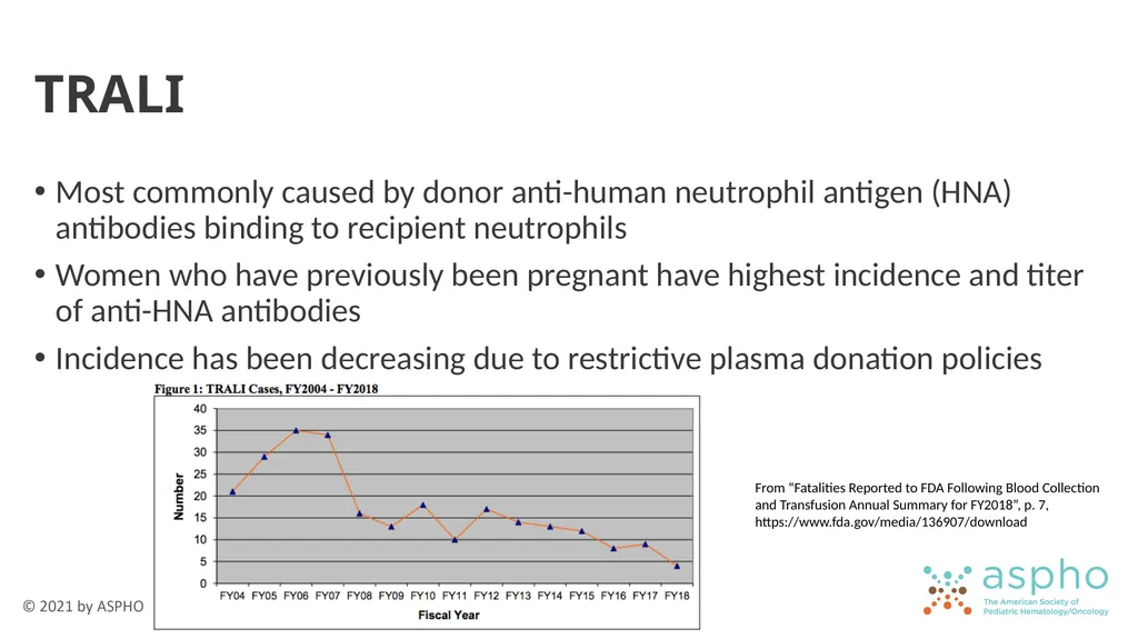
TRALI Most commonly caused by donor anti-human neutrophil antigen (HNA) antibodies binding to recipient neutrophils
Women who have previously been pregnant have highest incidence and titer of anti-HNA antibodies
Incidence has been decreasing due to restrictive plasma donation policies From “Fatalities Reported to FDA Following Blood Collection and Transfusion Annual Summary for FY2018”, p. 7, https://www.fda.gov/media/136907/download<br>
72
Transfusion-Associated Circulatory Overload (TACO)<br>
73
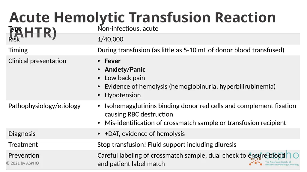
Acute Hemolytic Transfusion Reaction (AHTR)<br>
74
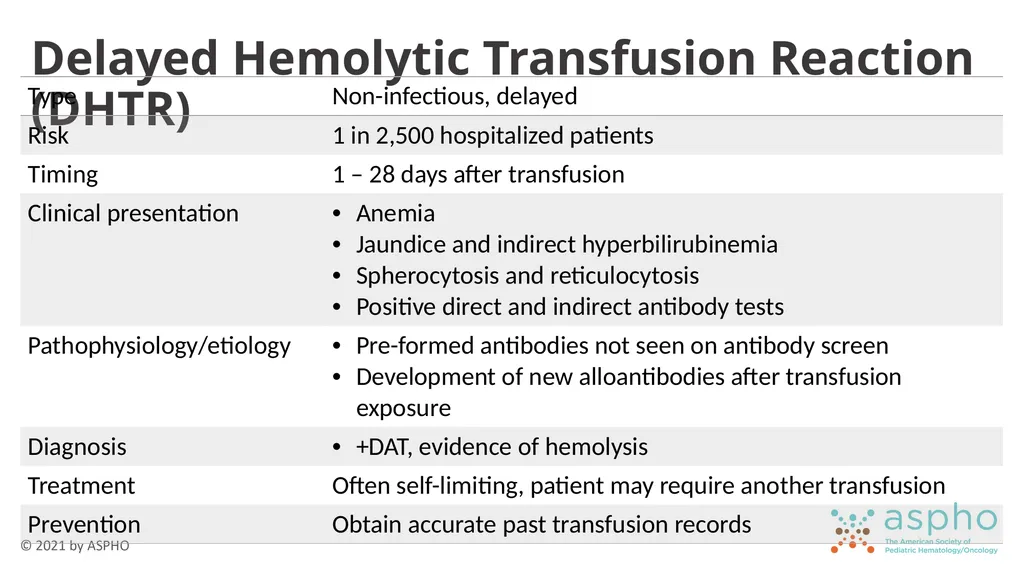
Delayed Hemolytic Transfusion Reaction (DHTR)<br>
75
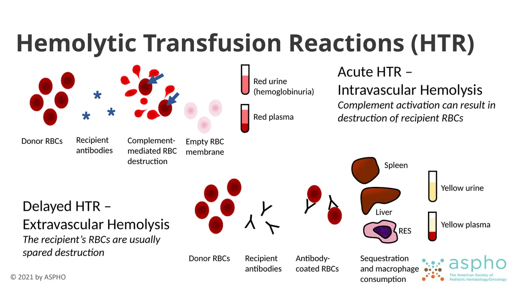
Hemolytic Transfusion Reactions (HTR) Acute HTR –
Intravascular Hemolysis
Complement activation can result in destruction of recipient RBCs Delayed HTR – Extravascular Hemolysis
The recipient’s RBCs are usually spared destruction<br>
Complement activation can result in destruction of recipient RBCs Delayed HTR – Extravascular Hemolysis
The recipient’s RBCs are usually spared destruction<br>
76
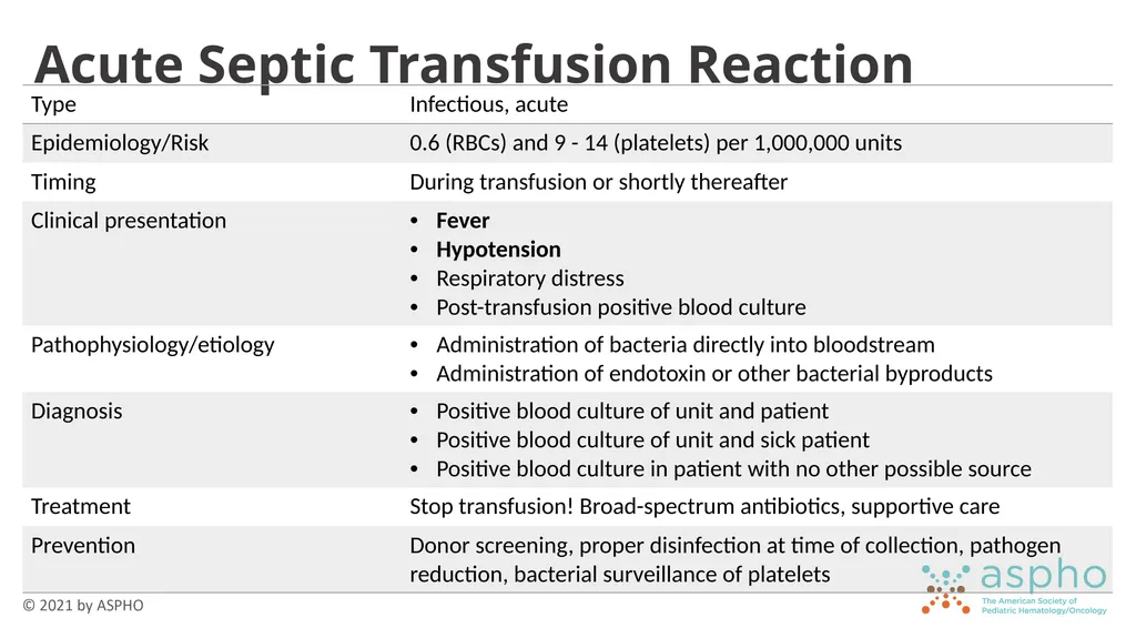
Acute Septic Transfusion Reaction<br>
77
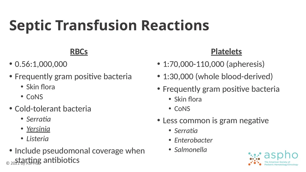
Septic Transfusion Reactions RBCs
0.56:1,000,000
Frequently gram positive bacteria
Skin flora
CoNS
Cold-tolerant bacteria
Serratia
Yersinia
Listeria
Include pseudomonal coverage when starting antibiotics Platelets
1:70,000-110,000 (apheresis)
1:30,000 (whole blood-derived)
Frequently gram positive bacteria
Skin flora
CoNS
Less common is gram negative
Serratia
Enterobacter
Salmonella<br>
78
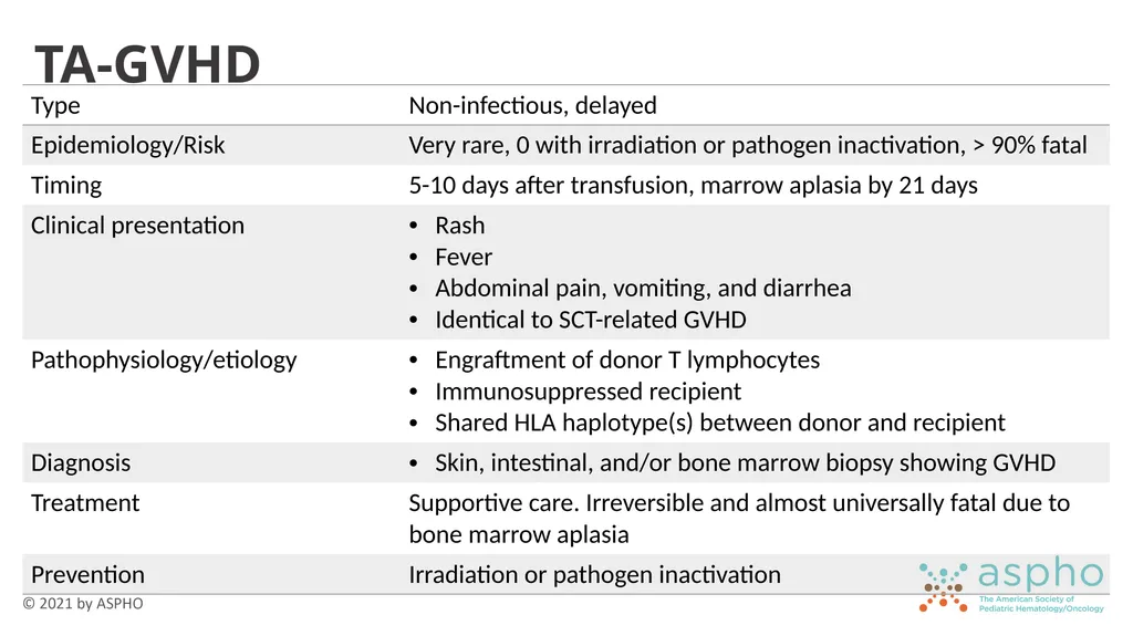
TA-GVHD<br>
79
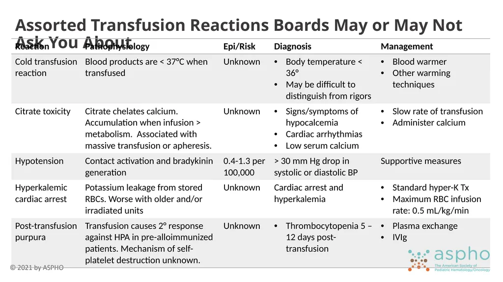
Assorted Transfusion Reactions Boards May or May Not Ask You About<br>
80
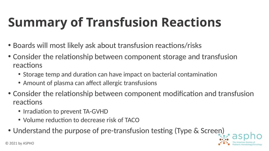
Summary of Transfusion Reactions Boards will most likely ask about transfusion reactions/risks
Consider the relationship between component storage and transfusion reactions
Storage temp and duration can have impact on bacterial contamination
Amount of plasma can affect allergic transfusions
Consider the relationship between component modification and transfusion reactions
Irradiation to prevent TA-GVHD
Volume reduction to decrease risk of TACO
Understand the purpose of pre-transfusion testing (Type & Screen)<br>
81
Good luck and pay attention to the Roman numerals.<br>