
and veins Fetal circulation Malformations Mark Kozsurek MD PhD markkozsurekhu ED I 19022019 httpgutbmjcomcontent577F1mediumgif septum secundum foramen ovale ID: 933259
Download Presentation The PPT/PDF document "Development of the arteries" is the property of its rightful owner. Permission is granted to download and print the materials on this web site for personal, non-commercial use only, and to display it on your personal computer provided you do not modify the materials and that you retain all copyright notices contained in the materials. By downloading content from our website, you accept the terms of this agreement.


Slide1
Development of the arteries and veinsFetal circulation. Malformations
Mark Kozsurek, M.D., Ph.D.mark@kozsurek.hu
ED I., 19/02/2019
http://gut.bmj.com/content/57/7/F1.medium.gif
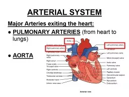
Slide2septum secundum
foramen ovale
septum primum
interventricular septum, muscular part
aorticopulmonary septum
interventricular septum, membranous part
Slide3Development of the veins
Slide41
2
3
Veins
draining
into
the sinus venosus:common cardinal vein (formed by the fusion of anterior and posterior
cardinal veins) umbilical veinvitelline vein
Slide5anterior aspect !!!
sinus venosus
ventricles
atria
Slide6sinus venosus
common cardinal vein
anterior cardinal vein
posterior cardinal vein
umbilical
vein
vitelline
vein
cut edge ot the sinu-atrial junction
Slide7sinus venosus
anastomosis between the right and left anterior cardinal veins
anterior cardinal vein
posterior cardinal vein
Slide8sinus venosus
anastomosis between the right and left anterior cardinal veins
anastomosis between the right and left posterior cardinal veins
anterior cardinal vein
posterior cardinal vein
Slide9sinus venosus
right and left brachiocephalic veins
superior vena cava
right and left common iliac veins
oblique vein of the left atrium
coronary sinus
posteriorcardinal vein
Slide10sinus venosus
right and left brachiocephalic veins
superior vena cava
right and left common iliac veins
oblique vein of the left atrium
coronary sinus
posteriorcardinal vein
right and left vitelline veins from the yolk sac
DUODENUM
Slide11sinus venosus
right and left brachiocephalic veins
superior vena cava
right and left common iliac veins
oblique vein of the left atrium
coronary sinus
posteriorcardinal vein
right and left vitelline veins from the yolk sac
DUODENUM
Slide12sinus venosus
right and left brachiocephalic veins
superior vena cava
right and left common iliac veins
oblique vein of the left atrium
coronary sinus
posteriorcardinal vein
right vitelline vein
DUODENUM
Slide13sinus venosus
right and left brachiocephalic veins
superior vena cava
right and left common iliac veins
oblique vein of the left atrium
coronary sinus
posteriorcardinal vein
right vitelline vein
DUODENUM
Slide14sinus venosus
right and left brachiocephalic veins
superior vena cava
right and left common iliac veins
oblique vein of the left atrium
coronary sinus
posteriorcardinal vein
placenta
umbilical veins
right vitelline vein
left umbilical vein
Slide15sinus venosus
supracardinal vein
subcardinal vein
kidney
superior vena cava
oblique vein of the left atrium
coronary sinus
placenta
right and left common iliac veins
right and left brachiocephalic veins
Slide16sinus venosus
kidney
superior vena cava
right and left brachiocephalic veins
oblique vein of the left atrium
coronary sinus
placenta
liver
supracardinal vein
subcardinal vein
right and left common iliac veins
Slide17sinus venosus
kidney
superior vena cava
right and left brachiocephalic veins
oblique vein of the left atrium
coronary sinus
placenta
liver
supracardinal vein
subcardinal vein
ductus venosus
right and left common iliac veins
Slide18sinus venosus
kidney
superior vena cava
right and left brachiocephalic veins
oblique vein of the left atrium
coronary sinus
liver
placenta
supracardinal vein
subcardinal vein
ductus venosus
right and left common iliac veins
Slide19sinus venosus
placenta
liver
azygos vein
accessory hemiazygos vein
hemiazygos vein
gonadal veins
portal vein
splenic vein
superior mesenteric vein
Slide20In the early stage of the embryonic development a pair of vitelline veins carry blood from the yolk sac, but later only the right one persists.
With the appearance and progressive development of the placenta the role of the vitelline veins is replaced by a pair of umbilical veins, but only the left one remains between the placenta and the primordial heart.
The main venous system draining the head and the caudal part of the embryo is represented by the anterior and posterior cardinal veins, respectively.
Supracardinal veins appear on the lateral side of the developing kidney. The supracardinal veins are connected with a horizontal anastomosis, resulting in a H-shaped structure.
Slide21On the medial sides of the primordial kidneys a new pair of veins develop: the subcardinal veins, which are also interconnected with a horizontal anastomosis.
The liver developing in the septum transversum devides the remaining right vitelline vein in three segments: prehepatic, intrahepatic and posthepatic parts can be distinguished.
Due to the growing liver the left umbilical vein is interrupted and looses its contact with the heart. The larger proportion of the blood arriving from the placenta enters the inferior vena cava on the surface of the liver via the ductus venosus, while the rest enters the hepatic sinuses.
Slide22On the left side the cardinal veins degenerate. The only notable persisting vessel is the coronary sinus.
The right anterior cardinal vein develops into the right brachiocephalic vein and the superior vena cava, while the anastomosis between the two anterior cardinal veins remains as the left brachiocephalic vein.
Supra- and subcardinal veins with the prehepatic segment of the right vitelline vein and the interconnecting anastomoses play an important role in formation of the inferior vena cava.
Prehepatic segment of the right vitelline vein develops into the portal vein.The major part of the supracardinal veins persists as the vena azygos/hemiazygos system.
Subcardinal veins are incorporated into the inferior vena cava or develop into the renal and gonadial veins.
Slide23Development of the arteries
Slide24Slide25Slide26Langman’s Medical Embryology
Slide27Slide28Fetal circulation
Postnatal adaptation of the circulatory system
Slide29Slide30Slide31Arterious blood is carried into the fetus by the umbilical vein. A smaller proportion of this blood passes through the liver and is collected by the IVC. The majority of the oxygeneted blood bypasses the liver through the ductus venosus and drains directly into the IVC. The Eustachian valve directs the blood toward the foramen ovale and the left atrium. This blood is then pumped into the aorta and supplies the tissues of the fetus. Finally returnes to the placenta via the two umbilical erteries arising from the internal iliac arteries.Venous blood derived from the head and neck regions as well as from the upper limb is collected by the SVC. This venous blood gets into the right atrium then into the right ventricle. As the lungs are collapsed the vascular resistance is very high: the blood may not flow toward the pulmonary arteries. This is why the ductus arteriosus is essential: it drains the venous blood of the pulmonary trunk into the aorta.Briefly: during the fetal period both of the ventricles eject blood into the aorta!!!
Slide32After birth no more arterious blood arrives through the umbilical vein. The lungs are inflated, the vascular resistance suddenly drops. There is no further reason for the blood ejected by the right ventricle to join the aorta. As more blood reaches the lungs, more returns through the pulmonary veins into the left atrium. The pressure increases here and the septum primum is pushed against the septum secundum: the foramen ovale closes, later the two septa completly fuse.Due to the increased oxygen levels smooth muscles of the ductus arteriosus contract and obliterate the lumen. (Otherwise the higher pressure in the aorta would result in a reversed flow through the ductus venosus.)Ductus venosus is slowly occupied by proliferating connective tissue and remains observable on the visceral surface of the liver as the venous ligament.
Slide33Malformations
1.
situs inversus totalis: all the viscera are mirrored. Prevalance is less than 1/10 000.
Slide342.
dextrocardia
: only the heart is swapped to the right side of the thorax (first seen and drawn by Leonardo da Vinci in 1452–1519)
Slide353.
atrial
and ventricular septal defect (ASD/VSD)
Slide364.
transposition
of
the
great
vessels: aorticopulmonary septum twists only 90 degrees.
Slide375.
Tetralogy
of
Fallot: a complex malformation
Slide38Thank you for your attention!