
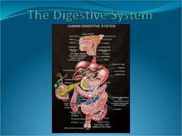
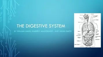
Digestive System Alimentary Canal Long muscular organ that includes the mouth oral cavity pharynx esophagus stomach small intestine large intestine amp anus Buccal Cavity Mouth receives food as it enters the body ID: 932010
Download Presentation The PPT/PDF document "Digestive System The Gastrointestinal Sy..." is the property of its rightful owner. Permission is granted to download and print the materials on this web site for personal, non-commercial use only, and to display it on your personal computer provided you do not modify the materials and that you retain all copyright notices contained in the materials. By downloading content from our website, you accept the terms of this agreement.

Slide1
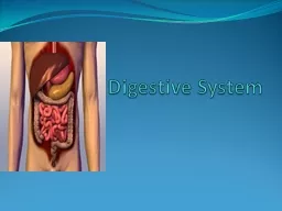
Digestive System
The Gastrointestinal System
Slide2Digestive System
Slide3Alimentary Canal
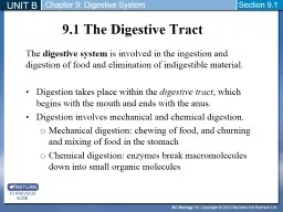
Long, muscular organ that includes the mouth (oral cavity), pharynx, esophagus, stomach, small intestine, large intestine & anus
Slide4Buccal Cavity
Mouth – receives food as it enters the body
Teeth - Food in the mouth is tasted, broken down physically by the teeth, digested, by saliva & swallowed
Mastication – Chewing & grinding of food by the teeth
Slide5Buccal Cavity
Tongue – Muscular organ with special receptors – taste buds
Hard Palate – bony structure that forms the roof of the mouth
Soft Palate – Separates the mouth from the nasopharynx
Uvula – hangs from the soft palate & keeps food from entering the nasopharynx during swallowing
Slide6Buccal Cavity
3 pairs of salivary glands
Parotid
Sublingual
Submandibular
Produce saliva to lubricates the mouth during speech & chewing and moistens food so that it can be swallowed easily
Slide7Salivary Glands
Saliva contains the enzyme salivary amylase (ptyalin)
Begins the chemical breakdown of carbohydrates (starches), into sugars that can be used by the body
Bolus – food that has been chewed & mixed with saliva
Slide8Pharynx
The Throat
A tube that carries the air to the trachea & food to the esophagus
When a bolus is being swallowed muscle action causes the epiglottis to close over the larynx, preventing the bolus from entering the respiratory tract causing it to enter the esophagus
Slide9Esophagus
Muscular tube dorsal to the trachea
Receives the bolus from the pharynx & carries it to the stomach
Relies on a rhythmic, wavelike involuntary movement of its muscles called peristalsis
Slide10Stomach
Enlarged part of the alimentary canal.
Receives food from the esophagus
Rugae – folds inside the stomach
Cardiac Sphincter – between esophagus & stomach
Pyloric sphincter – between stomach & small intestine
Slide11Stomach
Food in stomach 1-4 hours
Chyme – food is converted into a semi-fluid material
Gastric Juices – contain hydrochloric acid & enzymes
Slide12Stomach Enzymes
Hydrochloric Acid – kills bacteria, facilitates iron absorption & activates the enzyme pepsin
Lipase – starts the chemical break down of fats
Pepsin starts protein digestion
Rennin aids in the digestion of milk (found only in children)
Slide13Small Intestine
Coiled section of alimentary canal 20 feet long
Divided into the:
duodenum (9-10 in.) jejunum (8 ft.) ileum (12 feet)
Cecum – place that the small intestine connects to the large intestine.
Ileocecal valves – separates the ileum & cecum
Slide14Small Intestine
Slide15Enzymes in the Small Intestine
Duodenum – Bile (from the gallbladder & liver) and pancreatic juice enter through ducts
Process of digestion is completed in the small intestine
The products of digestion are absorbed into the blood stream for use by body cells
Slide16Small Intestine Enzymes
Maltase, sucrase & lactase break down sugars into simpler forms
Peptidases – completes the digestion of proteins
Steapsin – aids in the digestion of fats
Bile – from liver & gallbladder emulsifies fats
Pancreatic enzymes complete digestion
Pancreatic amylase or amylopsin acts on sugars
Trypsin and chymotrypsin act on proteins
Lipase or steapsin acts on fats
Slide17Absorption
After food is digested it is absorbed into the bloodstream
Walls of small intestine are lined with finger-like projections called villi
Villi contain blood capillaries & lacteals
Blood capillaries absorb the nutrients & carry them to the liver
Slide18Absorption
Slide19Large Intestine
5 ft. in length & 2 in. in diameter
Functions include: absorption of water & remaining nutrients, storage of indigestible materials before elimination
Synthesis of some B complex vitamins & vitamin K
Transportation of waste products from the body
Slide20Large Intestine
Cecum – first section & is connected to the ileum of the small intestine. It contains a small projection called the veriform appendix
Slide21Large Intestine
Colon has several divisions:
Ascending colon – up the right side of body from the cecum to the lower part of the liver
Transverse Colon – Extends across the abdomen, below the liver & stomach and above the small intestine
Slide22Large Intestine
Descending Colon – extends down the left side of the body. Connects with the sigmoid colon
Sigmoid Colon, an S shaped section that joins the rectum
Rectum – final 6 – 8 in. of the large intestine & is a storage area for indigestible & waste
The narrow anal canal opens at a hole called the anus
Fecal material or stool, the final waste product is expelled
Slide23Liver
Slide24Liver
Largest Gland
Secretes Bile which emulsifies fats & makes them water soluble
Stores Sugar in the form of glycogen
Stores Iron & Vitamins
Produces heparin, fibrinogen, prothrombin & Cholesteral
Detoxifies substances
Destroys bacteria taken into the blood from the intestine
Slide25Liver
Heparin: Prevents clotting
Fibrinogen: Blood Protein aids in clotting
Prothrombin: Blood Protein aids in clotting
Slide26Gallbladder
Small, muscular sac located under liver
Stores & concentrates bile
Bile needed to emulsify fats
Slide27Pancreas
Glandular organ
Produces pancreatic juices & enzymes
Produces insulin
Slide28Diseases
Appendicitis
– Acute inflammation or infections of the appendix
Symptoms: RLQ abdominal pain, nausea/vomting, mild fever, elevated WBC.
Danger- peritonitis
Slide29Diseases
Cholecystitis- inflammation of the gallbladder, characterized by formation of cholelithiasis (gallstones, which are crystallized cholesterol, bile salts, and bile pigments)
Symptoms- pain under the rib cage radiating to the right shoulder, indigestion, n/v occurs after eating fatty meal.
Slide30continued
Cholecystitis
- if a gallstone blocks the bile duct, it can rupture causing peritonitis
Treatment-low fat diet, lithotripsy, and possible cholecystectomy
Slide31Diseases
Constipation
-when fecal material stays in the large intestine too long causing excessive reabsorption of water. Stool becomes hard, dry, and difficult to eliminate.
Causes- low fiber diet; chronic laxative use makes the bowel “lazy”
Treatment- high fiber diet; fluids, and exercise (avoid laxatives if possible)
Slide32Diseases
Diarrhea-
frequent water stools caused by infection, stress, diet, irritated colon, or toxic substances. Can be dangerous in infants and small children.
Treatment is directed at eliminating the cause, modifying diet, and adequate fluids.
Slide33Diseases
Diverticulitis
- inflammation of the diverticula (sacs) that form in the lining of the intestine. Occurs when fecal material or bacteria become trapped in the “sacs”. Can result in abscess or rupture and cause peritonitis.
Symptoms- abdominal pain, gas, abdominal distention, constipation, diarrhea
Treatment-antibx, pain med, surgery
Slide34Diseases
Gastroenteritis-
inflammation of the mucosal membrane lining the GI tract.
Cause-food poisoning, infection, toxins
Symptoms- abdominal cramping, n/v, diarrhea
Treatment- replace fluids, rest, antibiotics, IV fluids if severe.
Slide35Diseases
Hemorrhoids
- painful dilated or varicose veins of the rectum or anus.
Symptoms- pain, itching, bleeding
Treatment- high fiber diet, stool softeners, sitz baths, warm moist compresses.
If severe, hemorrhoidectomy.
Slide36Diseases
Hepatitis
- inflammation of the liver
A- infectious hepatitis- transmitted in food or water that has been contaminated by feces of an infected person. Most benign type.
B- transmitted by all body fluids including breast milk, saliva, and urine. (vaccine)
C- also transmitted through blood and body fluids but more likely to cause cirrhosis.
Slide37Hepatitis continued
C-no vaccine
Symptoms- fever, fatigue, anorexia, n/v, mylagia, dark urine, clay colored stool, enlarged liver and jaundice.
Treatment- rest, diet high in protein and calories but low in fat. If severe, liver transplant.
Slide38Diseases
Hernia
- a rupture that occurs when an internal organ pushes through a weakening or natural opening in a body wall.
hiatal hernia- when the stomach protrudes through the diaphragm into the chest cavity through the opening for the esophagus.
If a hernia cannot be reduced, then a herniorraphy is performed.
Slide39Diseases
Ulcer
– open sore on the lining of the digestive tract. Peptic ulcers include stomach and duodenal ulcers.
Causes- H. pylori bacteria is most common cause (infection) by burrowing into the lining allowing gastric juices to create an ulcer.
Symptoms- burning pain, indigestion, melena, and hematemesis.
Slide40Ulcers cont.
Treatment- bland diet, decreased stress, avoiding irritants. Bismuth medications (pepto bismol is used) If H. Pylori is the cause, this is an infection that should be treated with antibiotics.
Slide41Diseases
GERD (gastroesophageal reflux disease
)- occurs when stomach contents and acid leak back into the esophagus and there are visible signs of irritation to the lining of the esophagus. Many people have acid reflux, heartburn, and indigestion but that doesn’t mean they have GERD.
Slide42GERD cont.
Symptoms- burning pain in your lower chest, tasting acid in your mouth, throat.
Treatment- dietary and lifestyle changes; avoiding irritation to the esophagus (chocolate, peppermint, alcohol, fatty foods, coffee, citrus fruits and juices)
Slide43Slide44