/
The Endocrine system Endocrine glands: The Endocrine system Endocrine glands:

The Endocrine system Endocrine glands: - PowerPoint Presentation

SereneBeauty
SereneBeauty . @SereneBeauty
Follow
342 views
Uploaded On 2022-08-03

The Endocrine system Endocrine glands: - PPT Presentation

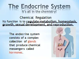
Source of hormone reached through circulation via ductless glands of internal secretion Composed of islands cords of secretory epithelial cells parenchyma with intervening supporting tissue stroma rich in blood and lymphatic capillaries prominent nuclei prolific cytoplasmic ID: 933635

gland cells hormone thyroid cells gland thyroid hormone blood secretory cytoplasm adrenal hormones pituitary pars secret small follicles nuclei

Share:

Link:

Embed:

Download Presentation from below link

Download Presentation The PPT/PDF document "The Endocrine system Endocrine glands:" is the property of its rightful owner. Permission is granted to download and print the materials on this web site for personal, non-commercial use only, and to display it on your personal computer provided you do not modify the materials and that you retain all copyright notices contained in the materials. By downloading content from our website, you accept the terms of this agreement.


Presentation Transcript

Slide1

The Endocrine system

Endocrine glands:

-

Source of hormone; reached through circulation via ductless glands of internal secretion.

-Composed of islands (cords) of secretory epithelial cells (parenchyma) with intervening supporting tissue (stroma) rich in blood and lymphatic capillaries; prominent nuclei; prolific cytoplasmic.

-Organelles (mitochondria, ER, Golgi bodies and secretory vesicles).

-The cells secrete hormone directly in to blood or lymph circulation, therefore they have rich blood supply.

Slide2

*Endocrine glands are either found as separate unite like:

1-Pituitary gland.

2-Thyroid gland.

3-Parathyroid gland.

4-Adrenal or suprarenal gland.

5-Pineal body.

*Or found as scattered masses within the exocrine gland-called mixer gland like:

1-Islet of pan crease.

2-Leydig cells of (interstitial cells of the testes).

3-Corpus luteum of the ovary.

Slide3

1-Pitutary gland (hypophysis):

1cm in diameter, weight 0.5, in female during pregnancy reach to 1 gm, have two lobes:

-Anterior pituitary:

develops as outgrowth of hypophyseal (Rathke´s) pouch, as up growth from the roof of the mouth.

Anterior lobe or adenohypophysis of 3 parts:

_pars distal is

: it is the largest subdivision constitute about 75% of anterior pituitary surrounded by collagen fibers.

_pars intermedium :

-thin zone tissue lying between the pars distalis and pars nervosa.

-cells basophilic, function in adult is not clear, but in the fetus the basophils may synthesizes and secretes melanocyte stimulating hormone (MSH), important for melanocyte activity.

Slide4

_pars tuberalis:

is a funnel-shaped region surrounding the Infundibulum of the neurohypophysis (most of the cells of this region are basophilic gonadotropic cells that secret FSH and LH).

*Hormones of interior pituitary:

-Growth hormone(GH) -Somatotrope cells.

-Prolactin(PRL) -lactotrope cells=

Mammotrope

.

-Thyroid stimulating H(TSH) -Thyrotrope cells.

-FSH &LH -Gonadotrope.

-Adrenocorticotropin (ACTH) -Corticotrope.

_Posterior lobe(Neurohypophysis):

-develop as down growth from the floor of the brain (Diencephalon).

-Consist of three parts:

1-Median eminence.

2-Infundibular stalk.

3-Infundibular process (pars nervosa).

Slide5

*Hypothalamus:

-control secretion of all pituitary hormones

.

-

located in small depression of sphenoid bone (Sella turcica).

*Hormones of posterior pituitary:

-Antidiuretic hormone(ADH)=synthesized mainly by neurons of hypothalamic supraoptic nucleus.

-Oxytocin =synthesized mainly by neurons of paraventricular of hypothalamus.

Slide6

2-Thyroid gland:

is surrounded by 2 capsules:

-

The outer is thick continuous with the deep cervical fascia.

-The inner, true, CT capsule sends in septa to partially enclose lobules.

In the lobules are rounded or bodies-follicles, in a loose stroma of ct with many blood vessels.

_Thyroid follicle:

-In man, they vary between 0.02 and 0.9 mm in diameter, a gland has several million follicles.

-filled with viscous fluid-thyroid colloid-variably acidophil.

-thyroid follicles are irregular, spherical structures; single layer range from squamous or cuboidal to columnar depending on the activity of the gland.

Slide7

-epithelial cells+basment membrane ;variable in size, contain homogeneous thyroid or thyroglobulin which consist of glycoprotein, mucoprotein and proteolytic enzyme.

-in active gland ;follicles small, colloid diminishes, cuboidal lining high, less active gland, lining cells flattened mostly lined by basophilic cuboidal follicular cells, a basal lamina outside which is an extensive plexus of blood capillaries with reticular fibers and fibroblast.

-follicular cells are surrounded the follicle lumen, the nucleus is central.

-thyroid stimulated by thyroid-stimulating hormone (thyrotropin) adenohypophysis.

Slide8

-thyroid follicles epithelial cells absorb iodide and concentrate it from blood by an iodide pump in basal plasma membrane ; iodide oxidized to iodine and transported in to follicular lumen; combines with thyroxin residues of thyroglobulin which remain bound to glycoprotein.

-polypeptide hormone calcitonin; lowers blood calcium by inhibiting rate of decalcification of bone by osteoclastic resorption and by stimulating osteoblastic activity ; regulated by blood calcium levels.

Para follicular cells (C or clear cells) present in follicle walls but not extending to colloid in interfollicular spaces; synthesize and secret calcitonin.

Slide9

3-Parathyroid gland:

Embedded in capsule of thyroid gland, thin fibrous capsule , delicate septa divide gland in to dense, cord-like of secretory cells, regulate serum calcium and phosphate levels via parathyroid hormone.

_Two types secretory cells:

1-Chief (principle) cells: secret parathyroid hormone; prominent nucleus and relatively little cytoplasm, actively secreting cells, represent about 20% of total.

2-Oxyphil cells: larger , less numerous, tend to clump among chief cells, smaller densely stained nuclei, strongly eosinophilic cytoplasm containing fine granules, function unknown.

Slide10

4-Adrenal glands(Suprarenal) :

-are paired organs that lie near the superior poles of the kidney, embedded in perirenal adipose tissue.

-They are flattened structures with a half –moon shape, about 4-6 cm long, 1-2 cm , 4-6 mm thick in adults.

-adrenal glands are each covered by a dense c.t capsule that send thin septa to the interior of the gland as trabeculae, the stroma consist mainly of a rich network of reticular fibers that support the secretory cells, the gland consist of two concentric layers ;a yellowish peripheral layer, the adrenal cortex and a reddish brown central layer, the adrenal medulla with prominent central vein.

Slide11

_Steroid secretory cells:

-mitochondria with unusual tubular cristae.

-secretes steroid hormones, structurally related to common precursor cholesterol, regulated by ACTH; 3 functional classes:

1-mineralocorticoids 2-glucocorticoids 3-sex hormones

_Adrenal Cortex :

-zona glomerulosa:

moderately appearance, secretory cells in rounded clumps, round strongly stained nuclei, little cytoplasm, some lipid droplets.

Secret mineralocorticoids (aldosterone) : regulation body sodium in renal tubules, blood pressure regulation, aldosterone controlled by juxtaglomerular apparatus.

Slide12

-Zona fasiculata:

parallel narrow cords of secretory cells arrange in column 1-2 cell thick, parallel to each other; cells large, abundant cytoplasm, large amount of lipid droplets appears as vacuoles which represent the steroid hormone.

Secret glucocorticoids (Cortisol), regulating glucose, fatty acid, metabolism and response to stress; controlled by ACTH also small androgenic sex hormones.

-Zona reticularis:

small closely-packed cells in irregular network of branching cords and clumps, cells smaller than those of Z. fasciculata, eosinophilic cytoplasm, few lipid droplets, cytoplasm stains more strongly, lipofuscin, probably secret small quantities androgens and glucocorticoids.

Slide13

_Adrenal medulla secretory cells:

-large granular nuclei, extensive, strongly basophilic cytoplasm.

-secretes catecholamine hormones: adrenaline and noradrenalin under direct preganglionic sympathetic nervous control; not secreted continuously, stored in cytoplasmic granules and released in response to nervous stimulation after chrome salt fixation, stored catecholamine granules from brown color chromaffin cells.

5-Pineal Body:

The seat of the soul-the third eye

-pineal evagination from posterior part of roof of third ventricle in midline, connected to brain via short stalk containing nerve fibers (from hypothalamus).

Slide14

-pineal sand (basophilic extracellular bodies) concentric layers of calcium and magnesium phosphate within an organic matrix.

-pinealocytes; pineal chief cells, highly modified neurons arranged in clumps and cords surrounded by rich net work of fenestrated capillaries, round, granular nuclei with prominent nucleoli and poorly stained cytoplasm; highly branched processes.

-contain variety of indole compounds including melatonin and its precursor serotonin.