
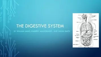
system is responsible for the physical and chemical breakdown of food so it can be taken into the bloodstream and used by body cells and tissues The Alimentary Canal Long muscular ID: 918789
Download Presentation The PPT/PDF document "Digestive System The digestive" is the property of its rightful owner. Permission is granted to download and print the materials on this web site for personal, non-commercial use only, and to display it on your personal computer provided you do not modify the materials and that you retain all copyright notices contained in the materials. By downloading content from our website, you accept the terms of this agreement.

Slide1
Digestive System
Slide2The digestive system is responsible for the physical and chemical
breakdown
of food so it can be taken into the bloodstream and used by body
cells
and
tissues.
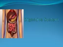
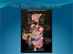
Slide3The Alimentary Canal
:
Long
muscular tubeBegins at the mouth and includes the pharynx, esophagus, stomach, small intestines, large intestines, and the anus.The Accessory Organs:The salivary glands, tongue, teeth, liver, gallbladder, and pancreas
The Digestive System includes:
Slide4Mouth,
buccal
, or oral cavity
A. Receives food as it enters the bodyB. Actions in the mouth1
.
Food is
tasted2. Broken down physically by the teeth3. Lubricated and partially digested by saliva4. Swallowed
Where does digestion start
?
Slide5Special structures in the mouth
2. Break down food physically by chewing and grinding the food, a process called
mastication
Teeth
Slide6Muscular organ
2. Contains special receptors called
taste
buds that allow person to taste sweet, salt, sour, and bitter sensations3. Also aids with chewing and swallowing of foodTongue
Slide7Hard palate
1. Bony structure that forms the
roof
of the mouth2. Separates the mouth from the nasal cavitiesWithin the mouth:
Slide8Soft palate
1. Located
behind
the hard palate2. Separates the mouth from the nasopharynx
Slide9Uvula
(a) Cone-shaped muscular
structure
(b) Hangs from the middle of the soft palate(c) Prevents food from entering the
nasopharynx
during
swallowing
Slide10Slide111.
Three
pairs of glands
Parotid, sublingual, and submandibular 2. Produce a liquid called saliva(a) Lubricates the mouth during speech and chewing(b) Moistens food so it can be swallowed easily(c) Also contains an enzyme called salivary amylaseaa. Substance speeding up a chemical reaction
bb. Begins the
chemical
breakdown of carbohydrates or starches into sugars that can be taken into the body Salivary Glands
Slide12Slide13A. After
the food is chewed and mixed with saliva, it is called a
bolus
and it enters the pharynx or throatB. Tube that carries both air and foodC. Carries the air to the trachea or windpipeD. Carries food to the esophagusPharynx or Throat
Slide14When bolus is swallowed, muscle action causes the
epiglottis
to close over the larynx
2. Prevents bolus from entering respiratory tract3. In this way, the bolus enters the esophagusIn the esophagus:
Slide15www.youtube.com/watch?v=1YfO11Pry6Y&feature=player_detailpage
Normal Swallow Animation - Thick and Easy Dysphagia - YouTube
Slide16Muscular tube
dorsal
to the trachea
Receives bolus from the pharynx and carries it to the stomachRelies on a rhythmic, wavelike involuntary movement of its muscles, called peristalsis, to move the food in a forward directionEsophagus
Slide17Slide18Enlarged
part of the alimentary canal
Receives the food from the
esophagusMucous membrane lining contains folds called rugae, which disappear as the stomach fills with food and expands Stomach
Slide19Slide20Circular
muscle between the esophagus and stomach
Closes
after food enters the stomachPrevents food from going back up into the esophagusCardiac Sphincter
Slide21Slide22Circular muscle between the stomach and small intestine
Keeps food in the
stomach
until it is ready to enter the small intestineFood usually remains in the stomach for about one to four hoursPyloric Sphincter
Slide23Slide24Produced
by glands in the stomach
Converts food into a semi fluid material called chymeGastric Juices
Slide25Juices
contain
hydrochloric acidKills bacteriaFacilitates the absorption of ironActivates the enzyme pepsinGastric Juices
Slide26Contain enzymes:
Lipase
, which begins the chemical breakdown of fats
Pepsin, which starts protein digestionIn an infant, the enzyme rennin is secreted1) Aids in the digestion of milk2) Not present in an adultGastric Juices
Slide27Coiled section of the alimentary canal about
20
feet long and 1 inch in
diameterReceives food, in form of chyme, from stomachSmall Intestine
Slide28There are three sections:
Duodenum—
first 9-10 inches
Jejunum—next 8 feetIleum—final 12 feetSmall Intestines
Slide29The first 9-10 inches
Bile
from the gallbladder and liver enter this section through ducts or tubes
Pancreatic juices from the pancreas also enter this section through ducts or tubes.Small Intestines--Duodenum
Slide30Eight
feet long
Forms the
middle section of the small intestineSmall Intestines--Jejunum
Slide31Final 12 feet
of the small intestine
Connects with the large intestine at the
cecumCircular muscle called the ileocecal valve separates the ileum and cecum and prevents food from returning to the ileum.Small Intestines--Ileum
Slide32Completes the process of digestion
Absorbs the products of digestion into the bloodstream for use by
body cells
Functions of the Small Intestines:
Slide33Produced by the small intestine
Contain the enzymes maltase,
sucrase
, and lactase, which break down sugars into simpler formsContain enzymes known as peptidases, which complete the digestion of proteinsContain the enzyme steapsin, which aids in the digestion of fatIntestinal Juices of the Small Intestines
Slide34Liquid that enters small intestine from
liver
and gallbladder
2. Emulsifies or physically breaks down fatsBile
Slide351.Liquid that enters small intestine from
pancreas
2. Contains
enzymes that complete the process of digestion A) Pancreatic amylase or amylopsin, which acts on sugars B) Trypsin and chymotrypsin, which act on proteins C) Lipase or steapsin, which acts on fatsPancreatic Juice
Slide36Fingerlike
projections that line wall of small intestine
Allow food to be absorbed or taken into
bloodstreamContain blood capillaries and lacteals Small Intestine--Villi
Slide37Blood capillaries absorb the digested
nutrients
and carry them to the liver where they are stored or released into general circulation for use by body cells
Small Intestine—Blood Capillaries
Slide38Lacteals
pick up most of the digested fats and carry them to the thoracic duct in the lymphatic system, which releases them into the circulatory system
Small Intestines--
Lacteals
Slide39When food has completed its passage through the small intestine only wastes, indigestible materials, and excess water remain
Small Intestines
Slide40Final
section of the alimentary canal
About
5 feet long and about 2 inches in diameterLarge Intestines
Slide41Absorption
of water and any remaining nutrients
Storage of indigestible materials before they are eliminated from the body Synthesis (formation) and absorption of some B-complex vitamins and vitamin K by bacteria present in intestine Transportation of the waste products out of the alimentary canalLarge Intestines--Functions
Slide42Cecum:
First
section
Connects with the ileum of the small intestineContains a small projection called the vermiform appendixLarge Intestines--Sections
Slide43Slide44Ascending colon:
continues up on the right side of the body from the cecum to the lower part of the
liver
Large Intestines--Sections
Slide45Transverse
colon:
extends across the abdomen, below the liver and stomach, but above the small intestineLarge Intestine--Sections
Slide46Descending colon:
extends down the left side of the body
Large Intestines--Sections
Slide47Sigmoid
colon:
Connects with
descending colonS-shaped section that joins with the rectumLarge Intestines--Sections
Slide48Rectum:
Final 6 to 8inches
Storage area for the
indigestibles or wastesHas a narrow canal called the anal canal, which opens at a hole called the anusFecal material or stool, the final waste product of the digestive processLarge Intestines--Sections
Slide49Liver:
Largest
gland in the body
Accessory organ for the digestive tractLocated under the diaphragm in the upper right quadrant of the abdomen Accessory Organs
Slide501. Secretes
bile
a. Used to emulsify or physically break up fats b. Also makes fats water soluble, which is necessary for absorption 2. Stores sugar in the form of glycogen a. Glycogen is converted to glucose
b. Released
into the bloodstream when additional blood sugar is needed
3. Stores iron and certain vitamins 4. Produces heparin, a substance that prevents clotting of the blood 5. Produces blood proteins such as fibrinogen and prothrombin, which aid in the clotting of blood
6. Produces
cholesterol
7.
Detoxifies
(renders less harmful) substances such as alcohol and pesticides, and destroys bacteria that have been taken into the blood from the intestine
Functions of the
Liver
:
Slide51Gallbladder:
1. Small muscular
sac2. Located under the liver and attached to it by connective tissue 3. Stores and concentrates bile, which it receives from the liver4. When the bile is needed in the digestive tract to emulsify fats, it contracts and pushes the bile through the common bile duct into the duodenumAccessory Organs
Slide52Pancreas:
1. Fish-shaped organ located behind the
stomach
2. Produces pancreatic juices a. Juices enter duodenum through pancreatic duct b. Contain
enzymes
to digest food
(1) Pancreatic amylase or amylopsin to break down sugars (2) Trypsin and chymotrypsin to break down proteins (3) Lipase or steapsin to act on fats3. Produces insulin a. Secreted into the bloodstream b. Regulates the metabolism or burning
of
carbohydrates to
convert
glucose
(
blood sugar) to energy
Accessory Organs