

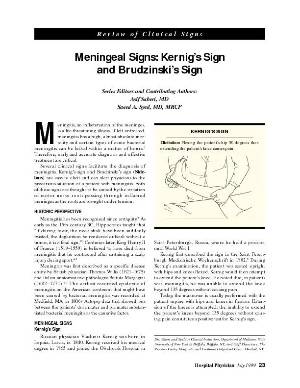
2 CNS Infection Meninges CNS infections most are due to sepsis Pathogenesis Hematogenous spread most common Traumatic implantation Local extension from nearby infection Ascent of peripheral nerve ID: 1045109
Download Presentation The PPT/PDF document "1 CNS Infection Meninges and Prions" is the property of its rightful owner. Permission is granted to download and print the materials on this web site for personal, non-commercial use only, and to display it on your personal computer provided you do not modify the materials and that you retain all copyright notices contained in the materials. By downloading content from our website, you accept the terms of this agreement.


1. 1CNS InfectionMeninges and Prions
2. 2CNS Infection - MeningesCNS infections: most are due to sepsisPathogenesis:Hematogenous spread (most common) Traumatic implantation Local extension from nearby infection Ascent of peripheral nerve
3. 3MeningitisInflammation of pia mater covering the brain Usually due to hematogenous spread; mechanism for bacterial meningitis:Adherence of bacteria to mucosa of nasopharynx Bacteremia Translocation through blood-brain barrier (BBB) Involves bacterial lysinsBacteria in subarachnoid space attract neutrophils. Acute meningitisRisk factors in children Undernutrition; otitis media Pneumonia; immunodeficiency Viral infection; sickle cell disease Craniofacial abnormality
4. 4Viral MeningitisPathogenesis:Most transmitted by fecal-oral route Respiratory route less commonClinical findings:Fever, nuchal rigidity, headache
5. 5Complications of meningitisSeizures; focal neurologic deficits Cranial nerve palsies Sensorineural hearing loss Communicating and noncommunicating hydrocephalus
6. 6Laboratory findings in viral meningitis (CSF)Increased CSF protein Due to increased vessel permeabilityIncreased total CSF leukocyte count Initially neutrophils but converts to lymphocytes in 24 hoursNormal CSF glucose
7. 7Laboratory findings in bacterial meningitis (CSF)Increased CSF protein Increased total CSF leukocyte count Decreased CSF glucoseUsage of glucose for bacterial and fungal growth (bacterial & fungal meningitis)
8. 8Chronic MeningitisTuberculous meningitisheadache, malaise, mental confusion, and vomitingmoderate increase in cellularity of the CSFmononuclear cells, or a mixture of polymorphonuclear and mononuclear cellsthe protein level is elevatedthe glucose content typically is moderately reduced or normalmay also lead towell-circumscribed intraparenchymal mass (tuberculoma)arachnoid fibrosis (hydrocephalus)
9. 9TB Meningitis - PathologyThe subarachnoid space contains a gelatinous or fibrinous exudatemost often at the base of the brainobliterating the cisterns and encasing cranial nerves There may be discrete white granules scattered over the leptomeningesArteries running through the subarachnoid space may show obliterative endarteritisinflammatory infiltrates in their wallsmarked intimal thickeningThe infection may spread through the CSF to the choroid plexuses and ependymal surfaceFlorid cases show well-formed granulomasoften with caseous necrosis and giant cellsSimilar findings are observed in tuberculomas within the brain
10. 10Chronic MeningitisMeningovascular neurosyphilisNeurosyphilis is a tertiary stage of syphilisoccurs in only about 10% of individuals with untreated infectionmajor manifestations is meningeal, called meningovascular neurosyphilisAs with other chronic infections, there can be parenchymal disease as wellParetic neurosyphilisTabes dorsalis
11. 11Meningovascular neurosyphilisinvolving the base of the brain and sometimes the cerebral convexities and the spinal leptomeningesassociated obliterative endarteritis with distinctive perivascular inflammatory reaction rich in plasma cells and lymphocytescerebral gummas (mass lesions rich in plasma cells) may also occur in relation to meninges and extend into the brain
12. 12Prion DiseasesThis group of diseases includes sporadic, familial, iatrogenic and variant forms of Creutzfeldt-Jakob disease (CJD)several animal diseases from this group are also knownincluding scrapie in sheep and goats and bovine spongiform encephalopathy in cattle ("mad cow" disease)all these disorders are associated with abnormal forms of a normal cellular protein, prion protein (PrPc)The abnormal form of this protein can act as an infectious agentit propagates itselfinjures the cells in which it is presentMost cases of prion disease are either sporadic orassociated with mutations in the gene that encodes PrPc
13. 13Pathogenesis of Prion DiseasesRelated to changes in the conformation of PrP from its native PrPc form to an abnormal configuration calledeither PrPsc (for scrapie) or PrPres (for protease resistant)in the abnormal conformation, the prion protein becomes resistant to protease digestionOnce formed, PrPsc can then initiatecomparable transformation of other PrPc moleculesThe infectious nature of PrPsc proteinability to propagate the pathologic conformational changeThe conformational change can occur spontaneously at an extremely low ratesporadic cases of prion diseaseIf there is a mutation in the gene encoding PrPc, then the change can occur at a higher ratefamilial forms of prion diseaseAccumulation of PrPsc in neural tissue causes cell injuryhow this material leads to the development of cytoplasmic vacuoles and eventual neuronal death is still unknown
14. 14Creutzfeldt-Jakob DiseaseCJD is a rare but well-characterized prion diseaseclinically as a rapidly progressive dementiaIt is sporadic in about 85% of cases (1 per million worlwide)familial forms also existThe disease has a peak incidence in the seventh decadeThere are well-established cases of iatrogenic transmissiondeep implantation electrodescontaminated preparations of human growth hormoneClinically, subtle changes in memory and behavior that rapidly progress to dementiaThe disease is uniformly fatal, with an average duration of only 7 months
15. 15Creutzfeldt-Jakob DiseaseThe progression of the dementia in CJD is usually rapid no or very little macroscopic evidence of brain atrophy On microscopic examination, the pathognomonic finding isa spongiform transformation of the cerebral cortex and deep gray matter (caudate, putamen)this consists of a multifocal process that results in the uneven formation of small, apparently empty, microscopic vacuoles of varying sizes within the neuropil and sometimes in the perikaryon of neurons In advanced cases, there is severe neuronal loss, reactive gliosis, and sometimes expansion of the vacuolated areas into cystlike spaces ("status spongiosus") No inflammatory infiltrate is presentIn all forms of prion disease, immunohistochemical staining demonstrates the presence of proteinase K-resistant PrPsc in tissueWestern blotting of tissue extracts after partial protease digestion allows detection of diagnostic PrPsc
16. 16Variant Creutzfeldt-Jakob DiseaseThey differed from typical CJD in several important respects:the disease affected young adultsbehavioral disorders figured prominently in the early stages of the diseaseneurologic syndrome progressed more slowly than in individuals with other forms of CJDThe neuropathologic findings and molecular features of these new cases were similar to those of CJDa close relationship between the two illnessesMultiple lines of evidence indicate that this new disease is a consequence of exposure tothe prion disease of cattle, bovine spongiform encephalopathyvCJD has a similar pathologic appearance with spongiform change and absence of inflammationthere are abundant cortical amyloid plaques, surrounded by spongiform change
17. 17Variant Creutzfeldt-Jakob DiseaseThey differed from typical CJD in several important respects:the disease affected young adultsbehavioral disorders figured prominently in the early stages of the diseaseneurologic syndrome progressed more slowly than in individuals with other forms of CJDThe neuropathologic findings and molecular features of these new cases were similar to those of CJDa close relationship between the two illnessesMultiple lines of evidence indicate that this new disease is a consequence of exposure tothe prion disease of cattle, bovine spongiform encephalopathyvCJD has a similar pathologic appearance with spongiform change and absence of inflammationthere are abundant cortical amyloid plaques, surrounded by spongiform change