
Marrow Transplantation Chapter 33 Tumor Immunology Figure 3301 Embryonic primary germ layers Redrawn from Larsen WJ Human embryology ed 3 Philadelphia 2001 Churchill Livingstone ID: 911841
Download Presentation The PPT/PDF document "Week 7 Immunology: Tumor Immunology, ..." is the property of its rightful owner. Permission is granted to download and print the materials on this web site for personal, non-commercial use only, and to display it on your personal computer provided you do not modify the materials and that you retain all copyright notices contained in the materials. By downloading content from our website, you accept the terms of this agreement.


Slide1
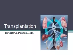
Week 7 Immunology: Tumor Immunology, Solid Organ Transplantation and Bone Marrow Transplantation
Slide2Chapter 33
Tumor Immunology
Slide3Figure 33-01.
Embryonic primary germ layers.
(Redrawn from Larsen WJ:
Human embryology,
ed 3, Philadelphia, 2001, Churchill Livingstone.)
Slide4Introduction
Oncology: study of neoplasms: benign or malignant
Benign Tumors:
encapsulated,
slow growing,
non-spreading
Low mitotic activity
Resembling parent tissue
Malignant Tumors
Slide5Malignant Tumors: carcinoma (glandular.-adenocarcinoma); squamous origin: squamous cell carcinoma
Increase in the number of cells that accumulate
Easily invades other tissues
Show nuclear and cytoplasmic characteristics
Adhesion molecules
Secretion of transforming growth factor alpha: promotes angiogenesis
recurrence
Slide6Figure 33-02.
Stem cell systems.
Normal tissues arise from a central stem cell that grows and differentiates to create progenitor and mature cell populations. Key properties of normal stem cells are the ability to self-renew (indicated by
curved arrow
), multilineage potential, and extensive proliferative capacity. Cancer stem cells arise by means of mutation in normal stem cells or progenitor cells and subsequently grow and differentiate to create primary tumors (
broken arrow
indicates that specific types of progenitors involved in the generation of cancer stem cells are unclear). As with normal stem cells, cancer stem cells can self-renew, give rise to heterogeneous populations of daughter cells, and proliferate extensively.
(Redrawn from Jordan CT, Guzman ML, Noble M:
N Engl J Med
355(12):1255, 2006.)
Slide7Epidemiology
Cancer in Adults
Higher incidence in men > women
Men: Prostate, lung, colorectal 54%
Women: breast, lung, colorectal 52%
Cancer in Children
Leukemia
Lymphoma
Central nervous system tumors
Risk Factors
Smoking
High fat, low fiber diet
Obesity
Family history (breast cancer)
Age
Slide8Etiologic Factors in Human Cancer
Environmental Factors
Chemicals, insecticides, fertilizers, herbicides
Radiation
Pollutants, air and water contamination, oil spills
Host Factors and Disease Associations
Immunodeficiencies
Immunosuppression
Neurofibromatosis
Cirrhosis
Chronic inflammatory bowel disease
Viruses
Epstein Barr Virus
Human Papilloma Virus
Hepatitis B
HIV
HTLV-I
Slide9Stages of Carcinogenesis
Box 33-1
The Process of Cancer
Cancer is a multistep process involving:
Initiation (
irreversible mutations involving
proto-
oncogenesis
)
Promotion (growth enhancement to pass on the
mutation
to other cells)
Progression (e.g., development of tumor heterogeneity for metastasis, drug resistance)
Slide10Cancer-Predisposing Genes
Cancer-predisposing genes may act in the following ways:
Affect the rate at which exogenous pre-carcinogens are metabolized to actively carcinogenic forms that can damage the cellular genome directly.
Affect’s a host’s ability to repair resulting damage to DNA
Alter the immune ability of the body to recognize and eradicate incipient tumors.
Affect the function of the apparatus responsible for the regulation of normal cell growth and associated proliferation of tissue
.
Slide11Proto-oncogenes
p53 Protein
Slide12Role Of Oncogenes
Mechanisms of Activation
Point mutations
Translocations
Gene amplification
Viral Oncogenes
Epstein Barr Virus
Human papilloma viruses
Tumor-Suppressing Genes
new discoveries of tumor suppressing
genes
Interferon IFN,
Transforming Growth Factor, TGF
Tumor Necrosis Factor, TNF
Help
downregulate
cell proliferation
Slide13Body Defenses Against Cancer
T Lymphocytes: Cytotoxic T Lymphocytes (CTL)
Natural Killer (NK) Cells: Antibody mediated cellular cytotoxicity
Macrophages: secrete TNF toxic to tumor vasculature
Antibodies: bind to tumor associated antigens and serve as target for NK cells
Slide14Tumor Markers
Categories of Tumor Antigens
Tumor-Specific Antigens: chemicals*
Tumor-Associated Antigens: viruses*
Oncofetal
Antigens: normally absent in adult tissue
Spontaneous Tumor Antigens
Specific Tumor Markers: CEA,,, PSA, AFP
Breast, Ovarian, and Cervical Cancer
Markers: CA
15-3,
CA
125, HER-2/
neu
Bladder Cancer Markers NMP-22, BTA
DNA microarray technology: signature patterns of certain cancers
Slide15Modalities for Treating Cancer
Chemotherapeutic Agents
Cell cycle active
Non-cell cycle active
Effects of Drug-Induced Immunosuppression
Risk of infections
Slide16Recent Advances
Immunotherapy
Mabs
-chemotherapy conjugates
Enhancement of Cell Mediated Immunity to tumors
Tumor Vaccines
Non-pathogenic virus carriers
Monoclonal antibodies (Herceptin,
Rituxan
,
Campath
, Erbitux
What’s New in Drug Therapy
Gleevec
Slide17Chapter 31
Solid Organ Transplantation
Slide18Introduction
Slide19Chromosome 6
Slide20Histocompatibility Antigens
Nomenclature of Human Leukocyte Antigen (HLA) Alleles
Role of Major Histocompatibility Complex (MHC) and HLAs
MHC Regions/Classes of HLA molecule
HLA Applications
Laboratory Evaluation of Potential Transplant Recipients and Donors
Slide21Nomenclature
The HLA alleles are classified based on their function in coding for a certain antigen or a null allele (non coder).
The designations are agreed upon during international workshops or conventions where the discovery of new antigens are proposed.
Slide22Role of MHC/HLAs
Between HLA ABC region and HLA-D region there is a group of genes (MHC class III genes) that code for complement proteins.
The LMP (large multifunctional protease) and the TAP (transporter associated with antigen presentation), are for antigen processing by the Antigen Presenting Cell (APC)
Slide23Figure 31-01.
Genetic organization of major histocompatibility complex
(MHC)
/HLA antigen.
LMP,
Large multifunctional protease;
TAP,
transporter associated with antigen presentation.
(From Nairn R, Helbert M:
Immunology for medical students
, ed 2, St Louis, 2007, Mosby.)
Slide24Role of MHC/HLAs
The major role of the MHC complex is to provide a vehicle that carries the foreign antigen and presents it to the macrophage or dendritic cell
The HLA A,B and C regions are class I MHC genes code for molecules that present antigens to CD8
+
T cells
HLA D region has the class II MHC genes that code for molecules that present antigens to CD4
+
T cells
Slide25CD4 T-helper cell
CD8 T suppressor cytotoxic
Slide26Applications
HLA typing: HLA A, B and DR are the ones considered. Each individual has two of each. The best match is 6/6, the poorest is 0/6.
A potential kidney recipient will be checked for
Recipients age*
Cause for kidney failure*
Pre-transplantation blood transfusions*
Slide27In bone marrow transplants, it is required to have a 6/6 match to reduce the chances for GVHD
A new approach removing the donor T cells, increases the chances for success greatly reducing the incidence of GVHD
Paternity testing: used to be ABO, Rh and MNS testing with a chance of 58% false positives.
Including HLA typing along with other RBC antigens MNS,
Kell
, Duffy, Kidd, have increase the certainty to 92%
Slide28Some HLA antigens have been associated with disease*
HLA-B27 Ankylosing Spondylitis
HLA-DR4 Rheumatoid Arthritis
HLA-DR3 Type I Diabetes
HLA-B8 Celiac Disease,
Sjogren’s
Syndr
.
HLA-DR5 Scleroderma
Slide29HLA Typing
Terasaki
microplates
Complement-Mediated Cytotoxicity.
Specific antisera + target cells + vital dye
Flow Cytometry
Target lymphocytes + antisera + anti-human IgG*
Microparticles
coated with recombinant HLA antigens
ELISA: antisera + recombinant HLA antigens
PCR: allele-specific primers
Slide30Figure 31-02.
Patients (recipients) requiring a solid organ transplant such as a kidney are human leukocyte antigen (HLA)–typed
(A)
and then placed on a transplant registry waiting list
(B).
(From Nairn R, Helbert M:
Immunology for medical students,
ed 2, St Louis, 2007, Mosby.)
Tranplantation
Slide31When a potential organ becomes available, the donor is HLA–typed
(C).
If the donor and recipient demonstrate a suitable match by computer, a blood sample is procured from the donor and cross-matched with the recipient’s blood to determine compatibility. If no HLA antibodies are detected against the donor cells, the organ is harvested and transplanted to the recipient
(D).
Slide32Facts about Solid Organ Transplant
Slide33Transplantation Terminology
Table
31-6
Transplantation Terms
Term
Definition
Autograft
Graft transferred from one position to another in the same individual
(e.g., skin, hair, bone).
Syngraft
Graft transplanted between different but identical
recipient
and donor
(e.g., kidney transplant between monozygous twins).
Allograft
(homograft)
Graft between genetically different recipient and donor of the same species; the grafted donor tissue or organ contains antigens not present
In the recipient.
Xenograft
(heterograft)
Graft between individuals of different species (e.g., pig heart valve to a human heart)
Slide34Types of Transplants
Kidney (high rate of success)
Heart Valves (Xenogeneic)
Heart 1967 Dr. Christian Barnard
Cornea (avascular)
Skin (high rate of rejection, immunosuppressive)
Liver
Lung (technically challenging)
Pancreas (limited success)
Bone
Slide35Figure 31-03.
People living with functioning graft (kidney, liver, heart, lung) at year end, 1996–2005.
(From OPTN/SRTR: 2006 annual report, Table 1.14.)
Slide36Graft-versus-Host Disease
Etiology:
Epidemiology
Signs and Symptoms:
Immunologic Manifestations:
Diagnostic Evaluation
Prevention
Slide37Etiology of GVHD
In an immunocompromised host, there is always a risk that the transplanted organ or bone marrow may have leukocytes that will recognize the hosts tissue as foreign and react against the host.
Instead of the host rejecting the graft, this is a situation that the graft is reacting against the host.
The lymphocytes coming with the graft undergo proliferation, cause inflammation, tissue damage and if not controlled it can cause death of recipient
.
The strongest the antigenic difference, the more severe the GVHD.*
Slide38Epidemiology of GVHD
There is a high risk of GVHD whenever immunologically competent lymphocytes are introduced into an immunocompromised host.
Chronic GVHD affects 20% to 40% of patients six months post transplant
Age of recipient
History of previous GVHD
Slide39Signs and Symptoms of GVHD
GVHD causes an inflammatory response
Similar to post transfusion symptoms, it starts in 3 to 30 days after transfusion
Lymphocytes infiltrate in the intestine, skin, liver; there is mucosal destruction, skin ulcerations, mouth lesions, diarrhea, liver destruction.
There is jaundice, fever, anemia, weight loss, skin rash, splenomegaly
Bone marrow GVHDD can occur in about 3 months post transplantation, starting with skin lesions, liver dysfunction, gastrointestinal tract inflammation.
Slide40Frequently, there are problems with CMV infection*
New infection
reactivation
Complications with
granulocytopenia
, fever, bacteremia, interstitial pneumonia.
There is diarrhea with abdominal pain and liver disease.
Chronic GVHD resembles collagen disease with skin changes, erythema and ulcerations
Slide41Immunologic Manifestations
Lymphocytes from transfusion or graft react with host tissue causing inflammation and systemic cell mediated damage
Slide42Diagnostic Evaluation
Evidence of immunosuppression or immunodeficiency seen by the decrease in lymphocytes suggests GVHD
CRP, ESR, CBC, CMP
Blood cultures to check for bacteremia
Viral antigen assays
Slide43Prevention
Elimination of mature lymphocytes from the graft with monoclonal antibodies helps reduce the risk.
Irradiation of blood products has been the most cost effective way to help prevent GVHD.
Slide44Factors in Graft Rejection
Immunologic Tolerance
T-cell tolerance
B-cell tolerance
Immune Response Gene-Associated Antigens
Manipulation of the
Ir
gene, responsible for interaction between T and B cells
Slide45Types of Graft Rejection
Hyperacute
Rejection*: mismatch
First set rejection: lymphocytes become sensitized to graft tissue antigens
Second set rejection: Lymphocyte cytotoxicity occurs faster, poor graft survival
Accelerated rejection: comparable to second set rejection
Acute Rejection: Host becomes sensitized to graft antigens, T lymphocytes begin the process and later antibody mediated damage occurs
Chronic Rejection: gradual rejection due to previous unsuccessful transplants; grafts may last years and they gradually become rejected.
Slide46Figure 31-03. Hyperacute rejection results from placement of tissue in an animal already possessing antibodies to antigens of grafted tissue. Second-set rejection is an accelerated first-set reaction and is seen in animals that have already rejected tissue at least once (Redrawn from Barrett JT: Textbook of immunology, ed 5, St Louis, 1988, Mosby.)
Slide47Mechanisms of Rejection
General Characteristics
Role of T Cells
Antibody Effects
Immunosuppressive Protocols
Post–Organ Transplant Complications
Xenotransplantation
Slide48Mechanisms of Rejection
General Characteristics: The expression of class II molecules and how much the APCs from the host have infiltrated the tissue influence the survival of the graft.
Role of T cells: recognition of class I molecules elicit cytotoxic activity. Activation of T-helper cells via class II molecules initiates antibody mediated reactions.
Antibody: Antibodies can bind to graft antigens and
Activate complement proteins
Opsonization
Antibody mediated cellular cytotoxicity
Slide49Immunosuppression
Immunosuppression is critical to the success of the transplant.
Drugs
Cyclosporin
A: inhibits induction of cytotoxic T-cells
Steroids: inhibits antigen-driven T-cell proliferation
Azathioprine: inhibits purine nucleotide
synthesis
RNA
Anti lymphocyte globulin: enhances
antiallogeneic
antibodies
Radiation
Slide50Post Transplant Complications
Infectious Disease
Cancer
Osteoporosis
Diabetes
Hypertension
Hypercholesterolemia
Slide51Xenotransplantation
Due to shortage of organs for transplant.
Pig heart valves for human hearts and pancreatic tissue to treat diabetes
Pig is the preferred source, similarities to human tissue, size of organs is similar, low risk of pig tissue to infect human organs
Slide52Biomarkers for Rejection
New molecular tools, gene profiling, etc., are progressing towards the development of better tools to diagnose tissue rejection.
Today it is still invasive by examining biopsy material.
Slide53Chapter 32
Bone Marrow Transplantation
Slide54Introduction
What is the goal of transplanting bone marrow or peripheral blood progenitor cells?
Slide55Cancers Treated with Progenitor Cell Transplants
Leukemia
Non-Hodgkin’s and
Hodgkin’s Lymphoma
Severe Combined
Immunodeficies
CML
Slide56What are Progenitor Blood Cells
?
Slide57Progenitor Stem Cells
Very early hematopoietic cells have the ability to become any blood cell depending on the present demand and microenvironmental factors
They express CD34
Some circulate the peripheral blood: PBSC
Slide58Types of Transplants
Table 31-4
Transplantation Terms
Term
Definition
Autograft
Graft transferred from one position to another in the same individual
(e.g., skin, hair, bone).
Syngraft
Graft transplanted between different but identical recent and donor
(e.g., kidney transplant between monozygous twins).
Allograft
(homograft)
Graft between genetically different recipient and donor of the same species; the grafted donor tissue or organ contains antigens not present
In the recipient.
Xenograft
(heterograft)
Graft between individuals of different species (e.g., pig heart valve to a human heart)
Slide59Traditional Treatment Options
Chemotherapy
Radiotherapy
Slide60Evaluation of Candidates for Peripheral Blood Stem Cell (PBSC)/Bone Marrow (BM)
Transplant
Slide61Obtaining Cells for Transplant
Bone Marrow (BM)
Peripheral Blood Progenitor (Stem) Cells (PBSCs)
Slide62Figure 32-02.
Bone marrow harvest from posterior iliac crest.
(Courtesy Bone Marrow Transplant Unit, Massachusetts General Hospital, Boston.)
Slide63Transplantation
Transplant-Related Complications
Infection
GVHD
Rejection & organ damage
First 100 days are critical
Manipulation and Storage of the Graft
Harvest and concentration of large volumes of marrow
Remove as much as the RBCs as possible in case recipient has antibodies to donor RBCs
Leukopheresis
machines
COBE
and Baxter
Cells are frozen with DMSO*
Infusion of Cells
DMSO in the infusion causes patients to feel a garlic-like taste
Infused cells travel to the bone marrow and there they will generate new cells
Slide64Figure 32-03.
COBE Spectra Apheresis System.
(Courtesy Gambro BCT, Lakewood, Colo.)
Slide65Figure 32-04.
Baxter Isolex 300i Magnetic Cell Selector.
(Courtesy Baxter, Deerfield, Ill.)
Slide66Transplants from Unrelated Donors
The National Marrow Donor Program (NAMDP)
Slide67Current Directions
Research: gene transfer, gene expression and hematopoietic manipulation.
ID and significance of minor HLA antigens.
Increase rate of transplant success
Donor leukocyte infusions.
Graft vs leukemia
Graft vs lymphoma
Slide68Future Directions
Methods to deliver specific cell populations.
New immunosuppressive drugs.
Highly specific M Abs against lymphoma cells.
Non-myeloablative approach to chemotherapy
.