
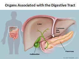
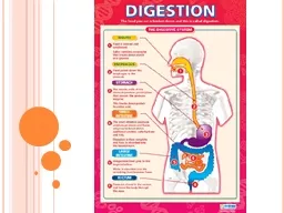
introduction The organs associated with the digestive tract include the major salivary glands the pancreas the liver and the gallbladder Products of these organs facilitate ID: 913471
Download Presentation The PPT/PDF document "Organs Associated with the Digestive Tra..." is the property of its rightful owner. Permission is granted to download and print the materials on this web site for personal, non-commercial use only, and to display it on your personal computer provided you do not modify the materials and that you retain all copyright notices contained in the materials. By downloading content from our website, you accept the terms of this agreement.


Slide1
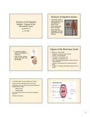
Organs Associated with the Digestive Tract
Slide2introduction
The
organs associated with the digestive tract include
the major salivary glands
,
the pancreas
,
the liver
, and the gallbladder
.
Products
of these organs facilitate
transport and
digestion of food within
the gastrointestinal
tract.
Slide3The liver
Anatomically :
The
liver is the largest internal organ, in adults
averaging about
1.5 kg or 2% of the body
weight
Located in
the right upper
quadrant of the abdomen just below the
diaphragm.
T
he
liver
has major
left
and right lobes with
two smaller
inferior lobes, most of which are covered by a thin capsule and mesothelium of the visceral peritoneum.
Slide4The
capsule thickens at the hilum (or
porta hepatis
) on the inferior side, where the dual blood supply from the
hepatic portal vein and hepatic artery enters
the organ and where the hepatic vein, lymphatics, and common hepatic (bile) duct exit.About 75% of the blood entering the liver is nutrient rich (but O2-poor) blood from the portal vein arising from the stomach, intestines, and spleen; the other 25% comes from the hepatic artery and supplies the organ’s O2.Histologically :It is a mixed gland organ , has both endocrine and exocrine functions ( compound tubular gland)
Slide5production of bile
:
this is
the
main digestive function of the
liver, the bile is a complex substance required for the emulsifcation, hydrolysis, and uptake of fats in the duodenum.The liver is the major interface between the digestive system and the blood, as the organ in which nutrients absorbed in the small intestine are processed before distribution throughout the body. Synthesis and endocrine secretion into the blood of the major plasma proteins, including albumins, fibrinogen, apolipoproteins, transferrin, and many others.
Conversion of amino acids into
glucose (
gluconeogenesis)Breakdown (detoxifcation) and conjugation of ingested toxins, including many drugs.Amino acid deamination, producing urea removed from blood in kidneys.
Functions of the liver
Slide6Storage of glucose
in glycogen granules and
triglycerides in
small lipid
droplets.
Storage
of vitamin A (in hepatic stellate cells) and other fat-soluble vitamins. Removal of effete erythrocytes (by specialized macrophages, or Kupffer cells). Storage of iron in complexes with the protein ferritin.
Slide7Structures of the liver
1.Stroma
Connective tissue capsule:
called
Glisson capsule
which is partially covered by mesothelium
Connective tissue septa : arise at the hilum (porta hepatis) , divide the liver into lobules, But the lobular organization is not evident in human .Reticular fiber : support the parenchyma
Slide8Structure of the liver
2.parenchyma:
Hepatocytes
:
the
key cells of this
organ, are among the most functionally diverse cells of the body.Arranged in anastomosing cords, radiating from central vein. These hepatocyte cords enclose between them bile canaliculi and are separated from each other by blood sinusoidsBlood sinusoids
Slide9I. Hepatocytes
LM
Shape:
Large
cuboidal or polyhedral epithelial
cells.
Nucleus: large, round central nuclei , The cells are frequently binucleated and about 50% of them are polyploid.Cytoplasm : eosinophilic cytoplasm( rich in mitochondria and sER) and pale (presence of glycogen and lipid droplets).
Slide10How hepatocyte adapt to its function
EM:
Abundant mitochondria(high metabolic activities)
multiple Golgi apparatus(secretory function)
Well developed rER(plasma proteins)
Abundant sER(bile acid synthesis, glycogen, detoxification)Numerous peroxisomes(oxidation of FA)Lysosomes and lipofuscin pigments(aged cells)
Glycogen and energy
Slide11The
hepatocyte
The hepatocyte has two functional surfaces:
1.Vascular surface:
Faces the perisinusoidal space
It shows long villi
2.The intercellular surface:Where bile canaliculi are enclosed betweenadjacent hepatocytesEach bile canaliculus shows short microvilliProjecting into its lumen and is bounded by tightJunction and desmosomes.
Slide12The blood sinusoids
the blood supply of the liver
The liver has dual blood supply
On entering the porta hepatis,
the portal vein and the hepatic artery
interlobular branches(in the portal canals) the sinusoidsthe central vein
hepatic veins
the inferior vena cava
Slide13II.The blood sinusoids
Function: exchange between blood and hepatocytes
Structure: lined by two types of cells:
Endothelial cell:
Flat cells
Numerous large fenestration without diaphragm
Surrounded by discontinuous basal laminaFunction: this allow free passage of plasma to perisinusoidal space for the exchange process to occur.
Slide14B.The kupffer cells
Origin
: from blood monocytes
Structures:
Large branching cells
Their pseudopodia are projecting into the lumen of the sinusoids
The cytoplasm contain many lysosomes containing hemosiderin pigments.Function: phagocytosis of aged RBCs
Slide15Space of Disse
Definition :
it is anarrow perisinusoidal space between the endothelial lining of the sinusoids and the hepatocytes.
Function
:it allows exchange of metabolites between the plasma and the hepatocytes
Contents
:1.Plasma filtered from the sinusoids2.Microvilli projecting from the hepatocytes3.Reticular fibers(support to the wall of sinusoids)4.Hepatic stellate cells (Ito cells) :vitamin A storing cells.
Slide16Hepatic stellate cells
In chronic inflammation of the liver,
these cells differentiate into
myofibroblasts
and deposit excess collagen
into the perisinusoidal space resulting in liver fibrosis.
Slide17The biliary tree
The hepatocytes
the bile canaliculi
(lined by cell membrane of adjacent hepatocyte
canals of Hering
(lined by cuboidal cells) the interlobular bile ducts ( in the portal canals , lined by cuboidal cells then columnar cells the Rt. And Lt. hepatic ducts.
Slide18Cells in the canal of Hering is consider as stem cells that paly arole in liver regeneration.
Slide19Hepatic lobulation
Three classification of hepatic lobules refer to different aspects of hepatocyte activities:
1.The classic hepatic lobule
2.Portal lobule
3.Liver acinus
Slide201.The classic hepatic lobule
It is a hexagonal mass of hepatocytes radiating around a central vein
Center:
central vein
Corner
: the portal canals, enclosing the portal traid, contain three structures:
1.An interlobular bile duct2.An interlobular branch of hepatic artery3.An interlobular branch of portal veinIn the classic hepatic lobule , blood and bile flow in opposite directions, the blood flow toward the central vein while the bile flow away from the central vein
Slide212.The Portal lobule
This is a
triangular
mass of hepatocytes from three adjacent
Classic hepatic lobules.
Center
: a portal canalAngles : the central veinEach portal lobule drain bile into the central bile duct.
Slide223.The liver acinus
It is
the functional unit
of the liver( correlate between metabolic activity, blood perfusion and liver pathology)
It is a
diamond shape
mass of two adjacent classic hepatic lobules.Center: vascular core formed of branches from portal vein and hepatic artery, present along the border between two classic lobules.
Slide233.The liver acinus
It is divided into
three zones
according to their blood supply:
Zone 1
:
The hepatocyte close to the vascular coreIt is richly supplied by bloodIts hepatocytes carry out oxidative metabolism and protein synthesis.Zone 2 : hepatocytes intermediate in location and their blood supplyZone 3 : hepatocytes farthest from the blood supply and close to the central veinIt has the poorest blood supplyIts hepatocytes carry out glycolysisIts cells are the first that suffer from degeneration and necrosis.
Slide24The gall bladder
Anatomically
: it is a hollow, pear shaped organ
a
ttached to the lower surface of the liver
Function
: storage, concentration of bile and release of bile whenneeded into the common bile duct
Slide25Structure of gall bladder
Three layers
:
1.
mucosa
:
Highly folded especially when the gall bladder is emptyEpithelium: simple columnar epithelium with apicalmicrovilli( cholangiocytes) (for salt and water absorption)Lamina propria: well vascularized2.Muscularis :smooth muscle fiber arranged in severalDirections and separated by fibroelastic connective tissue3.serosa/adventitia: according to anatomical surface
Slide26The biliary tract
The
bile produced by the hepatocytes flows through the
bile
canaliculi
, bile ductules, and
bile ductsThese structures gradually merge, forming a converging network that ultimately forms the common hepatic duct, which joins the cystic duct from the gallbladder and continues to the duodenum as the common bile ductThe hepatic, cystic, and common bile ducts are lined witha
mucous membrane
having a
simple columnar epithelium ofcholangiocytes.The lamina propria and submucosa are relatively thin, with mucous glands in some areas of the cysticduct, and surrounded by a thin muscularisThis muscle layer becomes thicker near the duodenum and finally, in the
duodenal papilla
, forms a sphincter that regulates bile flow into the
small bowel.
Slide27THANKS