PDF-Anatomy of the Digestive System Organs of the Alimenta
Author : trish-goza | Published Date : 2015-04-25
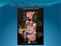
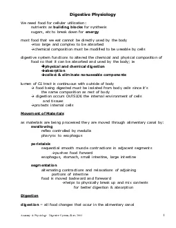
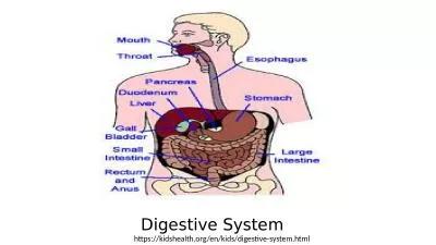
412423 Divisions of Digestive System 1 Alimentary Canal or Gastrointestinal Tract GIdigests and absorbs food coiled hollow tube with 2 openings mouth pharynx esophagus
Presentation Embed Code
Download Presentation
Download Presentation The PPT/PDF document "Anatomy of the Digestive System Organs o..." is the property of its rightful owner. Permission is granted to download and print the materials on this website for personal, non-commercial use only, and to display it on your personal computer provided you do not modify the materials and that you retain all copyright notices contained in the materials. By downloading content from our website, you accept the terms of this agreement.
Anatomy of the Digestive System Organs of the Alimenta: Transcript
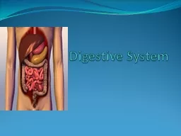
412423 Divisions of Digestive System 1 Alimentary Canal or Gastrointestinal Tract GIdigests and absorbs food coiled hollow tube with 2 openings mouth pharynx esophagus stomach small intestine large intestine 2 Accessory Organs assist in breakdown. . and Muscular. Systems . By Blandy , Esmeralda , Misty , Alexis , Idaly. Digestive system. The digestive tract (also known as the alimentary canal) is the system of organs within multicellular animals that takes in food, digests it to extract energy and nutrients, and expels the remaining waste.. Lesson5– . Digestive System Model. Objectives. :. Students will gain a full understanding of how the digestive and excretory system work in their own . bodies.. Students will be able to name the parts of the digestive system.. By Mr. Style. Parts of the Digestive System. The Mouth:. The . mouth is the beginning of the digestive system, and, in fact, digestion starts here before you even take the first bite of a meal. The smell of food triggers the salivary glands in your mouth to secrete saliva, causing your mouth to water. When you actually taste . Essential Question. What are the structures of the digestive system?. 2.07 Remember the structures of the . digestive . system. 2. Structures of the digestive system. Digestive system. Also known as:. A. Bacterial Diseases of the Upper Digestive Tract (Mouth & Stomach). 1. Tooth Decay (dental caries). A) This is an endogenous infection . 1) Most common infectious disease of humans . 2) Young are more susceptible than old. Embryonic Mouth Formation. Non-molting vs. Molting. Review. Animal Evolution. What’s this?. Deuterostomia. Protostomia. Embryo Formation. Deuterostomia. Protostomia. Embryo Formation. Non-organs vs. Organs. The digestive system is used for breaking down food into nutrients . which then pass into the circulatory system and are taken to where they are needed in the body.. Introduction. There . are four stages to food digestion:. 1. Digestion and absorption. It is the physical and chemical break down of food. Absorption. It is the passing of the digested food through the epithelial cells into the blood stream. 2. Digestive system. the human body systems . interact? . Today I will learn about the integumentary and lymphatic systems because both systems are responsible for protecting the human body from . disease. .. Fact or Fiction? (7 minutes). pharynx to esophagus all food changes that occur in the alimentary canalAnatomy Physiology Digestive System Ziser 2003breaking large molecules proteins fats starches etca thick coating of bicarbona Form and structure of the body and its parts. What things look like and where they are located. System of the body. Organs in the body. Job or function. Skeletal. bones. Protects vital. organs. Muscular. 1. What are the 2 groupings of the digestive organs?. The alimentary canal. The accessory digestive organs. 2. What are the functions of the alimentary canal?. It ingests and digests food, absorbs nutrients, and defecates wastes. to convert food into nutrients your body needs, and to rid the body of waste. . To do its job, the system requires the cooperation of a number of different organs throughout the body, including the mouth, stomach, intestines, liver and gallbladder. The digestive system has . three main . functions: . to convert food into nutrients your body . needs. Absorption. R. id . the body of . waste. To . do its job, the system requires the cooperation of a number of different structures and organs throughout the body, including the mouth, stomach, intestines, liver, pancreas and gallbladder..
Download Document
Here is the link to download the presentation.
"Anatomy of the Digestive System Organs of the Alimenta"The content belongs to its owner. You may download and print it for personal use, without modification, and keep all copyright notices. By downloading, you agree to these terms.
Related Documents