
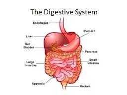
Stomach Jshaped organ Found in upper left portion of abdom cavity Can hold 1 Liter or more Contains thick folds in lining called RUGAE Functions Receives food from esophagus Mixes food with gastric juices ID: 915435
Download Presentation The PPT/PDF document "Day 2 Stomach-Liver Digestion" is the property of its rightful owner. Permission is granted to download and print the materials on this web site for personal, non-commercial use only, and to display it on your personal computer provided you do not modify the materials and that you retain all copyright notices contained in the materials. By downloading content from our website, you accept the terms of this agreement.


Slide1
Day 2 Stomach-Liver
Digestion
Slide2Stomach
J-shaped organ
Found in upper left portion of
abdom. cavityCan hold 1 Liter or moreContains thick folds in lining called RUGAEFunctions:Receives food from esophagusMixes food with gastric juicesInitiates protein digestionCarries on limited absorptionMoves food into small intestines
Slide3Parts of the Stomach
Four Parts
Cardia
Small area near esophageal openingFundusBalloons superior to cardia and is a temporary storage areaBodyMain part of stomach, found between fundus and pylorus
Pylorus
Near small intestine, narrows to form pyloric canal
Pyloric SphincterValve that controls gastric emptying
Slide4Gastric Secretions
Mucous membrane forms inner lining
Contains gastric pits which lead to gastric glands
Gastric Glands produce three types of secretions known as GASTRIC JUICEMucous cellsFound near opening of glands, produce mucusChief CellsFound deeper in glands, produce digestive enzymesParietal CellsFound deep in glands, produce HCL (Hydrochloric Acid)
Slide5Gastric Juices
Pepsin
Most important digestive enzyme
Produced by chief cellsForms when PEPSINOGEN contacts HCLBreaks down protein
Mucous Cells
*Forms a alkaline secretion that covers inner surface of stomach wall
*Prevents stomach from digesting itself
Slide6Gastric Juices Cont.
Intrinsic Factor
Secreted by parietal cells
Helps small intestine absorb vit. B12Regulation of Gastric SecretionProduced continuously, but rate variesControlled neurally and hormonallyThinking of food or food entering stomach causes stimulation of ACH and gastrin which increase
secretory
activity
Once food enters small intestine, hormone cholecystokinin stimulates decrease in gastric juice production
Slide7Gastric Absorption
Enzymes break down proteins but does not absorb much
What’s absorbed:
Only small amounts of water and certain salts are absorbedLipid-soluble drugsAlcohol
Slide8Mixing and Emptying Actions
Chyme
Produced after a meal
Consists of gastric juices and food particlesSemi fluid pastePeristaltic waves push Chyme towards pyloric sphincterLittle by little Chyme is pushed into small intestinesRate depends on type of food and fluidity of chyme
Liquids pass through stomach rapidly
Fatty foods remain for 3-6 hours after consumption
High protein foods are quicker than fats
Carbs
are faster than proteins
Slide9Vomiting
Results from a complex reflex that empties the stomach in the reverse of the normal direction. Irritation or distension in the stomach or intestines can trigger vomiting. Sensory impulses travel from the site of stimulation to the vomiting center of the Medulla, and motor responses follow. These include taking a deep breath, raising the soft palate and thus closing the nasal cavity, closing the opening to the trachea, relaxing the circular muscle fibers at the base of the esophagus, contracting the diaphragm so it presses downward over the stomach, and contracting the abdominal wall muscles to increase pressure inside the abdominal cavity. As a result, the stomach is squeezed from all sides, forcing its contents upward and out through the esophagus, pharynx, and mouth.
Slide10Accessory Organs
Once food leaves the stomach and enters the small intestine accessory organs add digestive juices
Pancreas
Lies horizontal across the posterior abdominal wallCells called pancreatic acinar produce pancreatic juiceSecretions in to small intestine are controlled by hepatopancreatic sphincter
Slide11Pancreatic Juice
Contains enzymes that digest
carbs
, fats, nucleic acids, and proteinsPancreatic amylaseSplits carbsPancreatic lipaseSplits fat moleculesNucleasesBreak down nucleic acid moleculesTrypsin, Chymotrypsin
,
Carboxypeptidase
Split proteins
Slide12Regulation of Pancreatic Secretions
Nervous and Endocrine systems regulate release of pancreatic juices
Hormone
Secretin Released when chyme enters small intestine to neutralize it
Slide13Liver
Found in upper right quadrant of abdominal cavity just inferior to diaphragm
Packed with blood vessels
StructureLarge right lobe, smaller left lobeHepatic lobules are the functional unitsHepatic sinusoidsVascular channels that receive newly absorbed nutrientsContains phagocytic cells called Kupffer Cells
Help remove bacteria and other foreign particles
Contains many hepatic ducts that merge to form common hepatic duct
Slide14Liver Function
Important metabolic
activites
Carb metabolismMaintains blood glucoseLipid metabolismFatty acid metabolismMost important is protein metabolismStores glycogen, iron, vitamins A, D, and B12Destroys damaged RBC’sRemoves toxic substances such as alcohol and drugs
Secretes Bile (Important to Digestion)
Slide15Liver Donations
The liver is unlike most organs in that it can regenerate. Up to 75% of a liver can be destroyed and the organ can regenerate and recover. For this reason, people can donate parts of their livers to people in liver failure, if the tissues of the donor and recipient are compatible.