/
Pelvic Diaphragm  Presented by Pelvic Diaphragm  Presented by

Pelvic Diaphragm Presented by - PowerPoint Presentation

blanko
blanko . @blanko
Follow
27 views
Uploaded On 2024-02-02

Pelvic Diaphragm Presented by - PPT Presentation

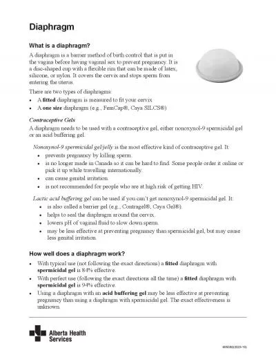
Msc Dr Reham Saad Kadhum Inferior Pelvic Wall or Pelvic Floor The floor of the pelvis supports the pelvic viscera and is formed by the pelvic diaphragm The pelvic floor stretches across the pelvis and divides ID: 1044098

pelvic artery sacral iliac artery pelvic iliac sacral nerve pelvis inferior supplies internal muscle rectal male nerves superior muscles

Share:

Link:

Embed:

Download Presentation from below link

Download Presentation The PPT/PDF document "Pelvic Diaphragm Presented by" is the property of its rightful owner. Permission is granted to download and print the materials on this web site for personal, non-commercial use only, and to display it on your personal computer provided you do not modify the materials and that you retain all copyright notices contained in the materials. By downloading content from our website, you accept the terms of this agreement.


Presentation Transcript

1. Pelvic Diaphragm Presented by Msc. Dr. Reham Saad Kadhum

2. Inferior Pelvic Wall or Pelvic FloorThe floor of the pelvis supports the pelvic viscera and is formed by the pelvic diaphragm.The pelvic floor stretches across the pelvis and divides it into the main pelvic cavity above, which contains the pelvic viscera, and the perineum below. Pelvic DiaphragmThe pelvic diaphragm is formed by the important levatores ani muscles and the small coccygeus muscles and their covering fasciae It is incomplete anteriorly to allow passage of the urethra in males and the urethra and the vagina in females.

3. Levator Ani MuscleThe levator ani muscle is a wide thin sheet that has a linear origin from the back of the body of the pubis, a tendinous arch formed by a thickening of the fascia covering the obturator internus, and the spine of the ischium . From this extensive origin, groups of fibers sweep downward and medially to their insertion as follows:1. Anterior fibers: The levator prostatae or sphincter vaginae form a sling around the prostate or vagina and are inserted into a mass of fibrous tissue, called the perineal body, in front of the anal canal. The levator prostatae support the prostate and stabilize the perineal body. The sphincter vaginae constrict the vagina and stabilize the perineal body.2. Intermediate fibers: The puborectalis forms a sling around the junction of the rectum and anal canal. The pubococcygeus passes posteriorly to be inserted into a small fibrous mass, called the anococcygeal body, between the tip of the coccyx and the anal canal.3. Posterior fibers: The iliococcygeus is inserted into the anococcygeal body and the coccyx.

4. Action: The levatores ani muscles of the two sides form an efficient muscular sling that supports and maintains the pelvic viscera in position. They resist the rise in intrapelvic pressure during the straining and expulsive efforts of the abdominal muscles (as occurs in coughing). They also have an important sphincter action on the anorectal junction, and in the female they serve also as a sphincter of the vagina .■■ Nerve supply: The perineal branch of the fourth sacral nerve and from the perineal branch of the pudendal nerve .

5. Coccygeus MuscleThis small triangular muscle arises from the spine of the ischium and is inserted into the lower end of the sacrum and into the coccyx ■■ Action: The two muscles assist the levatores ani in supporting the pelvic viscera.■■ Nerve supply : A branch of the 4th and 5th sacral nerves .Pelvic FasciaThe pelvic fascia is formed of connective tissue and is continuous above with the fascia lining the abdominal walls. Below, the fascia is continuous with the fascia of the perineum. The pelvic fascia can be divided into parietal and visceral layers.

6.

7.

8. Nerves of the PelvisSacral PlexusThe sacral plexus lies on the posterior pelvic wall in front of the piriformis muscle It is formed from the anterior rami of the 4th and 5th lumbar nerves and the anterior rami of the first, second, third, and fourth sacral nerves . The fourth lumbar nerve joins the fifth lumbar nerve to form the lumbosacral trunk. The lumbosacral trunk passes down into the pelvis and joins the sacral nerves as they emerge from the anterior sacral foramina.

9. Branches ( branches that supply the muscles of LL) ■■ 1. The sciatic nerve (L4 and 5; S1, 2, and 3), the largest branch of the plexus and the largest nerve in the body2. The superior gluteal nerve, which supplies the gluteus medius and minimus and the tensor fasciae latae muscles3. The inferior gluteal nerve, which supplies the gluteus maximus muscle4. The nerve to the quadratus femoris muscle, which also supplies the inferior gemellus muscle5. The nerve to the obturator internus muscle, which also supplies the superior gemellus muscle6. The posterior cutaneous nerve of the thigh, which supplies the skin of the buttock and the back of the thigh

10. Branches to the pelvic muscles, pelvic viscera, and perineum:1. The pudendal nerve (S2, 3, and 4), which leaves the pelvis through the greater sciatic foramen and enters the perineum through the lesser sciatic foramen 2. The nerves to the piriformis muscle3. The pelvic splanchnic nerves, which constitute the sacral part of the parasympathetic system and arise from the second, third, and fourth sacral nerves. They are distributed to the pelvic viscera.

11.

12. Arteries of the PelvisCommon Iliac Artery ( L4) Each common iliac artery ends at the pelvic inlet in front of the sacroiliac joint by dividing into the external and internal iliac arteries External Iliac ArteryThe external iliac artery runs along the medial border of the psoas muscle, following the pelvic brim , and gives off the : inferior epigastric and deep circumflex iliac branches. It leaves the false pelvis by passing under the inguinal ligament to become the femoral artery.Arteries of the True PelvisThe following arteries enter the pelvic cavity:■■ Internal iliac artery■■ Superior rectal artery■■ Ovarian artery■■ Median sacral artery

13. Internal Iliac ArteryThe internal iliac artery passes down into the pelvis to the upper margin of the greater sciatic foramen , where it divides into anterior and posterior divisions .Branches of the Anterior Division■■ Umbilical artery: From the proximal patent part of the umbilical artery arises the superior vesical artery, which supplies the upper portion of the bladder ■■ Obturator artery: This artery runs forward along the lateral wall of the pelvis with the obturator nerve and leaves the pelvis through the obturator canal.■■ Inferior vesical artery: This artery supplies the base of the bladder and the prostate and seminal vesicles in the male; Middle rectal artery: Commonly, this artery arises with the inferior vesical artery . It supplies the muscle of the lower rectum and anastomoses with the superior rectal and inferior rectal arteries.Middle rectal artery: Commonly, this artery arises with the inferior vesical artery . It supplies the muscle of the lower rectum and anastomoses with the superior rectal and inferior rectal arteries.

14. Internal pudendal artery: This artery leaves the pelvis through the greater sciatic foramen and enters the gluteal region . ■■ Inferior gluteal artery: This artery leaves the pelvis through the greater sciatic foramen below the piriformis muscle . It passes between the first and second or second and third sacral nerves.■■ Uterine artery: This artery runs medially on the floor of the pelvis and crosses the ureter superiorly . It passes above the lateral fornix of the vagina to reach the uterus. Here, it ascends between the layers of the broad ligament along the lateral margin of the uterus. It ends by following the uterine tube laterally,where it anastomoses with the ovarian artery. The uterine artery gives off a vaginal branch.■■ Vaginal artery: This artery usually takes the place of the inferior vesical artery present in the male. It supplies the vagina and the base of the bladder.

15. Branches of the Posterior Division■■ Iliolumbar artery: This artery ascends across the pelvic inlet posterior to the external iliac vessels, psoas, and iliacus muscles.■■ Lateral sacral arteries: These arteries descend in front of the sacral plexus, giving off branches to neighboring structures ■■ Superior gluteal artery: This artery leaves the pelvis through the greater sciatic foramen above the piriformis muscle. It supplies the gluteal region.Superior Rectal ArteryThe superior rectal artery is a direct continuation of the inferior mesenteric artery. The name changes as the latter artery crosses the common iliac artery. It supplies the mucous membrane of the rectum and the upper half of the anal canal.Ovarian Artery(The testicular artery enters the inguinal canal and does not enter the pelvis.) The ovarian artery arises from the abdominal part of the aorta at the level of the first lumbar vertebra .Median Sacral ArteryThe median sacral artery is a small artery that arises at the bifurcation of the aorta . It descends over the anterior surface of the sacrum and coccyx.

16.

17. Veins of the PelvisThe external iliac vein begins behind the inguinal ligament as a continuation of the femoral vein. It runs along the medial side of the corresponding artery and joins the internal iliac vein to form the common iliac vein . It receives the inferior epigastric and deep circumflex iliac veins.Internal Iliac VeinThe internal iliac vein begins by the joining together of tributaries that correspond to the branches of the internal iliac artery. It passes upward in front of the sacroiliac joint and joins the external iliac vein to form the common iliac vein . Median Sacral VeinsThe median sacral veins accompany the corresponding artery and end by joining the left common iliac vein.

18. Lymphatics of the PelvisThe lymph nodes and vessels are arranged in a chain along the main blood vessels. The nodes are named after the blood vessels with which they are associated. Thus, there are external iliac nodes, internal iliac nodes, and common iliac nodes.Joints Sacroiliac j (limited amount of movement )Symphysis pubis j (Almost no movement is possible at this joint. )Sacrococcygeal j (flexion and extension )

19.

20.

21. Sex Differences of the PelvisThe false pelvis is shallow in the female and deep in the male.■■ The pelvic inlet is transversely oval in the female but heart shaped in the male .The pelvic cavity is roomier in the female than in the male, and the distance between the inlet and the outlet is much shorter.■■ The pelvic outlet is larger in the female than in the male. In the female the ischial tuberosities are everted and in the male they are turned in.■■ The sacrum is shorter, wider, and flatter in the female than in the male.■■ The subpubic angle, or pubic arch, is more rounded and wider in the female than in the male.

22.