/
Pelvic Wall and Pelvic Diaphragm Pelvic Wall and Pelvic Diaphragm

Pelvic Wall and Pelvic Diaphragm - PowerPoint Presentation

daniella
daniella . @daniella
Follow
27 views
Uploaded On 2024-02-02

Pelvic Wall and Pelvic Diaphragm - PPT Presentation

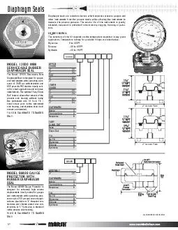
Presented by Msc Dr Reham Saad kadhum The Pelvis The bony pelvis is composed of four bones the two hip bones which form the lateral and anterior walls The sacrum ID: 1044093

sacral pelvic sacrum pelvis pelvic sacral pelvis sacrum anterior obturator wall posterior part coccyx lateral laterally symphysis form hip

Share:

Link:

Embed:

Download Presentation from below link

Download Presentation The PPT/PDF document "Pelvic Wall and Pelvic Diaphragm" is the property of its rightful owner. Permission is granted to download and print the materials on this web site for personal, non-commercial use only, and to display it on your personal computer provided you do not modify the materials and that you retain all copyright notices contained in the materials. By downloading content from our website, you accept the terms of this agreement.


Presentation Transcript

1. Pelvic Wall and Pelvic Diaphragm Presented by Msc. Dr. Reham Saad kadhum

2. The Pelvis The bony pelvis is composed of four bones: the two hip bones, which form the lateral and anterior walls, The sacrum and the coccyx, which are part of the vertebral column and form the back wall .The two hip bones articulate with each other anteriorly at the symphysis pubis and posteriorly with the sacrum at the sacroiliac joints. The bony pelvis thus forms a strong basin-shaped structure that contains and protects the lower parts of the intestinal and urinary tracts and the internal organs of reproduction.

3.

4. Orientation of the PelvisThe front of the symphysis pubis and the anterior superior iliac spines should lie in the same vertical plane.This means that the pelvic surface of the symphysis pubis faces upward and backward and the anterior surface of the sacrum is directed forward and downward.

5.

6. The pelvis is divided into two parts by the pelvic brim, which is formed by the sacral promontory (anterior and upper margin of the first sacral vertebra) behind, The iliopectineal lines (a line that runs downward and forward around the inner surface of the ileum) laterally, and the symphysis pubis (joint between bodies of pubic . bones) anteriorly. Above the brim is the false pelvis, which forms part of the abdominal cavity. Below the brim is the true pelvis.

7. False PelvisThe false pelvis is of little clinical importance. It is bounded behind by the lumbar vertebrae, laterally by the iliac fossae and the iliacus muscles, and in front by the lower part of the anterior abdominal wall. True PelvisKnowledge of the shape and dimensions of the female pelvis is of great importance for obstetrics, because it is the bony canal through which the child passes during birth. The true pelvis has an inlet, an outlet, and a cavity .

8. The pelvic inlet, or pelvic brim is bounded posteriorly by the sacral promontory, laterally by the iliopectineal lines, and anteriorly by the symphysis pubis■■ The pelvic outlet is bounded posteriorly by the coccyx, laterally by the ischial tuberosities, and anteriorly by the pubic arch The pelvic outlet has three wide notches. Anteriorly, the pubic arch is between the ischiopubic rami, and laterally are the sciatic notches.The pelvic cavity lies between the inlet and the outlet. It is a short, curved canal, with a shallow anterior wall and a much deeper posterior wall .

9.

10.

11. The sciatic notches are divided by the sacrotuberous and sacrospinous ligaments into the greater and lesser sciatic foramina . From an obstetric standpoint, because the sacrotuberous ligaments are strong and relatively inflexible, they should be considered to form part of the perimeter of the pelvic outlet. Thus, the outlet is diamond shaped, with the ischiopubic rami and the symphysis pubis forming the boundaries in front and the sacrotuberous ligaments and the coccyx forming the boundaries behind.

12. Structure of the Pelvic WallsThe walls of the pelvis are formed by bones and ligaments that are partly lined with muscles covered with fascia and parietal peritoneum. The pelvis has anterior, posterior, and lateral walls and an inferior wall or floor

13. Anterior Pelvic WallThe anterior pelvic wall is the shallowest wall and is formed by the bodies of the pubic bones, the pubic rami, and the symphysis pubis .

14. Posterior Pelvic WallThe posterior pelvic wall is extensive and is formed by the sacrum and coccyx and by the piriformis muscles and their covering of parietal pelvic fascia.

15. SacrumThe sacrum consists of five rudimentary vertebrae fused together to form a single wedge-shaped bone with a forward concavity. The upper border or base of the bone articulates with the fifth lumbar vertebra.The narrow inferior border articulates with the coccyx bone .Laterally, the sacrum articulates with the two iliac bones to form the sacroiliac joints The anterior and upper margins of the first sacral vertebra bulge forward as the posterior margin of the pelvic inlet—the sacral promontory which is an important obstetric landmark used when measuring the size of the pelvis.The vertebral foramina together form the sacral canal.The laminae of the 5th sacral vertebra, and sometimes those of the 4th, fail to meet in the midline, forming the sacral hiatus . The sacral canal contains the anterior and posterior roots of the lumbar, sacral, and coccygeal spinal nerves; the filum terminale; and fibrofatty material. It also contains the lower part of the subarachnoid space down as far as the lower border of the 2nd sacral vertebraThe anterior and posterior surfaces of the sacrum possess on each side four foramina for the passage of the anterior and posterior rami of the upper four sacral nerves

16.

17.

18. The sacrum is usually wider in proportion to its length in the female than in the male. The sacrum is tilted forward so that it forms an angle with the fifth lumbar vertebra, called the lumbosacral angle.CoccyxThe coccyx consists of four vertebrae fused together to form a small triangular bone, which articulates at its base with the lower end of the sacrum The coccygeal vertebrae consist of bodies only, but the first vertebra possesses a rudimentary transverse process and cornua. The cornua are the remains of the pedicles and superior articular processes and project upward to articulate with the sacral cornua

19. Piriformis MuscleThe piriformis muscle arises from the front of the lateral mass of the sacrum and leaves the pelvis to enter the gluteal region by passing laterally through the greater sciatic foramen . It is inserted into the upper border of the greater trochanter of the femur.■■ Action: It is a lateral rotator of the femur at the hip joint.■■ Nerve supply: It receives branches from the sacral plexus.

20. Lateral Pelvic WallThe lateral pelvic wall is formed by part of the hip bone below the pelvic inlet, the obturator membrane, the sacrotuberous and sacrospinous ligaments, and the obturator internus muscle and its covering fascia .Hip bone ( discussed in last year ) .

21. Obturator MembraneThe obturator membrane is a fibrous sheet that almost completely closes the obturator foramen, leaving a small gap, the obturator canal, for the passage of the obturator nerve and vessels as they leave the pelvis to enter the thighSacrotuberous LigamentThe sacrotuberous ligament is strong and extends from the lateral part of the sacrum and coccyx and the posterior inferior iliac spine to the ischial tuberosity .Sacrospinous LigamentThe sacrospinous ligament is strong and triangle shaped. It is attached by its base to the lateral part of the sacrum and coccyx and by its apex to the spine of the ischium . The sacrotuberous and sacrospinous ligaments prevent the lower end of the sacrum and the coccyx from being rotated upward at the sacroiliac joint by the weight of the body .

22.

23. Obturator Internus MuscleThe obturator internus muscle arises from the pelvic surface of the obturator membrane and the adjoining part of the hip bone The muscle fibers converge to a tendon, which leaves the pelvis through the lesser sciatic foramen and is inserted into the greater trochanter of the femur.■■ Action: It laterally rotates the femur at the hip joint.■■ Nerve supply: The nerve to the obturator internus, a branch from the sacral plexus

24.

25. Thanks