
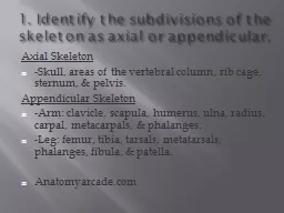
MBChB MSc PhD MRCPCH DCH UK The skeleton The skeleton can be divided into 2 subgroups Axial skeleton consists of bones of the Skull cranium Vertebral column Ribs and Sternum Thoracic Cage ID: 999578
Download Presentation The PPT/PDF document "Skeletal System Dr. Mohammed Hussein" is the property of its rightful owner. Permission is granted to download and print the materials on this web site for personal, non-commercial use only, and to display it on your personal computer provided you do not modify the materials and that you retain all copyright notices contained in the materials. By downloading content from our website, you accept the terms of this agreement.


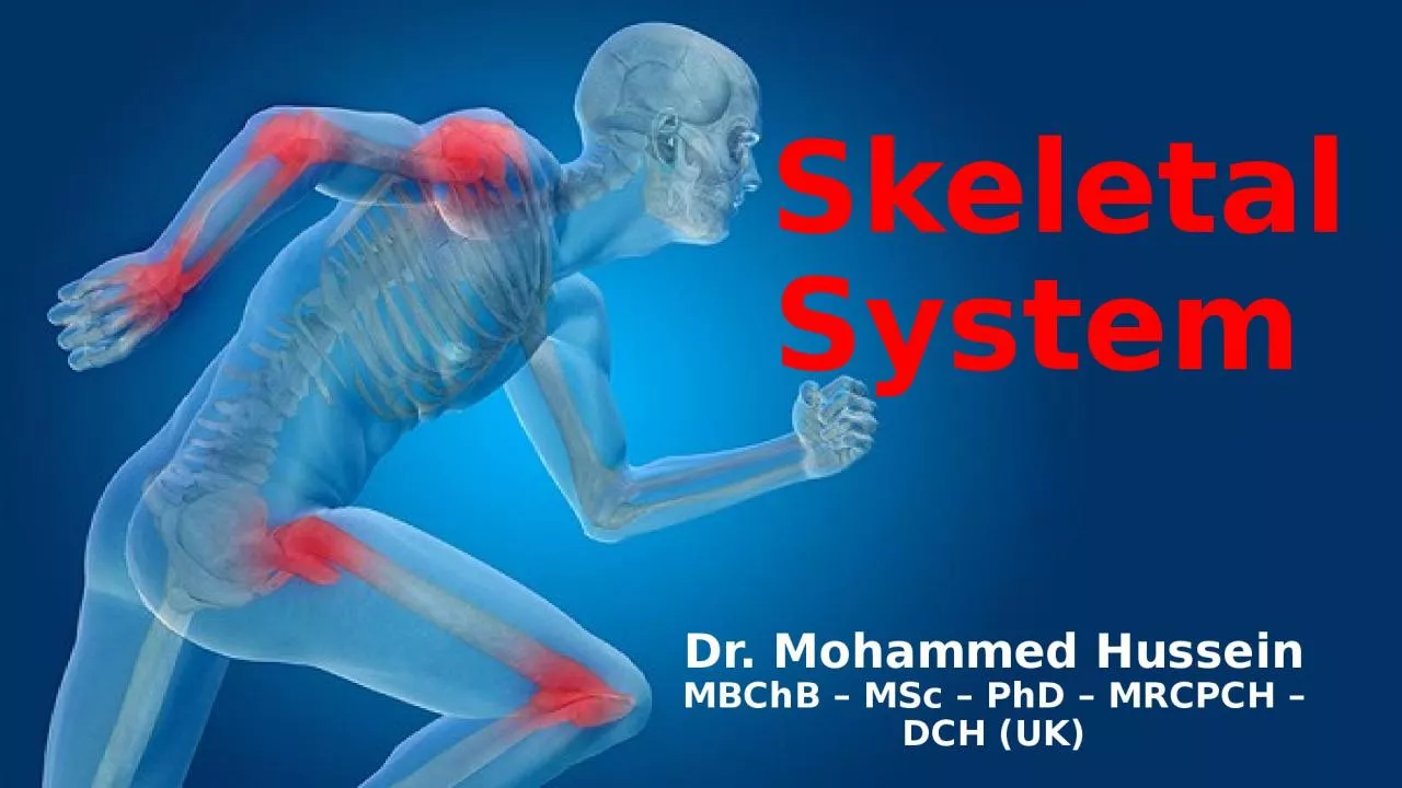
1. SkeletalSystem Dr. Mohammed HusseinMBChB – MSc – PhD – MRCPCH – DCH (UK)
2. The skeletonThe skeleton can be divided into 2 subgroups:Axial skeleton: consists of bones of the Skull (cranium) Vertebral column Ribs and Sternum (Thoracic Cage)Appendicular skeleton: consists of the bones of Upper and Lower Limbs
3. The skeletal systemThe skeletal system consists of Cartilages Bones
4. Cartilage It is avascular connective tissue consisting of cells and fibers embedded in a gel-like matrix.Fibers Cells Matrix
5. CartilageCartilage is nourished by diffusion from the surroundingsCartilage has:No blood vessels (avascular)No lymphaticsNo nerves
6. Types There are three types of cartilage:Hyaline cartilage: most common type (e.g. articular surfaces of bones). Fibrocartilage: e.g. symphysis pubis and intervertebral discs.Elastic cartilage: e.g. external ear auricle.
7. CartilageThe functions of cartilage are to: Support soft tissuesProvide a smooth, glide surface for bone articulation at jointsProvide a base for bone development and growth
8. BoneBone is a calcified, vascular lining connective tissue.
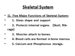
9. Bone Functions Protection of vital organsSupport and give shape of human bodyLevers on which muscles act for movementHousing of bone marrow (blood cells forming tissue)Reservoirs of minerals (calcium and phosphorus)
10. There are two basic types of bone: Compact ( Cortical ) Spongy ( Cancellous or Trabecular)
11.
12. BoneBones are classified based on their shape into: Long bones Short bones Flat bones Irregular bones Sesamoid bones
13. Long bones display a shaft located between two heads.Short bones have more or less the same width and length.Flat bones are flat, plate-like.Irregular bones have an irregular shape.Sesamoid bones develop within tendons.Long bone Short boneFlat boneIrregular boneSesamoid bone
14. DiaphysisMetaphysisEpiphysisMetaphysisEpiphysis
15. Blood and Nerve Supply Bones have their own blood vessels: provided by a nutrient artery that enters into bone cavity supplying the inner structures of the bone. Nerves accompanying the nutrient arteries into inner cavity of bone are vasomotor nerves that regulate blood flow.
16. Periosteum All bones are externally covered by fibrous connective tissue membrane called the periosteum
17. Periosteum The ends of bone (where joints are formed) not covered by periosteum, instead covered by a cartilage
18. Periosteum Periosteum contains:Blood vessels Nerves Stem cells
19. Periosteum Stem cells Are essential for forming of new bone after fracture of injury (A bone stripped of its periosteum will not survive)
20. Joints The sites where two skeletal elements come together, whether or not movement occurs between them, are termed joints
21. The two general categories of joints are Synovial joints: the skeletal elements are separated by a cavity Solid joints : the skeletal elements are held together by tissue (no cavity)Joint Classification
22. Synovial joints are connections between skeletal components where the elements involved are separated by a narrow articular (synovial) cavitySynovial joints
23. Synovial joints have a number of characteristic features: Articular cavity (synovial cavity) Articular cartilage (hyaline cartilage) Joint capsule Articular discs Bursae Tendon sheath Fat pads Synovial joints Additional StructuresUniversal Features
24. Hyaline cartilagesArticular cavity(Synovial cavity)Filled with synovial fluidJoint capsuleSynovial membraneFibrous membraneUniversal Features of a Synovial Joint
25. Articular discFat padTendon SheathBursaBone Bone Tendon Skin Additional Structures of a Synovial Joint
26. Description of Synovial JointsSynovial joints are described based on: The Range of Movementand The Shape
27. Description of Synovial JointsAccording to the rang of movement, synovial joints are described as:(movement in one plane)(movement in two planes)(movement in three planes)
28. Hinge joints Pivot joints Plane joints Condylar joints Bicondylar joints Saddle joints Ball and socket jointsDescription of Synovial JointsAccording to the shape, synovial joints are described as:UniaxialBiaxialMultiaxial
29. Hinge jointElbow jointUniaxial joint
30. Uniaxial jointAtlantoaxial jointPivot joint
31. Biaxial jointPlane (Flat or gliding) jointAcromioclavicular joint
32. Condylar (Ellipsoid) jointWrist jointBiaxial joint
33. Bicondylar jointFemurTibia Fibula Knee jointBiaxial joint
34. FemurTibia Fibula Bicondylar jointCondylar (Ellipsoid) joint
35. Biaxial joint?Saddle jointCarpometacarpal joint of thumbMultiaxial joint?
36. Ball & socket jointHip jointMultiaxial Joint
37. Solid joints are connections between skeletal elements where the adjacent surfaces are linked together either by: Fibrous connective tissue or by CartilageMovements at these joints are more restricted than at synovial jointSolid joints Fibrous jointCartilaginous joint
38. Solid joints Fibrous jointCartilaginous jointSuturesGomphosesSyndesmosesSymphysesSynchondroses
39. Fibrous joints
40. Cartilaginous joints
41. Stability of JointsThe stability of a joint depends on three main factors: The shape, size, and arrangement of the articular surfaces The ligaments The tone of the muscles around the joint
42. Shape of articular surfacesHip joint
43. LigamentsKnee joint
44. Foot jointsMuscle tone
45. Thank YouDr. Mohammed H. AssiMBChB – MSc – PhD – MRCPCH – DCH (UK)