
F UNCTION AND LESION DRTAJAYAN PROF amp HOD PM CNS influence the activity of skeletal muscle through two sets of neuron Upper motor neuron Lower motor neuron Upper motor neurons UMN are responsible for conveying impulses for voluntary motor activity through descending motor pat ID: 920127
Download Presentation The PPT/PDF document "UPPER AND LOWER MOTOR NEURON" is the property of its rightful owner. Permission is granted to download and print the materials on this web site for personal, non-commercial use only, and to display it on your personal computer provided you do not modify the materials and that you retain all copyright notices contained in the materials. By downloading content from our website, you accept the terms of this agreement.


Slide1
UPPER AND LOWER MOTOR NEURON
FUNCTION AND LESION
DR.T.AJAYAN
PROF. & H.O.D.
PM
Slide2CNS influence the activity of skeletal muscle through two sets of neuron
Upper motor neuronLower motor neuron
Slide3Upper motor neurons (UMN) are responsible for conveying impulses for voluntary motor activity through descending motor pathways that make up the upper motor neurons.UMN send fibers to the LMN, and that exert direct or indirect supranuclear control over the LMN of the cranial and spinal nerves..
UPPER MOTOR NEURON
Slide4WHERE THEY COME FROM ?
.
Slide5Axons from the cortical areas form the
corticospinal and corticobulbar tracts.1/3 from primary motor cortex (Betz’s cell axons -3-5%, and other 95% from small neurons)
1/3 from the somatic sensory cortex (areas 1, 2, and 3), and
adjacent temporal lobe region.
Slide6HOW UPPER MOTOR NEURON FUNCTION ?
Upper motor neuron control lower motor neuron through two different pathways.Pyramidal tractExtra pyramidal tract
Slide7PYRAMIDAL TRACTS corticospinal tract
EXTRAPYRAMIDAL TRACTS-Reticulospinal Olivospinal
Vestibulospinal
Tectospinal
Rubrospinal
tract
Corticobulbar
tract
Corticorubral
tract
Slide8Descending Tracts
Tract
Signal function
Corticospinal
(pyramidal)
Fine voluntary motor control of the limbs. The pathway also controls voluntary body posture adjustments.
Rubrospinal
Involved in involuntary adjustment of arm position in response to balance information; support of the body.
Reticulospinal
(1)
Pontine
Regulates various involuntary motor activities and assists in balance (leg extensors). Some pattern movements e.g. stepping (2) MedullaryInhibits firing of spinal and cranial motor neurons, control of antigravity muscles.Vestibulospinal (1) MedialIt is responsible for adjusting posture to maintain balance (neck muscles). (2) LateralIt is responsible for adjusting posture to maintain balance (body/lower limb).TectospinalControls head and eye movements, Involved in involuntary adjustment of head position in response to visual information.
Nerve pathways
Slide9Tracts
Slide10Descending pathways
Pyramidal systemLateral and anterior corticospinal tractsExtrapyramidal system
Tectospinal
tracts
Vestibulospinal
tracts
Rubrospinal
tracts
Anterior, medial, and lateral
reticulospinal
tracts
Slide11Descending Pathways
Pathway
Upper limb
Lower limb
Cortico/-pyramidal
This Tract functions to modulate the activity of Alpha or Gamma Motor Neurons as directed by the Motor Cortex.
Rubro-spinal
Stimulates flexors
Reticulo-spinal
Medullary inhibits extensors and excites flexors
Pontine excites extensors and inhibits flexors
(Generally upper limb)
Vestibulo-spinal
Doesn’t affect upper limbs but helps position head and neck in response to body tilting (medial)Stimulates extensors (lateral)Tecto-spinalControl of head, neck and eye movements.
Slide12Rubrospinal Tract
Slide13Ascending Pathway
Slide14Slide15UPPER MOTOR NEURON LESIONLoss of dexterity, voluntary skillful movements. (corticospinal
Babinski sign(corticospinal)Loss of superficial reflex (corticospinal).
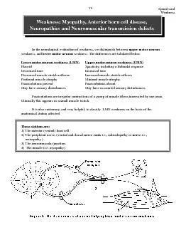
Slide16weakness with no muscle atrophySpasticity is hallmark of the UMN disease. Spasticity is a state of sustained increase in muscle tension in response to muscle lengthening, in particular, with passive movements.
hyperreflexia. deep tendon reflexPseudobulbar palsy is hallmark of the UMN disorder
Slide17PSEUDOBULBAR PALSY results from an upper motor neuron lesion to the corticobulbar pathways in the
pyramidal tract. It results from bilateral lesion of UMN’s of the muscles of the tongue (XII), face (VII), speech and swallowing (IX,X) Individuals with pseudobulbar palsy also demonstrate inappropriate emotional outbursts.
Slide18WHAT ARE LOWER MOTOR NEURONAll voluntary movement depend upon excitation of lower motor neuron by upper motor neuron These are the only neurons that innervate the skeletal muscle fibers, they function as the final common pathway, the final link between the CNS and skeletal muscles
Slide19WHERE THEY COME FROMMotor Neuron in spinal cord
Motor component of cranial nerve nuclei in brain stem (Those in cranial nerves innervate the skeletal muscles associated with the movements of the eyes, tongue, chewing, swallowing, vocalizing.)
Slide20CLASSIFICATION OF LMNLower motor neurons are classified based on the type of
muscle fiber they innervate:Alpha motor neurons (α-MNs) innervate extrafusal muscle fibers, the most numerous type of muscle fiber and the one involved in
muscle contraction
.
Gamma motor neurons
(γ-MNs) innervate
intrafusal muscle fibers
, which together with sensory afferents
compose
muscle spindles
. These are part of the system for sensing
body position
(proprioception)
Slide21LOWER MOTOR NEURON LESIONFlaccid paralesis
Muscle atrophy and HyporeflexiaMuscle hypotonicity Fasciculations
Slide22BULBAR PALSY is a similar disorder as psedobulbar palsy but is caused by lower motor neuron lesions
It consists of LMN signs in regions innervated by the facial (VII), glossopharyngeal (IX), Vagus (X) and hypoglossal (XII
Slide23Slide24Slide25The corticobulbar tract projects bilaterally to all the cranial motor nuclei except Part of facial nucleus that supply muscle of lower part of face receives
corticobulbar fibers from same hemisphere in UMN LESION muscle of lower part of face will paralyzed in LMN LESION all muscle of affected side will be paralyzed
Slide26Part of hypoglossal nucleus that supplies the genioglossus muscle receive corticobulbar fiber from opposite hemisphere in UMN LESION tongue will deviate to the side opposite to lesion in
LMN LESION tongue will deviate to the side of lesion