
8 Anal Canal Is about 4 cm in length Passes downwards and backwards from the rectal ampulla at the level of the prostate in the males to the anus Except during defecation its lateral walls are maintained in position by levatores ani muscles and anal sphincters ID: 908731
Download Presentation The PPT/PDF document "The Digestive System Lecture 5" is the property of its rightful owner. Permission is granted to download and print the materials on this web site for personal, non-commercial use only, and to display it on your personal computer provided you do not modify the materials and that you retain all copyright notices contained in the materials. By downloading content from our website, you accept the terms of this agreement.


Slide1
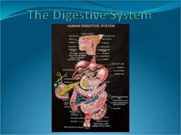
The Digestive System
Lecture 5
Slide28. Anal Canal
Is about
4 cm
in length
.
Passes downwards and backwards from the rectal ampulla (at the level of the prostate, in the males) to the anus.
Except during defecation, its lateral walls are maintained in position by levatores ani muscles and anal sphincters.
1
Slide3Relations
Anterior relations
In the male, it is separated from membranous urethra by the perineal body.
In the female, it is related to perineal body and lower part of vagina
.
Lateral
relations
It is separated from the fat of ischiorectal fossae by levator ani and external anal sphincter muscles.
Posterior relations
Related to anococcygeal raphe
.
2
Slide4Membranous urethra
Anal canal
Perineal body
Anterior relations of anal canal (male)
3
Slide5Anal canal
Perineal body
Vagina
4
Anterior relations of anal canal (female)
Slide6Lateral relations of anal canal
Levator ani
Obturator internus
Ischiorectal fossa
Anal canal
5
Slide7Anococcygeal Raphe
Levator ani
Posterior
relations of anal canal
6
Slide8The
upper half
of the anal canal is
lined by columnar epithelium.
The mucous membrane in this region exhibits
5
to
10
vertical folds, the anal columns, which are joined together at their lower ends by small semilunar folds called
anal valves.
The interval between a valve and the anal wall is called an anal sinus.
The function of the anal column and valves is not known.
7
Slide9The site of attachment of the valves forms the
pectinate line
, which indicates the level where the upper half of the anal canal joins the lower half.
The lower half of the canal is lined by stratified squamous epithelium, which gradually merges at the anus with the
perianal epidermis
.
In this region the mucous membrane has no vertical folds
.
8
Slide109
Slide11Internal Anal Sphincter
Consists of a thickening of the circular muscle of the gut wall which encloses the upper two-thirds of the anal canal.
It is enclosed by a layer of striped muscle that forms the voluntary external anal sphincter.
External Anal Sphincter
Consist three parts:
Subcutaneous part:
Encircles the lower end of the anal canal beneath the skin at the anal orifice, and has no bony attachments
.
9
Slide12Lies below the lower border of the internal anal sphincter and of the superficial part of the external anal
sphincter.
ii. Superficial part:
Is attached anteriorly to perineal body, and posteriorly to coccyx.
Lies deep to the subcutaneous part
.
iii. Deep part:
Encloses
the upper end of anal canal and has no bony
attachments.
10
Slide1311
Internal anal sphincter
Internal Anal Sphincter
Slide14External
Anal Sphincter
12
Slide15In addition to the sphincter, the lower part of rectum and upper part of anal canal are supported by
puborectalis
muscle, which passes around their lateral and posterior sides like a sling.
Contraction of puborectalis muscle causes the angle between rectum and anal canal to become more acute.
Thus its contraction is an important factor in preventing passage of feces from rectum to anal canal.
13
Slide16Tonic contractions of external and internal sphincters keep the anus and anal canal closed, and are inhibited during defecation
.
The contractions can, however, be overcome by strong contractions of the rectum.
The external sphincter is stronger than the internal, which appears to be unimportant for normal fecal continence since surgical division of internal sphincter does not result in
incontinence
.
14
Slide17If the external sphincter is paralyzed, sphincter control is lost.
At the anorectal junction, the internal sphincter, deep part of external sphincter, and puborectalis muscles form a distinct ring, called the
anorectal ring
, which can be felt on rectal examination
.
The longitudinal smooth muscle layer of anal canal is continuous above with that of rectum.
It forms a continuous layer around the canal and descends between internal and external sphincters
.
15
Slide18Some of the fibers are attached to the lining of the canal, while others pass laterally deep to the subcutaneous part of the external sphincter to become continuous with the septum of the ischiorectal fossa.
The attachment of the longitudinal fibers to the anal canal separates the internal rectal venous plexus from the external rectal venous plexus.
16
Slide19Blood Supply
Arterial supply:
Derived principally from superior rectal artery with contributions from middle and inferior rectal and median sacral arteries.
a. Superior rectal artery:
Is
direct continuation of
inferior mesenteric artery
.
Descends
in root of sigmoid mesocolon.
At the level of S3 vertebra (where the rectum commences) it divides into right and left branches, which descend on each side of rectum and subdivides into smaller branches.17
Slide20These branches pierce the muscular wall and supply the whole thickness of the rectal wall including the mucous membrane
.
They continue in the submucosa of the rectum and thence in the anal columns and end in a dense capillary plexus at the level of the anal valves, which anastomose with branches of the inferior rectal artery.
18
Slide21b. Middle rectal artery:
Is a branch of
internal iliac artery.
It is present in only one in five people.
It supplies only muscle of middle and lower portions of rectum.
c. Inferior rectal artery:
Is a branch of
internal pudendal artery
, in the perineum.
It supplies the internal and external anal sphincters, portion of anal canal below anal valves (lower half of the canal), and perineal skin.
19
Slide22d. Median sacral artery:
Supplies the posterior wall of anorectal junction, and of the anal canal.
20
Slide23Superior rectal
Middle rectal
Inferior rectal
Inferior mesenteric
Internal iliac
Median sacral
21
Slide24Venous Drainage:
The upper half of the canal drains by
superior rectal veins
(
about 6
in number), which begin in the
internal rectal venous plexus
(in the submucosa) and continues upwards in the submucosa. On the surface of the rectum they unite to form a superior rectal vein, which is continuous as the inferior mesenteric vein, a tributary of the portal circulation.22
Slide25The lower half of the canal drains by
inferior rectal veins
, which, on each side, arises from the
external rectal venous plexus
(lies immediately underneath the skin of the anal canal) and drains into internal pudendal vein (
systemic tributary
).
Communicating veins connect the external and internal plexuses, and so form an important connection between the
systemic and portal circulations
. 23
Slide26Much of the blood from the external plexus normally passes by these communicating veins into the internal plexus, and, in consequence of congestion or thrombosis in the internal plexus, may result in similar conditions in the external plexus
.
24
Slide2725
Slide28Lymph Drainage
:
The upper half of anal canal drains into
pararectal lymph nodes
and then
mesenteric lymph nodes
. The lower half of anal canal drains into medial group of superficial inguinal lymph nodes
. 26
Slide29Nerve Supply
A. Rectum
Sympathetic
fibers:
Are
derived from
inferior mesenteric plexus
, and accompanied inferior mesenteric and superior rectal arteries.
Parasympathetic
fibers: Are derived from S2, 3 and 4 by pelvic splanchnic nerves via
hypogastric plexus.
They are motor to rectal muscle.
27
Slide30Pain fibers
Accompany
both sympathetic and parasympathetic supplies.
Sensation of distension
I
s
conveyed by parasympathetic afferents.
28
Slide31B. Anal Canal
Mucous
membrane of upper half of the canal:
Is
sensitive to stretch and is supplied by sensory fibers from
hypogastric plexus
.
b. Lower half of the canal:
Is
sensitive to pain, temperature, touch, and pressure and is innervated by inferior rectal nerves.
c. The involuntary internal sphincter: Is supplied by
sympathetic fibers from hypogastric plexuses.
29
Slide32d. The voluntary external sphincter:
Is
supplied by
inferior rectal nerve
, a branch of pudendal nerve, and by perineal branch of the
S4 nerve
.
Portal-Systemic AnastomosisThe rectal veins form an important portal-systemic anastomosis because the superior rectal vein drains ultimately into the portal vein and the inferior rectal vein drains into the systemic system.
30
Slide33Internal Hemorrhoids (Piles)
Internal
hemorrhoids are varicosities of the tributaries of superior rectal (hemorrhoidal) vein and are covered by mucous membrane
.
The
tributaries of the vein, which lie in the anal column at the 3-, 7-, and 11- o'clock positions when the patient is viewed in the lithotomy position (commonly used for pelvic examinations of the female), are particularly liable to become varicosed.
31
Slide34Anatomically, a hemorrhoid is therefore a fold of mucous membrane and submucosa containing a varicosed tributary of superior rectal vein and a terminal branch of superior rectal artery.
Internal hemorrhoids are initially contained within the anal canal (
first degree
).
As they enlarge, they extrude from anal canal on defecation but return at the end of the act (
second degree
).
With further elongation, they
prolaps
on defecation and remain outside anus (
third degree
). 32
Slide35Since internal hemorrhoids occur in the upper half of the anal canal where the mucous membrane is innervated by autonomic afferent nerves, they are painless and are sensitive only to stretch.
The causes of internal hemorrhoids are many
.
They frequently occur in members of the same family, which suggests a
congenital weakness of the vein walls
.
Chronic constipation
, associated with prolonged straining at stool, is a common predisposing factor.
33
Slide36Pregnancy
hemorroids are common owing to pressure on the superior rectal veins by the gravid uterus.
Portal hypertension
as a result of cirrhosis of the liver can also cause hemorrhoids.
The possibility that
cancerous tumors of the rectum
are blocking the superior rectal vein must never be overlooked
.
34
Slide3735
Internal Hemorrhoids (Piles)
Slide3836
Internal Hemorrhoids (Piles)
Slide39External Hemorroids
External hemorrhoids are varicosities of the tributaries of inferior rectal (hemorrhoidal) vein as they run laterally from anal margin
.
They are covered by skin and commonly are associated with well-established internal hemorrhoids.
They are covered by mucous membrane of the lower half of anal canal or skin, and they are innervated by inferior rectal nerves.
37
Slide40They are sensitive to pain, temperatures, touch, and pressure, which explains why external hemorrhoids are painful.
Its
cause is unknown, although coughing or straining may produce distention of the hemorrhoid followed by stasis.
The presence of a small, actually tender swelling at anal margin is immediately recognized by the patient.
38
Slide41Anal Fissure
The
lower ends of the anal columns are connected by small folds called anal valves
.
In
people suffering from chronic constipation, the anal valves may be torn down to the anus as the result of the edge of the fecal mass catching on the fold of mucous membrane.
The
elongated ulcer so formed, known as an anal fissure, is extremely painful.
39
Slide42The fissure occurs most commonly in the midline posteriorly, or less commonly, anteriorly
.
40