
11 Ducts The exocrine pancreatic secretion is discharged into the duodenum through two pancreatic ducts the main pancreatic and the accessory pancreatic ducts The main pancreatic duct ID: 908740
Download Presentation The PPT/PDF document "The Digestive System Lecture" is the property of its rightful owner. Permission is granted to download and print the materials on this web site for personal, non-commercial use only, and to display it on your personal computer provided you do not modify the materials and that you retain all copyright notices contained in the materials. By downloading content from our website, you accept the terms of this agreement.


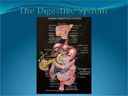


Slide1
The Digestive System
Lecture
11
Slide2Ducts
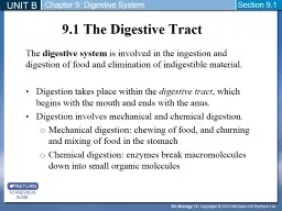
The exocrine pancreatic secretion is discharged into the duodenum through
two pancreatic ducts
, the
main pancreatic
, and the
accessory pancreatic ducts
.
The
main
pancreatic duct
commences at the
tail
of the gland by the union of small ducts; in its course towards the duodenum it receives numerous tributaries from lobules.
The duct terminates by joining the bile duct at the
hepatopancreatic ampulla
.
1
Slide3It opens into the second part of the duodenum at about its middle on the
major duodenal papilla
. Occasionally it may open independently into the duodenum.
An
accessory pancreatic duct
, which is much smaller than the main duct,
drains
the
front part of
the
head
of the pancreas and opens into the duodenum 2 cm proximal to the main duct on the minor duodenal papilla.
The accessory duct frequently communicates with the main duct.
2
Slide4Common bile duct
Main pancreatic duct
Hepatopancreatic ampulla
Accessory
pancreatic
duct
3
Figure 16.
Pancreatic ducts
Slide5Accessory pancreatic duct
Common bile duct
Major duodenal papilla
Main pancreatic duct
Head of pancreas
4
Figure 17.
Major and minor duodenal papillae
Slide6Structure
The pancreas is
enclosed
by a
thin collagenous fibrous capsule
, which sends
septa
into it, separating the pancreatic
lobules
.
The exocrine component
Secretes 1500–3000 ml
of isomotic alkaline fluid per day, consists of closely packed secretory acini, which drain into a
highly branched duct system. Pancreatic acinus is composed of
several serous cells surrounding a lumen.
5
Slide7The acinus cells
rest on a basement membrane
,
supported
by a
delicate sheath of reticular fibers
.
During secretory phase
the
lumen
is large and
distended with secretion
. 6
Slide8Figure 18. Exocrine and endocrine pancreas (sectional view). Stain: hematoxyline and eosin. Low magnification.
serous acini and
zymogenis
cells
intercalated duct
pancreatic islet
interlobular connective
tissue septa
blood vessel
interlobular
connective tissue
interlobular duct
pancinian
corpuscle
blood vessel
centroacinar cell
capillaries
pancreatic islet
centroacinar cell
7
Slide9Figure 19. Pancreatic islet.
Stain: hematoxyline and eosin. High magnification.
8
Slide10Figure 20. Pancreatic islet (special preparation). Stain: Gomori
’
s chrome alum hematoxyline and phloxine. High magnification.
9
Slide11intercalated
duct
connective
tissue
capsule
pancreatic islet
cenroacinar
ce
ll
secretory
acini
capillaries
Figure 21. Pancreatic endocrine (pancreatic islet) and exocrine region. Stain: peroxidase acid-schiff and hematoxiline. X 80.
10
Slide12During the
resting phase
zymogen granules
accumulate in the cells, which enlarge and encroach on the lumen.
The
acenus
cells have
round
or
spherical
nucleus
. The
basal part of the cell shows cytoplasmic basophilia, indicative of the
protein synthesis involved in enzyme manufacture.
Acinus secretion contains water,
ions
, and
several proteases
.
Pancreatic secretion
is
controlled
mainly through two hormones–
secretin
and
cholecystokinin
.
11
Slide13Both hormones
are
produced
by
enteroendocrine cells
of the small intestine (duodenum and jejunum).
Several acini join
an
intercalated duct lined
by
cuboidal
epithelium
.The initial
portions of intercalated ducts penetrate lumens of acini
. Nuclei, surrounded by a pale cytoplasm, belong to centroacinar cells
that constitute the intraacinar portion of the intercalated duct. 12A
Slide14Slide1512B
Slide1612C
Slide17Electron microscope shows that the cytoplasm of the a cinar cells contains numerous cisternae of
RER
, occupying the basal part of the cell, sugesting that these cells are involved with protein synthesis
.
Golgi complex
which occupies the apical pole of the nucleus is associated with several condensing vacules and numerous zymogen granules.
12D
Slide1812E
Slide19As compared with the
acinar
cell,
central cells
lack of
secretory granules
and
has very
scant
RER
.12F
Slide2012G
Slide21The
intercalated duct unite
to form larger ducts called
intralobular ducts
that, in turn,
unite
to form
larger interlobular ducts
line by
columnar epithelium
,
located within the connective tissue septa.
These ducts unite to form the main ducts
.12D
Slide22The endocrine component:
Includes the
islets of Langerhans
, which
lie
mainly within the
lobules
(
intralobular
) but it may
occasionally be interlobular.
The islet appear as spherical masses or
cords of cells, which stain faintly with hematoxylin and
eosin, in contrast to the exocrine acini which stain deeply.
The islets are usually separated from the acini by a
thin reticular membrane
, but in places appear to be in direct continuity with acinar cells.
13
Slide23Each islet has an
extensive blood supply
and the internal secretion from the cells passes directly into the blood stream.
Ultrastructural studies
reveal that the
islet cells
contain
small granules
that
concentrated
predominately in the
infranuclear region.
The majority of the
cells are β cells, known to secrete insulin, and
their granules have an oblong crystalline form. Of the remaining cells
, some are α cells that secrete glucagon, and have the more usual
spherical type of granule
.
14A
Slide2414B
Slide2514C
Slide26Vessels and Nerves of Pancreas
The
superior pancreaticodudenal
branch of the
gastroduodenal artery
and
inferior pancreaticoduodenal
branch of the
superior mesenteric artery
supply
the
head of the pancreas (including the uncinate process
). The rest
of the pancreas is supplied by the splenic branch of the celiac trunk.
There is free anastomosis
between these arteries.
Veins
from the
head
drain into the
superior and inferior pancreaticodudenal veins
.
15
Slide27The
rest
of the pancreas drains by the
splenic vein
.
Lymphatic vessels
from the pancreas pass to the
pancreaticosplenic nodes
(lie
along
the
superior border
of the pancreas) and aortic nodes
.
Sympathetic and parasympathetic nerves reach the gland from the coeliac plexus.
Pain fibers accompany the
sympathetic nerve
and enter the
central nervous system
via the
sympathetic trunk
.
16
Slide28Most Important
I. Mouth (Oral cavity)
a. Bulk of the lips.
b. Boundaries of the vestibule
c. Boundaries of the mouth proper.
d. Communication of mouth proper.
e. Walls of mouth proper.
f. Lining epithelium.
17
Slide29II. Salivary glands
Relations of parotid gland.
Relations of superficial part of submandibular gland.
Structures within the parotid gland.
Histology of salivary glands.
18
Slide30III. Tongue
Papillae and their distribution.
Location of anterior 2/3 and posterior 1/3 of tongue.
Sulcus terminalis and foramen caecum.
Contents of taste buds.
Muscles of tongue: Action.
Motor and sensory supply of tongue.
IV. Soft palate:
Its muscle and nerve supply.
19
Slide31V. Pharynx
a. Nasopharynx: Vertebral level of posterior wall; Tubal ridge; Communications, Level of pharyngeal isthmus; Extends.
b. Oropharynx: Palatoglossal and palatopha-ryngeal folds or arches; Communications; Extends.
c. Laryngopharynx: Extends; Communica-tions, Anterior wall.
d. Muscles of pharynx: Nerve supply, and actions.
20
Slide32e. Gaps
between superior and middle constrictors; and between middle and inferior constrictors, and structures pass through these gaps.
f. Gaps between skull and upper border of superior constrictor, and structures fill this gap.
g. Histology of the wall of
pharynx.
21
Slide33VI. Oesophagus
Relations of cervical and thoracic portions.
Level of deviation to the left.
Histology.
22
Slide34VII. Peritoneum
Falciform ligament.
Median, medial, and lateral umblical ligaments.
Mesentery of small intestine.
Transverse mesocolon.
Sigmoid mesocolon.
Mesoapendix.
Lesser and greater omentum.
23
Slide35Wall of lesser sac.
Innervation of parietal and visceral peritoneum.
Structure in the free (right) border of lesser omentum and the relations between them.
VIII. Stomac
h
Parts.
Relations and stomach bed.
Blood and nerve supply.
Lymphatic drainage.
24
Slide36IX. Duodenum
Relations of each part.
Blood supply.
X. Jejunum and ileum
a. Plicae circulares, peyre's patches, and the differences between jejunum and ileum.
b. Differences in the mesentery.
c. Blood supply, arcades, and straight arteries.
25
Slide37XI. Large intestine
Caecum: Location, relations, and blood supply.
Appendix: Location of its base; Differences in the position of its tip, and blood supply.
Ascending colon: Location, relations, and blood supply.
Transverse colon: Location and blood and nerve supply.
Descending colon: Location, relations and blood supply.
26
Slide38Sigmoid
colon:
Its
termination and blood supply.
Rectum
: Location, relations, and blood supply
.
Anal canal: Location, relations, Histology, blood and nerve supply, internal and external hemorrhoids, and anal fissure.
27
Slide39XII. Liver
Location.
Fissures.
Lobes.
Inferior (visceral) surface.
Boundaries of caudate and quadrate lobes and porta hepatis.
Histology of liver: Hepatocytes: Function of: Golgi complex, ESR, RER, Lysosomes, peroximase , and Hering's canal.
Functions of fat storing cells.
28
Slide40h. Function of Kupffer cells.
XIII. Gall bladder and common bile duct
Parts.
Location of fundus. Histology.
IX. Common bile duct
Locations.
Histology.
Termination.
29
Slide41Pancreas
Location
.
Relations
of head and body.
Its
relation to the transverse mesocolon.
Uncinate
process (part of what and
extends
In
which ligament the tail lies.
Ducts. Histology
of the exocrine portion.30