
Bones of the skeleton are organs that contain several different tissues Bones are dominated by bone tissue but also contain Nervous tissue Blood tissue and vessels Cartilage in articular cartilages Epithelial tissue lining the blood vessels ID: 928452
Download Presentation The PPT/PDF document "The Bones" is the property of its rightful owner. Permission is granted to download and print the materials on this web site for personal, non-commercial use only, and to display it on your personal computer provided you do not modify the materials and that you retain all copyright notices contained in the materials. By downloading content from our website, you accept the terms of this agreement.


Slide1
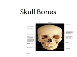
The Bones
Slide2Bones of the skeleton are organs that contain several different tissues
Bones are dominated by bone tissue but also contain Nervous tissue Blood tissue and vessels ,Cartilage in
articular
cartilages ,Epithelial tissue lining the blood vessels.
Slide3Bone function
Support
Protection
(protect internal organs)
Movement
(provide leverage system for skeletal muscles, tendons, ligaments and joints)
Mineral homeostasis
(bones act as reserves of minerals important for the body like calcium or phosphorus)
Hematopoiesis
: blood cell formation
Storage of adipose tissue: yellow
marrow
Slide4Classification of Bone
: Bones vary in shape and size
Bones are classified by
their shape
as long, short, flat, or irregular bone
Bones differ in the distribution of compact and spongy osseous tissues
Slide5Slide6Long bones
Long bones have a long shaft and two distinct ends
Classification is based on shape not size.
Short Bones
Short bones are roughly
cubelike
Thin compact bone layer surrounding spongy bone mass
Short bones are often carpal, tarsal and
sesamoid
bones
Flat bones
are thin, flattened and usually curved
Parallel layer of compact bone with spongy bone layer between Skull, sternum and ribs are example.
Irregular bones
don’t fit into the previous categories
Complicated shapes, Consist of spongy bone with a thin layer of compact
Examples are hip bones &
vertabrae
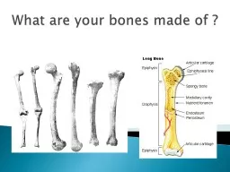
Slide7Bone anatomy
Diaphysis
:
long shaft of bone
Epiphysis:
ends of bone
Epiphyseal
plate:
growth plate
Metaphysis
:
b/w epiphysis and
diaphysis
Articular
cartilage:
covers epiphysis
Periosteum
:
bone covering (pain sensitive)
Sharpey’s
fibers:
periosteum
attaches to underlying bone
Medullary cavity:
Hollow chamber in bone
- red marrow produces blood cells
- yellow marrow is adipose
Endosteum
:
thin layer lining the medullary cavity
Slide8Slide9Blood and nerve supply of bone
Bone is supplied with blood by:
Periosteal
arteries
accompanied by nerves supply the
periosteum
and compact bone
Epiphyseal
veins
carry blood away from long bones nerves accompany the blood vessels that supply bones.
The
periosteum
is rich in sensory nerves sensitive to tearing or tension
Slide10Hematopoietic Tissue
The hematopoietic tissue, red marrow, is typically found within the cavities of spongy bone of long bones and in the
diploe
of flat bones
these cavities are referred to as red marrow cavities.
in infants the medullary cavity and all areas of spongy bone contain red bone marrow
In the adult the medullary cavity contains fat that extends into the epiphysis and there is little red marrow present in spongy bone cavities
blood cell production occurs only in the head of the femur and
humerous
most blood cell production occurs in the
diploe
areas of the sternum and hip
yellow marrow can revert to red marrow if the person becomes very anemic
Slide11There are two types of mature bone:
1. Compact
- which is found in the shafts of long bones (
diaphyses
). This makes up 80% of all bone, also called cortical bone, is the hard, stiff, smooth, thin, white bone
tissue
that surrounds all bones in the human body. It is also called osseous tissue or cortical bone and it provides structure and support for an
organism
as part of its
skeleton
, in addition to being a location for the storage of minerals like calcium. About 80% of the weight of the human skeleton comes from compact bone.
Slide12Slide132. Spongy (
cancellous
) bone
- which is found at the ends of long bones (in the epiphysis). This makes up 20% of all bone. This type of bone contains red bone marrow and a network of bony
trabeculae
.
Spongy bone is the tissue that makes up the interior of bones; compact bone is the tissue that forms the surface of bones. In long bones, spongy bone forms the interior of the epiphyses; the
diaphysis
(shaft) consists of compact bone surrounding the central marrow cavity.
Slide14Slide15Four cell types make up osseous tissue
Osteoprogenitor
cells,Osteoblasts,Osteocytes,Osteoclasts
.
Osteoprogenitor
cells
:
- derived from
mesenchyme
, all connective tissue is derived
- unspecialized stem cells, undergo mitosis and develop into
osteoblasts
, found on inner surface of
periosteum
and
endosteum
.
Osteoblasts
:
- bone forming cells, found on surface of bone (arrow), no ability to
mitotically
divide, collagen secretors
Osteocytes
:
mature bone cells. derived form
osteoblasts
, do not secrete matrix material, cellular duties include exchange of nutrients
and
waste with blood.
Osteoclasts
- bone
resorbing
cells, bone surface, growth, maintenance and bone repair
Slide16Bone formation
The process of bone formation is called
ossification
Bone formation occurs in four situations:
1) Formation of bone in an embryo
2) Growth of bones until adulthood
3) Remodeling of bone
4) Repair of fractures
Slide17Formation of Bone in an Embryo
Cartilage formation and ossification occurs during the sixth week of embryonic development
with two pattern
Slide18Intramembranous
Ossification
Flat bones of the skull and mandible are formed in this way “Soft spots” that help the fetal skull pass through the birth canal later become ossified forming the skull ,
intramembranous
ossification
: a group of
mesenchymal
cells within a highly
vascularized
area of the embryonic connective tissue proliferates and differentiates directly into
preosteoblasts
and then into
osteoblasts
.
Slide19Intramembranous
ossification follows four steps
: (a)
Mesenchymal
cells group into clusters, differentiate into
osteoblasts
, and ossification centers form. (b) Secreted
osteoid
traps
osteoblasts
, which then become
osteocytes
. (c) Trabecular matrix and
periosteum
form.
Slide20Slide21Endochondral
ossification
Most bones form by the process of
endochondral
ossification
.The mechanism responsible for the formation of all long bones of the axial skeleton. The replacement of cartilage by bone Most bones of the body are formed in this way including long bones.
This process occurs at three main sites: the
physis
, the epiphysis, and the cuboidal bones of the
carpus
and tarsus.
Process begins late in the second month of development
Process uses hyaline cartilage “bones” as the pattern for bone construction
During this process cartilage is broken down as ossification proceeds
Slide221)Formation
of a bone collar around hyaline cartilage model
2)
Osteoblasts
of the new
periosteum
secrete
osteoid
against the hyaline cartilage along the
diaphysis
Cartilage in the center of the
diaphysis
calcifies
Calcification of cartilage blocks nutrients and
chondrocytes
die
Matrix deteriorates and cavities develop
Bones stabilized by collar; growth occurs at epiphysis
3)Invasion
of the internal cavities by the
periosteal
bud and spongy bone
Bud contains nutrient artery & vein,
lymphatics
, nerve fibers, red marrow elements,
osteoblasts
and
osteoclasts
Slide23Slide244) Formation
of the medullary cavity as ossification continues
5)Secondary ossification
centers form in epiphyses
Cartilage in epiphyses calcifies and deteriorates opening cavities for entry of
periosteal
bud
Slide25Slide26Postnatal bone growth
During
infancy and youth bone growth occurs entirely by
interstitialgrowth
of the
epiphyseal
plates
bones grow in thickness by appositional growth
bones stop growing during adolescence or in early adulthood
some facial bones such as the nose or lower jaw continue to grow throughout life
Slide27Slide28