
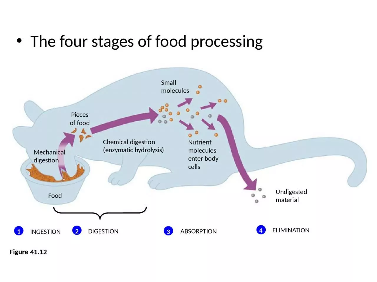
Figure 4112 Pieces of food Small molecules Mechanical digestion Food Chemical digestion enzymatic hydrolysis Nutrient molecules enter body cells Undigested material INGESTION 1 ID: 1032457
Download Presentation The PPT/PDF document "The four stages of food processing" is the property of its rightful owner. Permission is granted to download and print the materials on this web site for personal, non-commercial use only, and to display it on your personal computer provided you do not modify the materials and that you retain all copyright notices contained in the materials. By downloading content from our website, you accept the terms of this agreement.

1. The four stages of food processingFigure 41.12Piecesof foodSmallmoleculesMechanicaldigestionFood Chemical digestion(enzymatic hydrolysis)Nutrient moleculesenter body cellsUndigested materialINGESTION1DIGESTION2ELIMINATION4ABSORPTION3
2. Digestive CompartmentsMost animals process food In specialized compartments
3. Intracellular DigestionIn intracellular digestionFood particles are engulfed by endocytosis and digested within food vacuoles
4. Extracellular DigestionExtracellular digestionIs the breakdown of food particles outside cells
5. Animals with a more complex body planHave a digestive tube with two openings, a mouth and an anusThis digestive tubeIs called a complete digestive tract or an alimentary canal
6. The digestive tube can be organized into specialized regionsThat carry out digestion and nutrient absorption in a stepwise fashionEsophagusMouthPharynxCropGizzardIntestineAnusTyphlosoleLumen of intestineEsophagusAnusRectumMouthCropGastric cecaAnusIntestineGizzardCropStomachMouthEsophagusForegutMidgutHindgutEarthworm. The digestive tract ofan earthworm includes a muscular pharynx that sucks food in through themouth. Food passes through the esophagus and is stored and moistened in the crop. The muscular gizzard, whichcontains small bits of sand and gravel, pulverizes the food. Digestion and absorption occur in the intestine, which has a dorsal fold, the typhlosole, that increases the surface area for nutrient absorption.(b) Grasshopper. A grasshopper has several digestive chambers grouped into three main regions: a foregut, with an esophagus and crop; a midgut; and a hindgut. Food is moistened and stored in the crop, but most digestion occurs in the midgut. Gastric ceca, pouches extending from the midgut, absorb nutrients.(c) Bird. Many birds have three separate chambers—the crop, stomach, and gizzard—where food is pulverized and churned before passing into the intestine. A bird’s crop and gizzard function very much like those of an earthworm. In most birds, chemical digestion and absorption of nutrients occur in the intestine.Figure 41.14a–c
7. The mammalian digestive system consists of the alimentary canalAnd various accessory glands that secrete digestive juices through ducts
8. IIeumof small intestineDuodenum of small intestineAppendixCecumAscendingportion of large intestineAnusSmall intestineLarge intestineRectumLiverGall-bladderTongueOral cavityPharynxEsophagusStomachPyloricsphincterCardiacorificeMouthEsophagusSalivaryglandsStomachLiverPancreasGall-bladderLarge intestinesSmall intestinesRectumAnusParotid glandSublingual glandSubmandibular glandSalivaryglandsA schematic diagram of the human digestive systemPancreasFigure 41.15
9. Food is pushed along the digestive tract by peristalsisRhythmic waves of contraction of smooth muscles in the wall of the canal
10. The Oral Cavity, Pharynx, and EsophagusIn the oral cavity, food is lubricated and digestion beginsAnd teeth chew food into smaller particles that are exposed to salivary amylase, initiating the breakdown of glucose polymers
11. Organs Involved in Swallowing
12. Parts of the mammalian stomach
13. The region we call our throat is the pharynxA junction that opens to both the esophagus and the windpipe (trachea)The esophagusConducts food from the pharynx down to the stomach by peristalsis
14. From mouth to stomachEsophagusEpiglottis downTonguePharynxGlottisLarynxTracheaBolus of foodEpiglottisupTo lungsTo stomachEsophageal sphinctercontractedGlottis upand closedEsophageal sphincterrelaxedGlottisdown and openEsophageal sphinctercontractedEpiglottisupRelaxedmusclesContractedmusclesRelaxedmusclesStomachFigure 41.161 When a person is not swallowing, the esophageal sphincter muscle is contracted, the epiglottis is up, and the glottis is open, allowing air to flow through the trachea to the lungs. The swallowingreflex is triggeredwhen a bolus offood reaches thepharynx.2 The larynx, theupper part of therespiratory tract,moves upward andtips the epiglottisover the glottis,preventing foodfrom entering thetrachea.3 The esophagealsphincter relaxes,allowing thebolus to enter theesophagus.4After the foodhas entered theesophagus, thelarynx movesdownward andopens thebreathingpassage.5 Waves of muscularcontraction (peristalsis)move the bolus down the esophagus to the stomach.6
15. The StomachThe stomach stores foodAnd secretes gastric juice, which converts a meal to acid chymeGastric juiceIs made up of hydrochloric acid and the enzyme pepsin
16. The lining of the stomachIs coated with mucus, which prevents the gastric juice from destroying the cellsFigure 41.17Pepsin (active enzyme)HClParietal cellChief cellStomachFolds of epithelial tissueEsophagusPyloric sphincterEpitheliumPepsinogen321 Interior surface of stomach.The interior surface of the stomach wall is highly folded and dotted with pits leading into tubular gastric glands.Gastric gland. The gastric glands have three types of cells that secrete different components of the gastric juice: mucus cells, chief cells, and parietal cells.Mucus cells secrete mucus,which lubricates and protectsthe cells lining the stomach.Chief cells secrete pepsino-gen, an inactive form of thedigestive enzyme pepsin.Parietal cells secretehydrochloric acid (HCl).1Pepsinogen and HCIare secreted into thelumen of the stomach.2HCl convertspepsinogen to pepsin.3Pepsin then activatesmore pepsinogen,starting a chainreaction. Pepsinbegins the chemicaldigestion of proteins.5 µmSmall intestineCardiac orifice
17. Gastric ulcers, lesions in the liningAre caused mainly by the bacterium Helicobacter pyloriFigure 41.181 µmBacteriaMucuslayer of stomach
18. The Small Intestine The small intestineIs the longest section of the alimentary canalIs the major organ of digestion and absorption
19. Enzymatic Action in the Small IntestineThe first portion of the small intestine is the duodenumWhere acid chyme from the stomach mixes with digestive juices from the pancreas, liver, gallbladder, and intestine itselfFigure 41.19LiverBileAcid chymeStomachPancreatic juicePancreasIntestinaljuiceDuodenum of small intestineGall-bladder
20. The pancreas produces proteases, protein-digesting enzymesThat are activated once they enter the duodenumPancreasMembrane-boundenteropeptidaseTrypsinActive proteasesLumen of duodenumInactivetrypsinogenOther inactiveproteasesFigure 41.20
21. Enzymatic digestion is completedAs peristalsis moves the mixture of chyme and digestive juices along the small intestineFigure 41.21
22. Oral cavity,pharynx,esophagusCarbohydrate digestionPolysaccharides(starch, glycogen)Disaccharides(sucrose, lactose)Salivary amylaseSmaller polysaccharides,maltoseStomachProtein digestionNucleic acid digestionFat digestionProteinsPepsinSmall polypeptidesLumen of small intes-tine PolysaccharidesPancreatic amylasesMaltose and otherdisaccharidesEpitheliumof smallintestine(brushborder)DisaccharidasesMonosaccharidesPolypeptidesPancreatic trypsin andchymotrypsin (These proteasescleave bonds adjacent to certainamino acids.)SmallerpolypeptidesPancreatic carboxypeptidaseAmino acidsSmall peptidesDipeptidases, carboxypeptidase, and aminopeptidase (These proteases split off one amino acid at a time, working from opposite ends of a polypeptide.)Amino acidsDNA, RNAPancreaticnucleasesNucleotidesNucleotidasesNucleosidesNucleosidasesandphosphatasesNitrogenous bases,sugars, phosphatesFat globules (Insoluble inwater, fats aggregate asglobules.)Bile saltsFat droplets (A coating ofbile salts prevents small drop-lets from coalescing intolarger globules, increasingexposure to lipase.)Pancreatic lipaseGlycerol, fattyacids, glycerides
23. Hormones help coordinate the secretion of digestive juices into the alimentary canalFigure 41.22Amino acids or fatty acids in the duodenum trigger the release of cholecystokinin (CCK), which stimulates the release of digestive enzymes from the pancreas and bile from the gallbladder.LiverGall-bladderCCKEntero-gastroneGastrinStomachPancreasSecretinCCKDuodenumKeyStimulationInhibitionEnterogastrone secreted by the duodenum inhibits peristalsis and acid secretion by the stomach, thereby slowing digestion when acid chyme rich in fats enters the duodenum.Secreted by the duodenum, secretin stimulates the pancreas to release sodium bicarbonate, which neutralizes acid chyme from the stomach.Gastrin from the stomach recirculates via the bloodstream back to the stomach, where it stimulates the production of gastric juices.
24.
25.
26. Absorption of NutrientsThe small intestine has a huge surface areaDue to the presence of villi and microvilli that are exposed to the intestinal lumen
27. The enormous microvillar surfaceIs an adaptation that greatly increases the rate of nutrient absorptionEpithelialcellsKeyNutrientabsorptionVein carrying blood to hepatic portal vesselVilliLargecircularfoldsIntestinal wallVilliEpithelial cellsLymph vesselBloodcapillariesLactealMicrovilli(brush border)Muscle layers
28. The core of each villusContains a network of blood vessels and a small vessel of the lymphatic system called a lacteal
29. Amino acids and sugarsPass through the epithelium of the small intestine and enter the bloodstreamAfter glycerol and fatty acids are absorbed by epithelial cellsThey are recombined into fats within these cells
30. These fats are then mixed with cholesterol and coated with proteinsForming small molecules called chylomicrons, which are transported into lactealsFigure 41.24 Large fat globules are emulsified by bile salts in the duodenum.1 Digestion of fat by the pancreatic enzyme lipase yields free fatty acids and monoglycerides, which then form micelles.2 Fatty acids and mono-glycerides leave micelles and enter epithelial cells by diffusion.3Fat globuleLactealEpithelialcells ofsmallintestineMicelles madeup of fatty acids,monoglycerides,and bile saltsFat dropletscoated withbile saltsBile salts Chylomicrons containing fattysubstances are transported out of the epithelial cells and into lacteals, where they are carried away from the intestine by lymph.4
31. The Large IntestineThe large intestine, or colonIs connected to the small intestineFigure 41.25