
DR DIGVIJAY SHARMA DEPARTMENT OF PHYSIOTHERAPY UIHS KANPUR The ankle foot complex is structurally analogous to the wristhand complex of the upper extremity The bones of the ankle foot complex are traditionally divided into 3 functional segment ID: 997793
Download Presentation The PPT/PDF document "Ankle and Foot complex –" is the property of its rightful owner. Permission is granted to download and print the materials on this web site for personal, non-commercial use only, and to display it on your personal computer provided you do not modify the materials and that you retain all copyright notices contained in the materials. By downloading content from our website, you accept the terms of this agreement.


1. Ankle and Foot complex – DR. DIGVIJAY SHARMADEPARTMENT OF PHYSIOTHERAPYU.I.H.SKANPUR
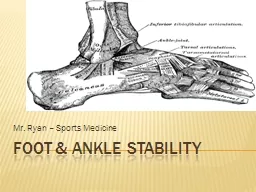
2. The ankle foot complex is structurally analogous to the wrist-hand complex of the upper extremity. The bones of the ankle - foot complex are traditionally divided into 3 functional segment.The hind foot / post segment, composed of talus and calcaneus.The mid foot / middle segment, composed of navicular, cuboid and 3 cuneiform.The fore-foot / anterior segment, composed of metatarsals and phalanges.
3. It is also refered as Talo-crural joint i.e. the articulation between the talus and distal tibia (tibio-talar surfaces) and the talus and fibula (talo- fibular surfaces ) .
4. Proximal articular surface – The proximal articular surface of the ankle is composed of the concave surface of distal tibia and of the tibial and fibular malleoli.These 3 facets forms are almost continuous forming the concave joint surface that extends more distally on the fibular side / lateral side and on the posterior margin of the tibia police. The structure of distal tibia and malleoli resembles and is referred to the mortise.The mortices of the ankle is adjustable relying on the proximal and distal tibiofemoral joint to both permit and control the changes of the mortise.
5. It is a plane synovial joint formed by the articulation of the head of the fibula with the posterolateral aspect of the tibia. A slight convexity to the tibial facet and slight concavity to the fibular facet seem to predominate .Each proximal joint is surrounded by a joint capsule and is reinforced by anterior and posterior tibiofibular ligament . Motion at the tibiofibular joint is variable but consistently small which can be known as superior and inferior sliding of fibular and as fibular rotation.
6. It is syndesmosis or fibrous union between the concave facet of the tibia and the convex facet of the fibula. There is no joint capsule.The joint is supported by strong ligaments are cural tibiofibular interosseous ligament, anterior and posterior tibiofibular ligament and the interosseous membrane. The interosseous membrane directly support both proximal and distal tibiofibular articulation.The joint is an extremely strong articulation.The function of the ankle joint depends on the stability of the tibiofibular mortise.
7. The distal articular surface of the ankle joint is formed by body of talus.The body of the talus has 3 articular surfaces. A large lateral facet, A smaller medial facet and A trochlear or superior facets.The ankle joint is the most congruent joint in the human body. The articular cartilage covering the trochlea is continuous with the cartilage covering the lateral and medial facets.The structural integrity is maintained throughout the range of motion of the joint by a number of important ligaments.
8. The capsule of the ankle joint is fairly thin and especially weak anteriorly and posteriorly.The ligaments that support the proximal and distal tibiofibular joints are important for stability of mortises.There are cural-tibio-fibular interosseous ligament , anterior and posterior tibio-fibular ligament and the interosseous membrane.Two other major ligament are medial and lateral collateral ligaments.
9. The medial collateral ligament is most commonly called the deltoid ligament and it is a fan shaped ligament having both superficial and deep fibres arising from the borders of the tibial malleolus and insert in a continuous line on the Navicular anteriorly and on the talus and calcaneus distally and posteriorly.The deltoid ligament as a whole is extremely strong ligament .This ligament does helps to control medial distraction stresses on the joint but also helps check motion at the extremes of joint range.
10. It is composed of 3 separate band and commonly referred to as separate ligament.These are the anterior and posterior talofibular ligament and calcaneofibular ligament.Lateral collateral ligament are weaker and more prone to injury then the MCL. The LCL helps to maintain varus stresses resulting in lateral distraction of the joint and check extremes of joint range of motion.The other ligament that contribute stability to the ankle joint are- Inferior extensor retinaculum Superior extensor retinaculumLateral extensor retinaculum
11. The ankle joint is a synovial joint with 1° of freedom of movement. The primary motion is dorsi and plantar flexion.However, the ankle joint is capable of some rotation of the talus within the mortise in both the transverse plane and a vertical axis called talar rotation or abduction/adduction and in frontal plane around AP axis called talar tilt or inversion/eversion.
12. Dorsiflexion- 20° from neutral ( 0 - 20 degree)Plantar flexion- 30° - 35° from neutral (0 – 30/50 degree) Medial rotation 7°Lateral rotation 10°Talar tilt 5° average
13. The talocalcaneal or subtalar joint is a articulation between talus superiorly and the calcaneus inferiorly.
14. There are 3 separate plane articulations between the talus and calcaneus. The smaller anterior and middle talocalcaneal articulation are formed by 2 convex facets on the inferior body and neck of the talus and 2 concave facets on the calcaneum. The posterior talocalcaneal articulation is the largest and formed by a concave facet on the under surface of the body of talus and a convex facet on the body of the calcaneum.
15. The posterior articulation has its own capsule, the anterior and middle articulation shares a capsule with the talonavicular joint.The subtalar joint is a stable joint that rarely dislocates or undergoes degenerative changes. It receive ligamentous support from ligamentous structure that support the ankle as well as from ligamentous structure that cross the subtalar joint.A number of structure contribute to the lateral support of the subtalar joint. These included from superficial to deep - the calcaneofibular ligament , lateral talocalcaneal ligament, the cervical ligament and the interosseous talocalcaneal ligaments.The cervical ligament is strongest of the talocalcaneal structure.
16.
17. The subtalar joint is composed of 3 articulations , the alternating convex-concave facet that limits the potential mobility as well as making the motion complex and distinct , the results is a tri-planar motion of the talus around a single oblique joint axis . The subtalar joint is a uniaxial joint with 1 degree of freedoms. Motions available are supination and pronation.
18. It is classically considered as a part of transverse tarsal joint. The talocalcaneonavicular joint name lies together that navicular and the subtalar (talocalcaneal) joints that are both anatomically and functionally related . The talonavicular articulation is formed proximally by the anterior portion of the head of talus and distally by the concave posterior navicular . The talus head however also articulates inferiorly with the anterior and medial facets of the calcaneus. The TCN joint is ball and socket joint where the large concavity of the head of talus is received by a large socket formed by convexity of navicular.
19. The important ligaments are- Plantar calcaneonavicular ligamentDeltoid ligament medially andBifurcate ligament laterally
20. The TCN joint like its subtalar component that contributes to it , It is tri planar joint with 1º of freedom.Supination / pronation
21. It is a compound joint formed by the talonavicular and calcaneocuboid joints because the talonavicular joint is classically considered to be part of transverse tarsal joint, it belongs to 2 joint complexes i.e. the TCN and the Transverse tarsal joint.The other component of transverse tarsal joint is the calcaneocuboid joint . The 2 joints together present an s shape joint line that intersects the foot horizontally dividing the hind foot from mid and fore foot.
22. The calcaneocuboid joint is formed proximally by the anterior calcaneus and distally by the posterior cuboid. The articular surface of both the calcaneus and cuboid are complex being reciprocally concave / convex across both dimensions.CAPSULE AND LIGAMENT – The joint has its own capsule that is reinforced by several major ligaments. These are lateral band of the bifurcate ligament , dorsal calcaneocuboid ligament and the planter calcaneocuboid ( short ) ligament and the long planter ligament.The long plantar ligament is the most important of these ligaments.
23. Transverse tarsal joint functions around two independent Axis. Longitudinal axis is nearly horizontal . The transverse tarsal ligament produces motion around tri planar axis such as supination / pronation as seen at the subtalar / TCN joints.Around AP axis the tarsal joint produces inversion/ eversion.
24. Supination TwistExtreme pronation of the foot is accompanied by the adduction of the head of talus, eversion of the calcaneus, pronation of the transverse tarsal joint .If the forefoot is to remain in ground, there is supination of Tarsometatarsal joint, which is referred to as supination twist.Pronation TwistExtreme supination of the foot is accompanied by abduction of the head of talus, inversion of the calcaneus and mandatory supination of the transverse tarsal joint. In this condition to keep the foot on ground, there is pronation of tarsometatarsal joint ,this is referred to as Pronation Twist.
25. Supination Pronationtwist twist
26. The TMT are plane synovial joints formed by the distal tarsal row proximaly and by the bases of the metatarsal anteriorly. The first TMT is the articulation between the base of 1st metatarsal and the medial cuneiform.It has its own articular capsule . The second TMT joint is the articulation between the base of the second metatarsal with mortise formed by the middle cuneiform and the side of the medial and lateral cuneiforms. This joint is sets back more posteriorly than other TMT joint. It is stronger and its motion more restricted.
27. Third TMT joint formed by the third metatarsal and the lateral cuneiforms shares a capsule with second TMT joint.The fourth and fifth TMT joints are formed by the bases of the fourth and fifth metatarsal with cuboid. These two joints also share a joint capsule.Each TMT joints is reinforced by numerous dorsal planter and interosseous ligaments . Additionally stability is contributed by the deep transverse metatarsal ligaments, similar to that found in hand that prevent a splaying of the metatarsal heads at the TMT joint.AXES - Each TMT joint is considered to have a unique although not fully independent axis of motion , the axis of foot passes through the 2nd metatarsal .
28. The function of TMT joint is primarily is a continuation of the function of the transverse tarsal joint is attempt to regulate the position of metatarsals and phalanges relatives to the weight bearing surface . The motions of the TMT joints are somewhat interdependent it also contribute hollowing and flattening of the foot. An active dorsiflex force across the non weight bearing TMT will simultaneously extend the 5 metatarsals while also creating inversion at the 1st two rays and eversion at the last 2 rays .The opposite motions accompanying TMT dorsiflex flattened the contour of the planter of foot and active plantarflex force.
29. Summary of motions of the rays of the foot Dorsiflexion Plantarflexion1st ray Inversion Eversion Slight Adduction Slight Abduction2nd ray Slight Inversion Slight Eversion3rd ray4th ray Slight Eversion Slight Inversion5th ray Eversion Inversion Slight Abduction Slight Adduction
30. The 5 MTP joints condyloid synovial joints with 2° of freedom , flexion/Extension and abduction / adduction .MTP joint structure – The MTP joint are formed proximally by the head of the metatarsals and distally by the bases of the proximal phalanges .Each joint has its own capsule . The stability of the MTP joint is provided by the plantar plates or plantar pads , the collateral ligament and the deep transverse metatarsal ligament.
31. MTP joints are condyloid joints with 2° of freedom , flexion / extension and abduction/ adduction . The MTP joints serves primarily to allow the foot to hinge at the toes so that the heel may rise of the ground while still maintaining the small but dynamic base of support (BOS) afforded by the toes and the toe musculature dysfunction is enhance by 2 structural aspects of the MTP joints , the metatarsal break and the effect of the MTP extension on the plantar aponeurosis.
32. It refers to the single oblique axis for MTP flexion and extension that lies through 2nd to 5th metatarsal heads . It may range from 55° to 70°. It is called break because It is the point where the foot Hinges as heel rises in weight bearing. There is active contraction of plantar flexors muscles and the head of the metatarsals glides, plantarily on proximal phalanx.
33. These are 5 proximal interphalangeal joint and four distal interphalangeal joints , each phalanx is vertically identical in structure to its counter part in hand although substantially shorter in length.The toes function to smooth height shift to opposite foot in gait and help to maintain stability by pressing against the ground both in static posture when necessary in gain. each interphalangeal is formed by the head of the proximal phalanx and base of the distal phalanx.Functions- The interphalangeal joint of the toes are synovial hinge joints with 1° degree of freedom flexion/ extension.
34.