
The organ include A Salivary glands function of them are 1 To wet and lubricate ingested food and oral mucosa 2 initiate the digestion of carbohydrates and lipids with amylase and ID: 1032443
Download Presentation The PPT/PDF document "Organs associated with digestive system ..." is the property of its rightful owner. Permission is granted to download and print the materials on this web site for personal, non-commercial use only, and to display it on your personal computer provided you do not modify the materials and that you retain all copyright notices contained in the materials. By downloading content from our website, you accept the terms of this agreement.


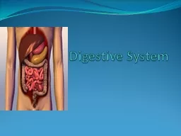


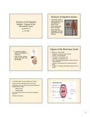

1. Organs associated with digestive system (Accessory organ)
2. The organ include: A- Salivary glands: function of them are:1- To wet and lubricate ingested food and oral mucosa.2- initiate the digestion of carbohydrates and lipids with amylase and lipase.3- to secrete innate immune components such as lysozyme and lactoferrin.
3. B- Pancreas: Secretes digestive enzymes that act in the small intestine and hormones important for the metabolism of the absorbed nutrientsC- The Liver:1- Produces bile for digestion and absorption of fats2- Plays a major role in carbohydrate and protein metabolism3- inactivates many toxic substances and drugs4- synthesizes most of blood plasma proteins
4. SALIVARY GLANDS Exocrine glands in the mouth produce saliva, which has digestive, lubricating, and protective functions. With a normal pH of 6.5-6.9.There are three pairs of large salivary glands: The parotidSubmandibular Sublingual glands
5.
6. Histology - A capsule of connective tissue surrounds each major salivary gland. - The parenchyma of each consists of secretory end pieces and branching duct separated by connective tissue
7. Parotid glands, located in each cheek near the ear, are branched acinar glands with secretory part composed of serous cells secrete α-amylase that initiates hydrolysis of carbohydrates and proline (antimicrobial protein)Submandibular glands, which produce two-thirds of all saliva, are branched tubuloacinar glands, serous and mucus cells. In addition to α-amylase and proline-rich proteins, serous cells of the submandibular gland secrete lysozyme for hydrolysis of bacterial walls.Sublingual glands, the smallest of the major glands, are branched tubuloacinar glands. The main product of the gland is mucus. Formed of serous and mucus cells.
8. Pancreas The pancreas is a mixed exocrine-endocrine gland that produces both digestive enzymes and hormones.A thin capsule of connective tissue cover the pancreas The secretory acini are surrounded by a basal lamina that is supported only by a delicate sheath of reticular fibers with a rich capillary network.The digestive enzymes are produced by cells of exocrine portion of the pancreasHormones are synthesized in endocrine epithelial cells known as pancreatic islets (islets of Langerhans)Exocrine pancreas secretes 1.5-2L of fluid per day, rich in bicarbonate ions and digestive enzymes.
9.
10. LiverThe liver is the second biggest organ in the body, in adults averaging about 1.5 kg. Located in the right upper quadrant of the abdomen just below the diaphragm.Liver is an interface between the digestive system and blood Most blood in liver 80% comes from the portal vein arising from intestine, stomach and spleen.
11. Histology The liver is covered by a thin fibrous capsule of connective tissue that becomes thicker at the hilum.The portal vein and the hepatic artery and the duct are surrounded by connective tissue all the way to their termination.
12. Hepatic lobulesLiver cells (Hepatocytes) are epithelial cells grouped in an interconnected plateLiver cells arranged of thousands of small polyhedral hepatic lobules.Hepatocytes like bricks of wall arranged radially around the central vein
13. Hepatocytes branch and anastomose freely, forming spongy like structure Liver sinusoids are surrounded and supported by delicate sheath of reticular fibers and endothelial cellsTwo cell types are associated with1- Stellate macrophage (Kupffer cells) their role to breakdown aged RBC, free heme for re-use, remove bacteria and act as antigen- pressenting cells 2- Stellate fat storing cells with small lipid droplets with vitamin A
14. Hepatic lobule structure and functionA- secretion of protein factors in blood (endocrine)B- secretion of bile components (Exocrine)C- removal of oxygen and small compounds of all kinds from blood- liver has strong capacity for regeneration despite its slow rate of cell renewal- loss of hepatic tissue from the action of toxic substance triggers the remaining healthy hepatocytes begin to divide - the bile produced by hepatocytes flow through the bile ducts and hepatic duct- the bile duct and hepatic duct are lined by mucous membrane having a simple columnar epithelium.
15.
16. Thank you