/
Muscles ,  fasciae  and Muscles ,  fasciae  and

Muscles , fasciae and - PowerPoint Presentation

finley
finley . @finley
Follow
65 views
Uploaded On 2024-01-29

Muscles , fasciae and - PPT Presentation

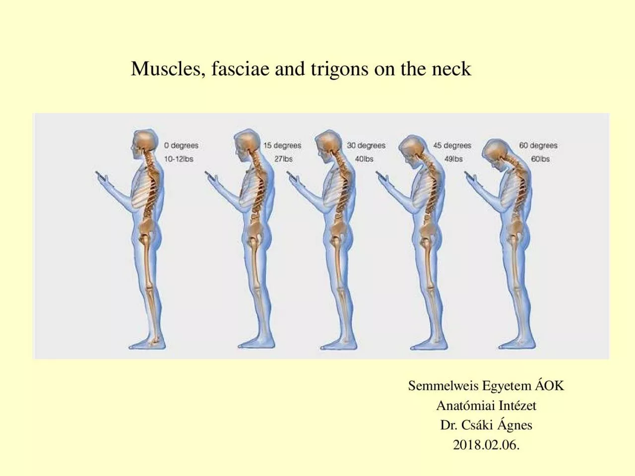
trigons on the neck Semmelweis Egyetem ÁOK Anatómiai Intézet Dr Csáki Ágnes 20180206 Deep prevertebral muscles of the neck M longus colli M longus ID: 1042627

muscles layer prevertebral hyoid layer muscles hyoid prevertebral fascia bone neck pretracheal carotid deep triangle scalene superficial sheath cervical

Share:

Link:

Embed:

Download Presentation from below link

Download Presentation The PPT/PDF document "Muscles , fasciae and" is the property of its rightful owner. Permission is granted to download and print the materials on this web site for personal, non-commercial use only, and to display it on your personal computer provided you do not modify the materials and that you retain all copyright notices contained in the materials. By downloading content from our website, you accept the terms of this agreement.


Presentation Transcript

1. Muscles, fasciae and trigons on the neckSemmelweis Egyetem ÁOKAnatómiai IntézetDr. Csáki Ágnes2018.02.06.

2. Deep, prevertebral muscles of the neckM. longus colliM. longus capitisM. rectus capitis anteriorM. rectus capitis lateralisActions: flex the head and neck anteriorly Innervation: ventral rami of spinal nervesScalenus muscles

3. NameOriginInsertionFunctionm. anterior scalene scalene tubercle on the 1. ribanterior tubercles of the transverse processes of C3-C6The three muscles:ipsilateral contraction causes ipsilateral lateral flexion of the neck, and bilateral contraction causes anterior flexion of the neck in case of forced respiration act as accessory muscles of respirationm. middle scalenebehindthe subclavian groove on the 1. ribposterior tubercles of the transverse processes of C2-C7m. posterior scalene2. ribposterior tubercles of the transverse processes of C5-C7anteriorposteriorScalene musclesinnervation: ventral rami of spinal nerves C3-C8

4. Suprahyoid muscles The suprahyoid muscles are a group of four muscles, located superiorly to the hyoid bone of the neck. They all act to elevate the hyoid bone – an action involved in swallowing, sucking, drinking, opening of mouth.Hyoid boneorigin insertion m. mylohyoidN. V/3Mylohyoid line of mandibuleMylohyoid raphe and body of hyoid bone(2) m. digastricAnterior belly - inner side of the mentum /N.V/3Posterior belly - mastoid process /N.V IITendon is connected to the hyoid bone(3) m. stylohyoidN. VIIStyloid processBody and greater horn of the hyoid bone around the tendon of the digastric (4) m. geniohyoidN. XII Inner side of the mentum (genu of mandible)Body of hyoid bone

5.

6. Infrahyoid muscles The infrahyoid muscles are a group of four muscles that are located inferiorly to the hyoid bone in the neckDepress the hyoid bone take parts in swallowing, sucking, drinking, opening of mouthorigin insertion (1) m. sternohyoid Inner side of sternum andcapsule of the sternoclavicular joint Body of the hyoid bone (2) m. sternothyroid Inner side of sternum andI. rib cartilage Linea obliqua of the thyroid cartilage (3) m. thyrohyoid linea obliqua of the thyroid cartilage Body and greater horn of the hyoid bone (4) m. omohyoid Inferior belly: sup.transverse scapular lig.superior belly: body and greater horn of the hyoid bone Tendon is connected to the sternoclavicular joint and veins Innervation: Ansa cervicalis profunda (Cervical plexus C1- C3)

7. Lateral view of supra- and infrahyoid muscles

8. Superficial neck musclesSternocleidomastoid m.Originates from the manubrium sterni and clavicle, inserts to the mastoid process of the skullipsilateral contraction causes ipsilateral lateral flexion and rotation of the head („ear comes near to the shoulder”,bilateral contraction causes protraction of the headInnervation: IX cranial nerve (with the trapezius m.)

9. Triangles of the neckANTERIOR AREA:(according to the scm m.)Submandibular triangle: two bellies of the digastric and base of mandibule Base of the trigon: the mylohyoid m.Content: submandibular gland2. Submentale triangle: anterior bellies of the two digastric m., hyoid bone Base of the trigon: the mylohyoid m.Content: lymph nodes3. Carotid triangle: sternocleidomastoid, omohyoid and posterior belly of digastric m.Content: carotid sheeth (bifurcation of common carotid, internal jugular vein, vagus nerve)Puls!Posterior area antero-lateral area

10. LATERAL AREA: supraclavicular triangle - divided by the omohyoid m. intoOmotrapezoid triangle : bordered by sternocleidomastoid, omohyoid and trapezius Base of the triangle: deep neck muscles covered by the prevertebral fasciaContent: cervical plexus (Erb point-cervical pl.) (and brachial plexus trunks)Omoclavicular triangle: sternocleidomastoid, omohyoid and clavicleContent: scalene hiatus (gap) with the subclavian artery and trunks of the brachial plexus

11. Cervical fasciasFascia is a layer of fibrous tissue that surrounds muscles, vessels and nerves.In the neck, there are several layers of fascias, which act to support and compartmentalise the structures present

12. https://www.slideshare.net/indiandentalacademy/compartments-of-the-head-and-neck-kiran-nx-powerlite

13.

14.

15.

16. SUPERFICIAL CERVICAL FASCIA Subcutan connective tissue and the platysma onlyDEEP CERVICAL FASCIA : spf layer (investing layer) pretracheal layer prevertebral layer

17. Superficial or investing layer (yellow)Superficial layerFom the base of the mandible Sheathes the sternocleidomastoid and the trapeziusAt the angle of the mandibule fuses with the parotideomasseteric fascia („parotid nest”)

18. Middle or pretracheal layer (green) Sheathes the infrahyoid muscles In the midline forms a strong, aponeurosis like layer Connects the tendon of the omohyoid to the sternoclavicular joint Forms the carotid sheath! Pretracheal layer

19. Deep or prevertebral layer (violet) In the midline behind the pharynx is atteched strongly to the longus capitis and longus colli Laterally forms the scalene „tent”Prevertebral layerCarotid sheath

20. Deep cevical fascia- Spf. layer or investing layer- pretracheal layer (visceral part) (carotid sheath)- prevertebral layerThe investing layer of fascia in highlighted in blue. The fascia completely envelopes the sternocleidomastoid and trapezius.Transverse section of the neck.

21. Deep cevical fascia- Spf. Layer or investing layerpretracheal layer(muscular) (visceral) (carotid sheath)- prevertebral layerThe trachea, esophagus, and infrahyoid muscles are enclosed by the pretracheal layer . It can be anatomically divided into two parts:Muscular – encloses the infrahyoid muscles(purple)Visceral  – encloses the trachea and oesophagus and forms the carotid sheath

22. Deep cevical fascia- Spf. Layer or investing layer- pretracheal layer (visceral part) (carotid sheath)- prevertebral layerThe prevertebral layer surrounds the vertebral column and its associated muscles (scalene, longus colli, longus capitis m., and deep muscles of the back)

23. Scalene tentLower section – scalene tent

24. Pretracheal-Buccopharyngeal fasciaAlar fasciaFASCIAL SPACES Superficial Retropharyngeal Danger PrevertebralParapharyngeal space

25. The prevertebral fascia consists two layers: prevertebral and alar fasciaThe alar fascia fuses anteriorly with the buccopharyngeal fascia at the level of the 4th thoracic vertebra.Behind the alar fascia the danger space continuos downward to the thorax.

26. The superficial layer and muscular part of pretracheal layer end at the sternum, but the visceral layer and the prevertebral layer continous downward to the thorax!! - inflammation

27. Superficial space - anterior jugular veinsNumb. 2 spf layerNumb. 3 pretracheal layer

28.

29.

30. Irodalom:images.MD, 2006 Current Medicine LLC Kahle W, Leonhardt H, Platzer W: Color Atlas/Text of Human Anatomy, 1992, Thieme, Stuttgart Putz R, Pabst R (editors): Sobotta Atlas of Human Anatomy, 1993, Urban & Schwarzenberg, MünchenRomanes GJ (editor): Cunningham’s Textbook of Anatomy, 1991, Oxford University Press, OxfordSzentágothai J, Réthelyi M: Funkcionális anatómia, 2002, Medicina, BudapestVízkelety T: Az ortopédia tankönyve, 1995, Semmelweis Kiadó, Budapesthttp://teachmeanatomy.info/the-basics/https://www.youtube.com/watch?v=F2sSVWEdjWQhttps://www.slideshare.net/indiandentalacademy/compartments-of-the-head-and-neck-kiran-nx-powerliteThank you!