
As a result of cephalocaudal and lateral folding of the embryo a portion of the endodermlined yolk sac cavity is incorporated into the embryo to form the primitive gut Two other portions of the endodermlined cavity the yolk sac and the allantois remain outside the embryo ID: 777428
Download The PPT/PDF document "Digestive System DIVISIONS OF THE GUT TU..." is the property of its rightful owner. Permission is granted to download and print the materials on this web site for personal, non-commercial use only, and to display it on your personal computer provided you do not modify the materials and that you retain all copyright notices contained in the materials. By downloading content from our website, you accept the terms of this agreement.


Slide1
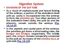
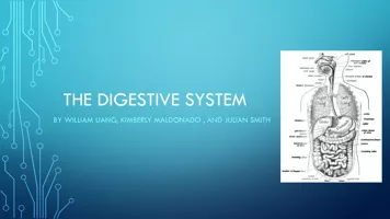
Digestive System
DIVISIONS OF THE GUT TUBE
As a result of
cephalocaudal
and lateral folding of the embryo, a portion of the endoderm-lined yolk sac cavity is incorporated into the embryo to form the
primitive gut
. Two other portions of the endoderm-lined cavity, the yolk sac and the allantois, remain outside the embryo (
Fig. 14.1A-D
).
In the cephalic and caudal parts of the embryo, the primitive gut forms a blind-ending tube, the
foregut and hindgut,
respectively. The middle part, the
midgut
, remains temporally connected to the yolk sac by means of the
vitelline
duct
, or yolk stalk (
Fig. 14.1D
).
Slide2Slide3Development of the primitive gut and its derivatives is usually discussed in four sections:
(a) The
pharyngeal gut
, or pharynx, extends from the
oropharyngeal
membrane
to the respiratory diverticulum and is part of the foregut;
(b) The remainder of the foregut lies caudal to the pharyngeal tube and extends as far caudally as the
liver outgrowth
.
(c) The
midgut
begins P.210 caudal to the liver bud and extends to the junction of the right two-thirds and left third of the transverse colon in the adult.
d) The
hindgut
extends from the left third of the transverse colon to the
cloacal
membrane
Slide4Endoderm forms the
1-epithelial
lining
of the digestive tract
and 2-
gives rise to the specific cells (the
parenchyma
) of glands, such as hepatocytes and the exocrine and endocrine cells of the pancreas.
The
stroma
(connective tissue) for the glands is derived from
visceral mesoderm.
Muscle, connective tissue, and peritoneal components of the wall of the gut also are derived from
visceral mesoderm
.
Slide5MESENTERIES
Portions of the gut tube and its derivatives are suspended from the dorsal and ventral body wall by mesenteries,
double layers of peritoneum
that enclose an organ and connect it to the body wall. Such organs are called
intraperitoneal
,
whereas organs that lie against the posterior body wall and are covered by peritoneum on their anterior surface only (e.g., the kidneys) are considered
retroperitoneal.
Peritoneal ligaments are double layers of peritoneum (mesenteries) that pass from one organ to another or from an organ to the body wall. Mesenteries and ligaments provide pathways for vessels, nerves, and
lymphatics
to and from abdominal viscera (
Figs. 14.3
and
14.4
Slide6Initially the foregut, midgut
, and hindgut are in broad contact with the mesenchyme of the posterior abdominal wall (
Fig. 14.3
). By the fifth week, however, the connecting tissue bridge has narrowed, and the caudal part of the foregut, the
midgut
, and a major part of the hindgut are
suspended from the abdominal wall by the
dorsal mesentery
(
Figs. 14.3C
and
14.4
), which extends from the lower end of the esophagus to the
cloacal
region of the hindgut. In the region of the
stomach
, it forms the
dorsal
mesogastrium
or greater
omentum
; in the region of the
duodenum
, it forms the
dorsal
mesoduodenum
; and in the region of the colon, it forms the
dorsal
mesocolon
.
Dorsal mesentery
of the
jejunal
and
ileal
loops forms the
mesentery proper
Slide7Slide8Ventral mesentery
which exists only in the region of the terminal part of the esophagus, the stomach, and the upper part of the duodenum (
Fig. 14.4
), is derived from the
septum
transversum
. Growth of the liver into the mesenchyme of the septum
transversum
divides the ventral mesentery into
(a)
the lesser
omentum
, extending from the lower portion of the esophagus, the stomach, and the upper portion of the duodenum to the liver and
(b)
the
falciform
ligament
, extending from the liver to the ventral body wall .
Slide9FOREGUTL
Esophagus
When the embryo is approximately 4 weeks old, the respiratory diverticulum (lung bud) appears at the ventral wall of the foregut at the border with the pharyngeal gut (
Fig. 14.5
). The
tracheoesophageal
septum gradually partitions this diverticulum from the dorsal part of the foregut (
Fig. 14.6
). In this manner, the
foregut divides into a ventral portion, the respiratory
primordium
, and a dorsal portion, the esophagus
At first, the esophagus is short (
Fig. 14.5A
), but with descent of the heart and lungs, it lengthens rapidly (
Fig. 14.5B
). The muscular coat, which is formed by surrounding splanchnic mesenchyme, is
striated
in its upper two-thirds and innervated by the
vagus
; the muscle coat is
smooth
in the lower third and is innervated by the splanchnic plexus.
Slide10Slide11Slide12Stomach
The stomach appears as a fusiform dilation of the foregut in the
fourth week
of development (
Fig. 14.8
). During the following weeks, its appearance and position change greatly as a result of the different rates of growth in various regions of its wall and the changes in position of surrounding organs. Positional changes of the stomach are most easily explained by assuming that it rotates around a
longitudinal and an
anteroposterior
axis (
Fig. 14.8
).
Slide13Slide14The stomach rotates 90° clockwise around its longitudinal axis, causing its left side to face anteriorly and its right side to face posteriorly (
Fig. 14.8A-C
). Hence, the left
vagus
nerve, initially innervating the left side of the stomach, now innervates the
anterior wall
; similarly, the right nerve innervates the
posterior wall
. During this rotation, the original posterior wall of the stomach grows faster than the anterior portion, forming the
greater and lesser curvatures
Slide15The cephalic and caudal ends of the stomach originally lie in the midline, but during further growth, the stomach rotates around an
anteroposterior
axis
, such that the caudal or pyloric part moves to the right and upward, and the cephalic or cardiac portion moves to the left P.215
and slightly downward (
Fig. 14.8D,E
). The stomach thus assumes its final position, its axis running from above left to below right.
Slide16Since the stomach is attached to the dorsal body wall by the
dorsal
mesogastrium
and to the ventral body wall by the
ventral
mesogastrium
(
Figs. 14.4
and
14.9A
), its rotation and disproportionate growth alter the position of these mesenteries. Rotation about the longitudinal axis pulls the dorsal
mesogastrium
to the left, creating a space behind the stomach called the
omental
bursa (lesser peritoneal sac
) (
Figs. 14.9
and
14.10
). This rotation also pulls the ventral
mesogastrium
to the right. As this process continues in the fifth week of development, the spleen
primordium
appears as a mesodermal proliferation between the two leaves of the dorsal
mesogastrium
(
Figs. 14.10
and
14.11
). With continued rotation of the stomach, the dorsal
mesogastrium
lengthens, and the portion between the spleen and dorsal midline swings to the left and fuses with the peritoneum of the posterior abdominal wall (
Figs. 14.10
and
14.11
).
Slide17The posterior leaf of the dorsal mesogastrium
and the peritoneum along this line of fusion degenerate. The spleen, which remains
intraperitoneal
, is then connected to the body wall in the region of the left kidney by the
lienorenal
ligament and to the stomach by the
gastrolienal
ligament and
Lengthening and fusion of the dorsal
mesogastrium
to the posterior body wall also determine the final position of the pancreas. Initially, the organ grows into the dorsal
mesoduodenum
, but eventually its tail extends into the dorsal
mesogastrium
(
Fig. 14.10A
). Since this portion of the dorsal
mesogastrium
fuses with the dorsal body wall, the tail of the pancreas lies against this region (
Slide18Once the posterior leaf of the dorsal mesogastrium
and the peritoneum of the posterior body wall degenerate along the line of fusion, the tail of the pancreas is covered by peritoneum on its anterior surface only and therefore lies in a retroperitoneal position. (Organs, such as the
pancreas
, that are originally covered by peritoneum, but later fuse with the posterior body wall to become retroperitoneal, are said to be
secondarily retroperitoneal.)
Slide19As a result of rotation of the stomach about its anteroposterior
axis, the dorsal
mesogastrium
bulges down (
Fig. 14.12
). It continues to grow down and forms a double-layered sac extending over the transverse colon and small intestinal loops like an apron (
Fig. 14.13A
). This P.217
double-leafed apron is the greater
omentum
; later, its layers fuse to form a single sheet hanging from the greater curvature of the stomach (
Fig. 14.13B
). The posterior layer of the greater
omentum
also fuses with the mesentery of the transverse colon (
Fig. 14.13B
).
Slide20The lesser omentum
and
falciform
ligament form from the ventral
mesogastrium
, which itself is derived from mesoderm of the septum
transversum
. When liver cords grow into the septum, it thins to form
(
a) the peritoneum of the liver;
(
b) the
falciform
ligament, extending from the liver to the ventral body wall; and
(
c) the lesser
omentum
, extending from the stomach and upper duodenum to the liver (
Figs. 14.14
and
14.15
). The free margin of the
falciform
ligament contains the
umbilical vein
(
Fig. 14.10A
), which is obliterated after birth to form the
round ligament of the liver
(
ligamentum
teres
hepatis
).
Slide21The free margin of the P.218 lesser
omentum
connecting the duodenum and liver (
hepatoduodenal
ligament
) contains the bile duct, portal vein, and hepatic artery (portal triad). This free margin also forms the roof of the
epiploic
foramen of Winslow
, which is the opening connecting the
omental
bursa (lesser sac) with the rest of the peritoneal cavity (greater sac) (
Fig. 14
Slide22Duodenum
The terminal part of the foregut and the cephalic part of the
midgut
form the duodenum. The junction of the two parts is directly distal to the origin of the liver bud (
Figs. 14.14
and
14.15
). As the stomach rotates, the duodenum takes on the form of a C-shaped loop and rotates to the right. This rotation, together with rapid growth of the head of the pancreas, swings the duodenum from its initial midline position to the right side of the abdominal cavity (
Figs. 14.10A
and
14.17
). The duodenum and head of the pancreas press against the dorsal body wall, and the right surface of the dorsal
mesoduodenum
fuses with the adjacent peritoneum. Both layers subsequently disappear, and the
duodenum and head of the pancreas
become fixed in a retroperitoneal position.
Slide23The entire pancreas thus obtains a retroperitoneal position. The dorsal
mesoduodenum
disappears entirely except in the region of the pylorus of the stomach, where a small portion of the duodenum (duodenal cap) retains its mesentery and remains
intraperitoneal
.
During the second month, the lumen of the duodenum is obliterated by proliferation of cells in its walls. However, the lumen is
recanalized
P.219
shortly thereafter (
Fig. 14.18A,B
). Since the foregut is supplied by the celiac artery and the
midgut
is supplied by the superior mesenteric artery, the duodenum is supplied by branches of both arteries (
Fig. 14.14
).
Slide24Slide25Liver and Gallbladder
The liver
primordium
appears in the middle of the third week as an outgrowth of the endodermal epithelium at the distal end of the foregut (
Figs. 14.14
and
14.15
). This outgrowth, the hepatic diverticulum, or liver bud, consists of rapidly proliferating cells that penetrate the septum
transversum
, that is, the mesodermal plate between the pericardial cavity and the stalk of the yolk sac (
Figs. 14.14
and
14.15
). While hepatic cells continue to penetrate the septum, the connection between the hepatic diverticulum and the foregut (duodenum) narrows, forming the
bile duct
. A small ventral outgrowth is formed by the bile duct, and this outgrowth gives rise to the
gallbladder and the cystic duct
Slide26During further development, epithelial liver cords intermingle with the vitelline
and umbilical veins, which form hepatic sinusoids. Liver cords differentiate into the parenchyma (liver cells) and form the lining of the biliary ducts. Hematopoietic cells,
Kupffer
cells, and connective tissue cells are derived from mesoderm of the septum
transversum
.
Slide27When liver cells have invaded the entire septum
transversum
, so that the organ bulges caudally into the abdominal cavity, mesoderm of the septum
transversum
lying between the liver and the foregut and the liver and the ventral abdominal wall becomes membranous, forming the lesser
omentum
and
falciform
ligament, respectively. Together, having formed the peritoneal connection between the foregut and the ventral abdominal wall, they are known as the ventral mesentery (
Fig. 14.15
).
Mesoderm on the surface of the liver differentiates into visceral peritoneum except on its
P.220
cranial surface (
Fig. 14.15B
). In this region, the liver remains in contact with the rest of the original septum
transversum
. This portion of the septum, which consists of densely packed mesoderm, will form the central tendon of the diaphragm. The surface of the liver that is in contact with the future diaphragm is never covered by peritoneum; it is the bare area of the liver (
Fig. 14.15
).
Slide28PANCREAS
The pancreas is formed by two buds, dorsal and ventral, originating from the endodermal lining of the duodenum (
Fig. 14.19
). Whereas the dorsal pancreatic bud is in the dorsal mesentery, the ventral pancreatic bud is close to the bile duct (
Fig. 14.19
). When the duodenum rotates to the right and becomes C-shaped, the ventral pancreatic bud moves dorsally in a manner similar to the shifting of the entrance of the bile duct (
Fig. 14.19
). Finally, the ventral bud comes to lie immediately below and behind the dorsal bud (
Fig. 14.20
). Later, the parenchyma and the duct systems of the dorsal and ventral pancreatic buds fuse (
Fig. 14.20B
). The ventral bud forms the
uncinate
process and inferior part of the head of the pancreas. The remaining part of the gland is derived from the dorsal bud.
Slide29The main pancreatic duct (of
Wirsung
) is formed by the distal part of the dorsal pancreatic duct and the entire ventral pancreatic duct (
Fig. 14.20B
). The proximal part of the dorsal pancreatic duct either is obliterated or persists as a small channel, the accessory pancreatic duct (
of
Santorini
). The main pancreatic duct, together with the bile duct, enters the duodenum at the site of the major papilla; the entrance of the accessory duct (when present) is at the site of the minor papilla. In about 10% of cases, the duct system fails to fuse, and the original double system persists.
Slide30Slide31Slide32Slide33In the third month
of fetal life, pancreatic islets (of Langerhans) develop from the
parenchymatous
pancreatic tissue and scatter throughout the pancreas.
Insulin
secretion begins at approximately the
fifth month
.
Glucagonand
somatostatin
-secreting cells also develop from parenchymal cells. Visceral mesoderm surrounding the pancreatic buds forms the pancreatic connective tissue.
Slide34MIDGUT
In the 5-week embryo, the
midgut
is suspended from the dorsal abdominal wall by a short mesentery and communicates with the yolk sac by way of the
vitelline
duct or yolk stalk (
Figs. 14.1
and
14.15
). In the adult, the
midgut
begins immediately distal to the entrance of the bile duct into the duodenum (
Fig. 14.15
) and terminates at the junction of the proximal two thirds of the transverse colon with the distal third. Over its entire length, the
midgut
is supplied by the superior mesenteric artery (
Fig. 14.24
).
Slide35Development of the midgut
is characterized by rapid elongation of the gut and its mesentery, resulting in formation of the primary intestinal loop (
Figs. 14.24
and
14.25
). At its apex, the loop remains in open connection with the yolk sac by way of the narrow
vitelline
duct (
Fig. 14.24
).
The cephalic limb
of the loop develops into the distal part of the duodenum, the jejunum, and part of the ileum.
The caudal limb
becomes the lower portion of the ileum, the cecum, the appendix, the ascending colon, and the proximal two thirds of the transverse colon.
Slide36Physiological Herniation
Development of the primary intestinal loop is characterized by rapid elongation, particularly of the cephalic limb. As a result of the rapid growth and expansion of the liver, the abdominal cavity temporarily becomes too small to contain all the intestinal loops, and they enter the
extraembryonic
cavity in the umbilical cord during the sixth week of development (physiological umbilical herniation) (
Fig. 14.26
).
Slide37Rotation of the
Midgut
Coincident with growth in length, the primary intestinal loop rotates around an axis formed by the superior mesenteric artery (
Fig. 14.25
). When viewed from the front, this rotation is counterclockwise, and it amounts to approximately 270° when it is complete (
Figs. 14.24
and
14.25
). Even during rotation, elongation of the small intestinal loop continues, and the jejunum and ileum form a number of coiled loops (
Fig. 14.26
). The large intestine likewise lengthens considerably but does not participate in the coiling phenomenon. Rotation occurs during herniation (about 90°), as well as during return of the intestinal loops into the abdominal cavity (remaining 180°) (
Fig. 14.27
).
Slide38Retraction of Herniated Loops
During the 10th week, herniated intestinal loops begin to return to the abdominal cavity. Although
P.224
the factors responsible for this return are not precisely known, it is thought that
regression
of the
mesonephric
kidney
,
reduced growth of the liver, and
expansion
of the abdominal cavity play important roles.
Slide39The proximal portion of the jejunum
, the first part to reenter the abdominal cavity, comes to lie on the left side (
Fig. 14.27A
). The later returning loops gradually settle more and more to the right.
The
cecal
bud
, which appears at about the sixth week as a small conical dilation of the caudal limb of the primary intestinal loop, is the last part of the gut to reenter the abdominal cavity. Temporarily, it lies in the right upper quadrant directly below the right lobe of the liver (
Fig. 14.27A
). From here, it descends into the right iliac fossa, placing the ascending colon and hepatic flexure on the right side of the abdominal cavity (
Fig. 14.27B
). During this process, the distal end of the
cecal
bud forms a narrow diverticulum,
the appendix (
Fig. 14.28
).
Slide40HINDGUT
The hindgut gives rise to the distal third of the transverse colon, the descending colon, the sigmoid, the rectum, and the upper part of the anal canal. The endoderm of the hindgut also forms the internal lining of the bladder and urethra (see
Chapter 15
).
The terminal portion of the hindgut enters into the posterior region of the cloaca, the primitive
anorectal
canal; the allantois enters into the anterior portion, the primitive urogenital sinus
Slide41The cloaca
itself is an endoderm-lined cavity covered at its ventral boundary by surface ectoderm. This boundary between the endoderm and the ectoderm forms the
cloacal
membrane (
Fig. 14.36
). A layer of mesoderm, the
urorectal
septum
, separates the region between the allantois and hindgut. This septum is derived from the merging of mesoderm covering the yolk sac and surrounding the allantois (
Figs. 14.1
and
14.36
). As the embryo grows and caudal folding continues, the tip of the
urorectal
septum comes to lie close to the
cloacal
membrane
Slide42At the end of the seventh week, the cloacal
membrane ruptures, creating the anal opening for the hindgut and a ventral opening for the urogenital sinus. Between the two, the tip of the
urorectal
septum forms the
perineal
body (
Fig. 14.36C
). The upper part (two-thirds) of the anal canal is derived from endoderm of the hindgut; the lower part (one-third) is derived from ectoderm around the
proctodeum
(
Fig. 14.36B,C
).
Ectoderm
in the region of the
proctodeum
on the surface of part of the cloaca proliferates and
invaginates
to create the anal pit (Fig. 13.36D). Subsequently, degeneration of the
cloacal
membrane (now called the anal membrane) establishes continuity between the upper and lower parts of the anal canal.
Slide43Since the
caudal part
of the anal canal originates from ectoderm, it is supplied by the
inferior rectal arteries
, branches of the internal
pudendal
arteries. However, the
cranial part
of the anal canal originates from endoderm and is therefore supplied by the
superior rectal artery
, a continuation of the inferior mesenteric artery, the artery of the hindgut. The junction between the endodermal and ectodermal regions of the anal canal is delineated by the
pectinate
line
, just below the anal columns. At this line, the epithelium changes from columnar to stratified squamous epithelium.
P.231
.
Slide44Slide45