

body 16 of body weight 1223 square meter It is composed from the epidermis and dermis Based on the thickness of the epidermis there are thick and thin skin The junction of dermis and epidermis is irregular the projection of the dermis ID: 916046
Download Presentation The PPT/PDF document "Skin The skin is the first heaviest orga..." is the property of its rightful owner. Permission is granted to download and print the materials on this web site for personal, non-commercial use only, and to display it on your personal computer provided you do not modify the materials and that you retain all copyright notices contained in the materials. By downloading content from our website, you accept the terms of this agreement.


Slide1
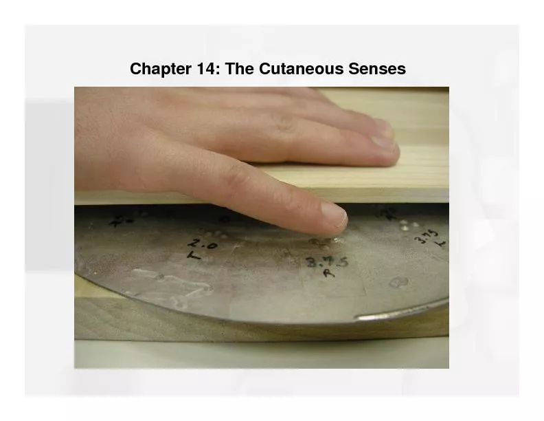
Skin
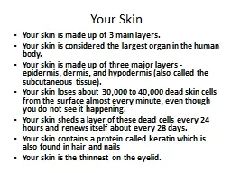
Slide2The skin is the first heaviest organ in the
body
16
% of body weight,
1.2-2.3 square meter.
It
is composed from the
epidermis
and
dermis
.
Based
on the thickness of the epidermis there are thick and thin skin.
The
junction of dermis and epidermis is irregular, the projection of the dermis
(papillae)
interdigitate
with epidermal
evagination
(epidermal ridge).
Slide3The skin functions are:
1
.
provide continuous communication with the environment (as receptor organ
)
2
. has protective action against ultraviolet rays (UV) by the action of Melanin pigment
3.
Thermoregulator
by the action of blood vessels ,glands of the skin and the adipose tissue
4
.
Execretion
of waste product and
5
.
Vit.D
synthesis
.
Slide4Upon close observation, human skin show ridge and groove in the tips of the finger and
volar
surfaces of the hands and feet (palm and sole) called
Dermatoglyphics
, they are unique for each individual, appearing as loops, arches etc--- which are used for personal identification.
Slide5Epidermis:
The
epidermis consists of
a
stratified
squamous
keratinized
epith
with three less abundant cell types:
Melanocytes
,
Langerhans
cells and
Merkel's cells
.
Slide6The epidermis consists of five layers:
1- Stratum
Basale
(
St.Germinativum
)
:
Is
a single layer of columnar OR
cuboidal
basophilic cells resting on the basal lamina.
Desmosomes
and
Hemidesmosomes
High mitotic
activity, this layer with the next layer responsible for constant renewal of epidermal cells (which occur every 15-30 days).
The
cells contain
keratin intermediate
filaments. As the cells progress upward, the number of filaments increases until it represent half of the protein in last layer.
Slide72-St.
Spinosum
(prickle layer)
:
St.
spinosum
consist of
cuboidal
cells with centrally located nuclei, the cytoplasm filled with bundles of keratin filaments (
Tonofilament), these bundles terminated in the spiny cytoplasmic projection, the cells of this layer firmly bound together by cytoplasmic projections and Desmosomes. St. Basale and St. spinosum also called (malpigian layer)
Slide8St.
spinosum
Slide93-St.
Granulosum
:
Consist
of 3-5 layers of polygonal cells,
Their
cytoplasm are filled with basophilic granules called
Keratohyalline granules which account for their basophilia. Presence of lamellar granules which are surrounded by membrane, these granules fuse with the cell membrane and discharge their content into the intercellular spaces to form the intercellular cement which act as a barrier to prevent the penetration of foreign materials and provide important sealing effect in the skin.
Slide104-St
.
lucidum
:
Is
more apparent in thick skin, is translucent consist of
eosinophilic
flattened cells,
The organelles and nuclei are no longer evident The cytoplasm consist of densly packed keratin filaments.
Slide115- St.
corneum
:
Consist
of 15-20 layers of flattened non-nucleated keratinized cells their cytoplasm are filled with filamentous
scleroprotein
called keratin
.
Tonofilaments are packed together in a matrix contributed by the keratohyalline granules. After keratinization, the cells consist of fibrillar proteins and thickened plasma membrane, they are called Horny cells. During keratinization, lysosomal hydrolytic enzymes digest the organelles.
Slide12In psoriasis,
there
is an increase in the number of proliferating cells in the stratum
basale
and the stratum
spinosum
as well as a decrease in the cycle time of these cells. This result in greater epidermal thickness and more rapid renewal of epidermis.
Slide13Slide14Melanocytes
:
The
colour
of the skin is depends on the pigment called Melanin, the number of blood vessels and the color of the blood flowing in
them.
Melanin
is a dark brown pigment produced by the
melanocyte found between the cells of St. basale and the hair follicles. They have rounded cells bodies with long processes extend between St. basale and spinosum Melanin is synthesized in the melanocytes. Melanin granules once formed migrate by the cytoplasmic processes of melanocytes into the cells of St. basale
and
spinosum
, then the melanin granules are injected into
keratinocytes
,
Slide15Melanin granules accumulate
in
supranuclear
area of the cytoplasm, to protect the nuclei from the deleterious effect of solar UV radiation
.
Melanocytes
are not distributed in a random way, they are about 1000 melnocytes/square mm in the skin of the thigh ,and about 2000/square mm in the skin of scrotum. Sex and race does not influence the number of melanocytes/square mm, difference in skin color is due to the difference in the number of melanin granules in the keratinocytes. Darkening of the skin color after exposure to sun light (Tanning) is due to 2 steps:1- Rapid release of pigment into the keratinocytes 2- Acceleration of the rate of melanin pigment synthesis
Slide16In human lack of
cortisol
from the adrenal cortex causes overproduction of
adrenocorticotropic
hormone, which increases the pigmentation of skin. An example of this is
Addison disease
, which is caused by dysfunction of the adrenal glands.
Slide17Albinism
, a hereditary inability of the
melanocyte
to synthesize melanin, is caused by the absence of
tyrosinase
activity. As a result the skin is not protected from solar radiation by melanin, and there is a greater incidence of basal and
squamous
cell carcinoma (skin cancer). The degeneration and disappearance of entire
melanocytes
result in a depigmentation disorder called Vitiligo
Slide18Langerhans's
cells:
Are star shaped cells found in the St.
spinosum
, they are bone marrow derived, they are capable of binding, processing and presenting antigens to T lymphocytes to stimulate them. So these cells are
Ag-presenting cells
.
Slide19Merkel's cells:
They are present in thick skin of palm and soles, there are free nerve endings are present at the base of these cells, so, these cells serve as sensory
mechanoreceptors.
Slide20Thank you