/
DISEASES OF GI TRACT DISEASES OF GI TRACT

DISEASES OF GI TRACT - PowerPoint Presentation

miller
miller . @miller
Follow
342 views
Uploaded On 2022-06-15

DISEASES OF GI TRACT - PPT Presentation

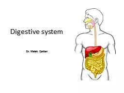
VMD603PG Dr Bipin Kumar Assistant Professor Veterinary Medicine BVC Bihar Animal Sciences University Patna DISEASES OF GI TRACT ORAL CAVITY Dental disease Foreign bodies ID: 918650

include gastric esophageal ulceration gastric include ulceration esophageal acid ulcer signs clinical result mucosa mucosal horses obstruction colic typically

Share:

Link:

Embed:

Download Presentation from below link

Download Presentation The PPT/PDF document "DISEASES OF GI TRACT" is the property of its rightful owner. Permission is granted to download and print the materials on this web site for personal, non-commercial use only, and to display it on your personal computer provided you do not modify the materials and that you retain all copyright notices contained in the materials. By downloading content from our website, you accept the terms of this agreement.


Presentation Transcript

Slide1

DISEASES OF GI TRACT VMD-603(PG)Dr Bipin KumarAssistant Professor, Veterinary MedicineBVC, Bihar Animal Sciences University, Patna

Slide2

DISEASES OF GI TRACTORAL CAVITYDental disease, Foreign bodies, Neoplasia, Infectious diseases (mostly vesicular stomatitis), and ulceration from NSAIDs or locally caustic agents.

Slide3

DysphagiaIt can be classified anatomically as prepharyngeal, pharyngeal, or postpharyngeal (esophageal).

Prepharyngeal

dysphagia is expressed clinically by dropping food (quidding) or water from the mouth, hypersalivation, reluctance to chew, or difficulty in prehension.

Pharyngeal or esophageal dysphagia is characterized predominantly by coughing and nasal discharge consisting of food, water, or saliva.

Dysphagia can be caused by pain, muscular, neurologic, or obstructive disorders.

Neurologic causes of dysphagia include diseases affecting the forebrain, brainstem, or peripheral cranial nerves controlling prehension (

Vm

, Vs, VII, and XII), transfer of the food bolus to the pharynx (Vs and XII), or swallowing (IX and X).

Slide4

SialadenitisSalivary glands in the horse are paired and include the parotid, mandibular, and polystomatic sublingual glands. The parotid gland is the largest and can secrete saliva at up to 50 ml/minute, with daily secretion up to 12 L in a 500 kg horse. Parotid secretion occurs only during mastication and can be blocked with atropine or anesthesia of the oral mucosa. Parotid saliva is relatively hypotonic, with average daily ion concentrations (

mEq

/L) as follows: Na+ 55, K+ 15, Cl- 50, HCO3- 52.

Salivary diversion will typically not result in acid-base or electrolyte abnormalities for several days, at which point horses will become slightly hyponatremic (day 3), markedly

hypochloremic

(day 3), mildly hypokalemic (day 10), and mildly

alkalemic

(day 10).

Concentrations of sodium, chloride, and bicarbonate ions increase at high rates of flow

Slide5

DISEASES OF THE ESOPHAGUSThe equine esophageal mucosa consists of keratinized stratified squamous mucosa with no digestive or absorptive function. The submucosa confers elasticity to the esophageal wall. Parasympathetic fibers of the vagus innervate the smooth muscle; sympathetic innervation of the esophagus is minimal.

During swallowing, the upper esophageal sphincter closes immediately, the lower esophageal sphincter (LES) opens, and food passage is propelled via esophageal peristalsis.

Gastric distention causes mechanical constriction of the LES while also triggering a vagal reflex that increases LES tone.

These factors normally prevent spontaneous reflux.

Slide6

Esophageal obstruction (Choke)Primary impaction with feed (typically roughage), bedding, or other material. Predisposing factors include poor dentition, prior esophageal trauma, gulping food, dry or improperly moistened feeds. Secondary impaction can result from intraluminal (foreign body), extramural (neoplasia, vascular ring anomaly, granuloma), intramural (abscess, granuloma, neoplasia, cyst, diverticulum, stenosis), or functional (dehydration, exhaustion, pharmacologic, primary

megaesophagus

, esophagitis, autonomic dysautonomia, vagal neuropathy, sedation) disorders.

Clinical signs are dysphagia, including nasal regurgitation of feed, saliva, or water.

Other signs include coughing, gagging or retching, ptyalism, nervousness, and an extended neck carriage.

Slide7

Miscellaneous esophageal disordersEsophagitis involves inflammation of the esophagus with or without ulceration. Reflux esophagitis typically occurs secondary to functional esophageal disorders or delayed gastric emptying (severe gastric ulceration, ileus, functional gastric outflow obstructions, etc.). Other causes of esophagitis include trauma, infection, and chemical injury. For reflux esophagitis, reduction of gastric acid is critical.

Sucralfate may enhance mucosal healing, but these effects have not been proven.

Megaesophagus is typically acquired in the horse and can occur following repeated obstruction, extramural masses, neurologic disorders resulting in vagal neuropathy, or pleuropneumonia (also resulting in vagal neuropathy).

Slide8

Other causes include equine dysautonomia, myasthenia gravis (not yet reported definitively in the horse), or systemic disorders. Idiopathic megaesophagus has also been reported in young horses. Endoscopy and contrast radiography can aid the diagnosis. Treatment is aimed at the underlying disorder. Medical management is warranted for at least 60 days with anti-inflammatory and antimicrobial therapy in addition to dietary management. Surgical management can include esophagostomy with fenestration, resection and anastomosis,

esophagomyotomy

, and patch grafting.

Slide9

DISEASES OF THE STOMACH EQUINE GASTRIC ULCER SYNDROME. Peptic ulcer disease is defined as erosions or ulcers of any portion of the gastrointestinal tract normally exposed to acid. Mucosal damage can include inflammation, erosion (disruption of the superficial mucosa), or ulceration (penetration of the submucosa). In severe cases, full-thickness ulceration can occur, resulting in perforation. The oral portion of the equine stomach is lined by stratified squamous mucosa similar to that lining the esophagus. The aboral portion of the stomach is lined with glandular mucosa, and the distinct junction between the two regions is deemed the

margo

plicatus

.

Ulceration can occur in either or both gastric regions although different clinical syndromes and pathophysiologic mechanisms apply.

As a result, the broad term “Equine gastric ulcer syndrome” (EGUS) has been used to encompass the wide array of associated clinical syndromes.

Slide10

Pathophysiology: An imbalance between inciting and protective factors in the mucosal environment can result in ulcer formation. The major intrinsic factors promoting ulcer formation include hydrochloric acid (HCl), bile acids, and pepsin, with HCl the predominant factor. Various intrinsic factors protect against ulcer formation such as the mucus-bicarbonate layer, mucosal perfusion, mucosal prostaglandin E2 and epidermal growth factor production, and gastroduodenal motility. Excess acid exposure is the predominant mechanism responsible for squamous ulceration, although many details remain unclear. HCl is secreted by parietal cells in the gastric glands via a hydrogen-potassium adenosine triphosphatase (H+,K+-ATPase) pump on the luminal side.

Slide11

The predominant stimuli to hydrochloric acid secretion are gastrin, histamine, and acetylcholine. Gastrin is released by G cells within the antral mucosa, while histamine is released by mast cells and enterochromaffin-like (ECL) cells in the gastric gland. Histamine binds to type 2 receptors on the parietal cell membrane, causing an increase in cAMP, resulting in activation of the proton pump. Gastrin and acetylcholine can also stimulate histamine release directly, and histamine is the primary stimulant of acid production. Gastric acid secretion by parietal cells is primarily inhibited by somatostatin, released by fundic and antral D cells. Gastric acid secretion also is inhibited by epidermal growth factor (EGF), a peptide produced in saliva.

Slide12

Clinical syndrome in Yearlings and adult horses Clinical signs attributable to EGUS in older horses are variable and classically include anorexia, and chronic or intermittent colic of varying severity. Many horses with endoscopic evidence of disease may appear to be clinically normal or have vague signs that include decreased consumption of concentrates, postprandial episodes of colic, poor performance, poor quality haircoat, and decreased condition or failure to thrive. Diarrhea is not typically associated with gastric ulceration in adult horses, although ulceration can occur concurrently with other causes of diarrhea. Bleeding from ulcers in the gastric squamous mucosa is typically not associated with anemia or hypoproteinemia

Slide13

Diagnosis only current method of confirmation is via gastroscopy, which can easily be performed in the standing horse or foal with mild sedation. Urine and blood sucrose absorption testing have recently been evaluated as a measure of gastric mucosal permeability in horses with EGUS. Also, serum α1-antitrypsin was detectable more frequently in foals with gastric ulceration than normal foals. Duodenoscopy is used to diagnose duodenal ulceration or inflammation.

Slide14

TreatmentAnti-ulcer therapy centers on acid suppression. The principal therapeutic options for ulcer treatment include H2 antagonists (cimetidine, ranitidine, famotidine, nizatidine),Proton pump inhibitors (PPI; omeprazole, pantoprazole, rabeprazole, esomeprazole).The mucosal adherent sucralfate, and antacids.

Slide15

Pyloric stenosis and delayed gastric emptyingPyloric stenosis is a structural resistance to gastric outflow. Congenital pyloric stenosis has been reported in foals and one yearling and results from hypertrophy of the pyloric musculature. Acquired pyloric stenosis can result from neoplasia or duodenal ulceration. Clinical signs are dependent upon the degree of obstruction but include abdominal pain, salivation, and teeth grinding. Complete or near complete obstruction can result in gastric reflux and reflux esophagitis

Measurement of gastric emptying can aid the diagnosis.

Slide16

Several methods of measurement are currently available, including nuclear scintigraphy, acetaminophen absorption, and post-consumption octanoic acid blood or breath testing. If complete obstruction is not present, medical therapy with a prokinetic such as bethanecol can increase the rate of gastric emptying. Phenylbutazone and cisapride have also been shown to attenuate the delay in gastric emptying caused by endotoxin administration. Surgical repair is necessary for definitive treatment of complete or near-complete obstruction and consists of either gastroenterostomy or pyloroplasty.

Slide17

Gastric dilatation and ruptureGastric dilatation can be classified as primary, secondary, or idiopathic. Causes of primary gastric dilatation include gastric impaction, grain engorgement, excessive water intake after exercise, aerophagia, and parasitism. Secondary gastric dilatation occurs more commonly and can result from primary intestinal ileus or small or large intestinal obstruction. Clinical signs of gastric dilatation include those associated with acute colic and, in severe cases, nasal discharge.

Associated laboratory abnormalities include hemoconcentration, hypokalemia, and hypochloremia.

Regardless of the initiating cause, gastric rupture usually occurs along the greater curvature.

Gastric rupture is usually fatal due to widespread contamination of the peritoneal cavity, septic peritonitis and septic shock.

Surgical repair is thus limited.

Slide18

Gastric impactionGastric impaction can result in either acute or chronic signs of colic. Although a specific cause is not always evident, ingestion of coarse roughage (straw bedding, poor quality forage), foreign objects (rubber fencing material), and feed that may swell after ingestion or improper mastication (persimmon seeds, mesquite beans, wheat, barley, sugar beet pulp) have been implicated. Possible predisposing factors include poor dentition, poor mastication and rapid consumption of feedstuffs, and inadequate water consumption. Clinical signs can vary from anorexia and weight loss to acute colic. In severe cases, spontaneous reflux may occur. In cases presenting with acute colic, a diagnosis is often made via ultrasonography or during exploratory celiotomy.

In addition to pain management, specific treatment consists of gastric lavage via nasogastric intubation or massage and injection of fluid to soften the impaction during laparotomy.

Slide19

Thank you