
wi f p ma y o t c n vo s ys m Rem ove the sk in fro m the re gion of the sku ll that extends betw een the ey es a nd ears and posterio rly to the back of the sku ll Scrap e aw ay m uscle and conn ective tissu e so you can see the su tures betw een t ID: 42838
Download Pdf The PPT/PDF document "NERVOUS SY STEM The nerv ous sy stem is ..." is the property of its rightful owner. Permission is granted to download and print the materials on this web site for personal, non-commercial use only, and to display it on your personal computer provided you do not modify the materials and that you retain all copyright notices contained in the materials. By downloading content from our website, you accept the terms of this agreement.


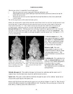
45 NERVOUS SYSTEM The nervous system is composed of several major parts: The central nervous system composed of the brain and spinal cord The peripheral nervous system made up of myelinated sensory and motor neurons leaving the central nervous system The autonomic nervous system composed of non-myelinated nerves that control glands and visceral organs. We will focus primarily on the central nervous system. Remove the skin from the region of the skull that extends between the eyes and ears and posteriorly to the back of the skull. Scrape away muscle and connective tissue so you can see the sutures between the bones of the skull (Fig. 7.1 ). Also remove the muscles that attach the skull to the vertebrae in the occipital region. Using heavy forceps crack the skull along the ridge where the occipital and interparietal bones meet. Working anteriorly and being careful not to apply pressure to the brain lift away pieces of the bones that cover the brain cavity. Stop just before you reach the nasal bones. Now bend the head ventrally an carefully cut through the axisto reveal the spinal cord. Use Figures 7.1-7.3 to locate the following: Cerebral hemispheres (1). The large cerebral hemispheres lie under the frontal (1a) and parietal bones (1b). They are separated by a deep fissure. The cerebral hemispheres are associated with integration. Olfactory bulb. The small olfactory bulbs lie anterior to the cerebral hemispheres and just caudal to the nasal bones (arrow). This region of the brain is associated with smell. Cerebellum (2). The cerebellum lies under the interparietal bone (2) and posterior to the cerebral hemispheres. It has three lobes: two lateral hemispheres and a medial vermis. The cerebellum controls coordination and equilibrium. Medulla oblongata (3). The medulla oblongata lies between the cerebellum and the spinal cord. It regulates heart rate, blood pressure, respiration and hormonal secretions. Spinal cord. The spinal cord extends from the brain through the canals in the vertebrae. Spinal nerves are given off at each region of the spinal cord. Brains which have been removed from the cranial cavity are shown in figures 7.3 and 7.4. They illustrate several of the above structures in a slightly different view. If you have time, you may want to remove the brain and try to identify some of the cranial nerves. igure 7.1. Surface of the cranium after removing the skin, muscle and connective tissue. The arrow is pointing to the suture between the nasal bones. Figure 7.2. Rat brain lying in the cranial cavity and covered with dura mater. The olfactory bulbs appear as light ovals under the nasal bones (arrow). 46 Optional - Removing the brain from the cranial cavity Carefully chip away bone on one of the lateral surfaces of the skull and toward the ventral surface. You will need to remove the bone that protects the inner. This bone lies between the cerebellum and the cerebrum. Cut the medulla oblongata below the point that it has become the spinal cord. Lift the spinal cord and cut the nerves and blood vessels that attach to the brain, leaving them as long as possible.. Remove it from the skull. If time permits you may want to try to identify some of the nerves originating from the ventral surface e the brain is removed you will see more detail if it is viewed under a dissecting microscope. Table 7.1 Cranial nerves of the rat. eFunction(s)NerveFunction(s) I. Olfactory SmellVII. Facial*Jaw muscles II. OpticVisionVIII. Auditory*Hearing III. Oculomotor Eye movementIX. Glossopharyngeal*Pharynx and tongue IV. TrochlearEye movementX. Vagus*Larynx, heart, lungs, diaphragm, stomach V. TrigeminalSkin, jaw muscles, tongue, teeth XI. Accessory*Neck muscles, pharyngeal organs VI. Abducens*Eye movementXII. Hypoglossal*Tongue * These seven nerves originate from the medulla oblongata Figure 7.3. Rat brain in a dorsal view with the protective dura mater removed. Figure 7.4. Rat brain in ventral view with several of the spinal nerves identified