
Epidermoid Cyst Common affecting young and middle age adults Usually seen on the head neck and trunk Often can identifiy a punctum Cyst contains keratin Superficial Epidermal cyst 5 cm Fluid filled ID: 1032787
Download Presentation The PPT/PDF document "Skin Anatomy Skin Appendages" is the property of its rightful owner. Permission is granted to download and print the materials on this web site for personal, non-commercial use only, and to display it on your personal computer provided you do not modify the materials and that you retain all copyright notices contained in the materials. By downloading content from our website, you accept the terms of this agreement.


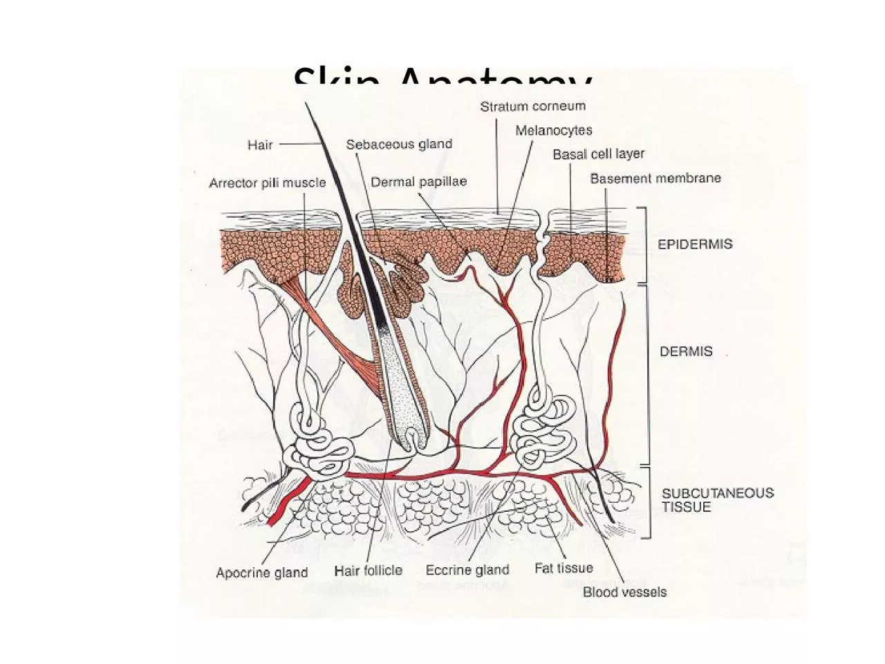
1. Skin Anatomy
2. Skin Appendages
3. Epidermoid CystCommon, affecting young and middle age adultsUsually seen on the head, neck and trunkOften can identifiy a punctumCyst contains keratin
4. Superficial Epidermal cyst5 cmFluid filledFluctuant Orifice or "pore" near the center
5. Skin Lesions and Diagnosis
6. Pilar Cysts
7. Strawberry hemangiomaRedPatch5 cmsElevatedBlanch with pressureCapillary angiomas composed of small, superficial vessels
8. Nevus A benign cluster of melanocytic cells arising as a result of proliferation of melanocytes at the dermo-epidermal junction. These may all remain in contact with the basal layer (the junctional nevus) or may become dettached from the basal layer and lie free in the dermis (the compound and intradermal nevus).
9. Blue NevusAn area of blue-black dermal pigmentation produced by an aberrant collection of pigment producing melanocytesThe brown pigment absorbs the longer wavelength of light and scatters blue light (Tyndall effect)Extend into the deep dermis, often occur on extremities and the dorsum of the hand
10. Junctional NeviNevi begin as small, flat, pigmented macules = junctional neviA smooth, hairless, light to dark brown macule, varying in size from 1 –6 mmOccurs on any site, especially on palms, soles, scrotumDuring adolescence some will become compound or intradermal It is characterized by single melanocytes, or theques of them in the lower epidermis
11. Nevus SpilusPigmented, light brown or tan macule, varied diameter, speckled with smaller, darker-colored macules or papulesLower extremity & trunk frequentlyMay be <1cm or largeMay follow a dermatomal distribution when largeUsually they do not cross the midline
12. Epidermal Nevus
13. Nevus Comedonicus
14. KeloidAn exaggerated reparative fibroblastic response to injury of the skinGenetic tendencyMost commonly found on the ears, neck and trunk
15. Keloid (after piercing) Very firm Rubbery lesions Reddish or darkly colored Occur after trauma sometimes very minor trauma May itch.
16. DermatofibromaCan occur on any part of the body, most common on the lower extremities, to a lesser degree on the upper extremities and trunkMay be single or multipleUsually pink or brownCommonly 6mm or lessHard consistency
17. Hidrocystoma
18. Lipoma
19. Premalignant Tumors
20. Actinic KeratosisHyperkeratotic lesions occurring in sun exposed adult skinMay exist in a premalignant state for yearsOften begin as an area of increased vascularity with the surface becoming roughMay progress to squamous cell carcinoma
21. Actinic keratosis
22. Actinic KeratosisA pre-cancerous condition of thick, scaly patches of sun-damaged skin.
23. Types of Skin CancerPre-CancerousActinic keratosis CancerousSquamous cell carcinomaBasal cell carcinomaMelanomaOthers
24. Malignant Tumours of The SkinBasal Cell Carsinoma (BCC)Squamous Cell Carsinoma (SCC)Malignant Melanoma (MM)
25. Skin CancerBasal CellSquamous CellMelanoma
26. Basal Cell Carcinoma Basal cell carcinoma is the most common malignancy of whites, arising from cells of the basal layer of the epithelium or from the external root sheath of the hair follicle
27. Basal Cell CarcinomaSkin cancer that arises from basal cells, small round cells found in the lower part of the skin (epidermis). Over 75% of all skin cancers Slow-growingGenerally treatable
28. Basal Cell CarcinomaMost common form of skin cancerUsually appears on sun damaged skinPearly appearance with superficial telangiectasiasRarely metastasizesDerived from basal cell layer of the epidermis
29. Classification of BCC(ACKERMAN):A- LOCALIZED1- Nodular2- Nodulocystic3-PigmentedB- SUPERFICIAL1-Superficial spreading2-MultifocalC-INFILTRATIVE1- Mopheic
30. Nodular basal cell carcinoma
31. Basal Cell Carcinoma
32. Superficial BCCOften multipleUpper trunk or shoulders commonest site but can appear anywherePink or red scaly patch with raised edge on close examinationSlowly growing over months or yearsBleed or ulcerate easily
33. Superficial basal cell carcinoma
34. Superficial Basal Cell Carcinoma
35. Sclerosing Basal Cell Carcinoma
36. Basal Cell Cancer Warning SignsSmall, smooth, pale, or waxy shiny lumpFirm, red lumpA lump that bleeds or develops a crust
37. Pigmented Nodular Basal Cell Carcinoma
38. Squamous Cell Carcinoma Squamous cell carcinoma originating from the keratinizing or malpighian (spindle) cell layer of the epithelium, is seen primarily in older patients, mostly men
39. Squamous Cell CarcinomaBegins in squamous cells which are found in the surface of the skin They are thin, flat cells that look like fish scalesApproximately 20% of all skin cancers
40. Squamous Cell Carcinomamost common below the line “tragus-comissura”the primary etiological factor is solar radiation. chronic ulcers including chronic lesions, burn wound, osteomyelitis, cytotoxic drugs, immunosuppressant drug treatment, a wide variety of dermatoses, discoid lupus erythematosus, and hidradenitis suppurativa play a significant role in the development of the relatively small number of these skin cancers. Seen most often in black skined peopleRadioresistanceFive percent to 10% of squamous cancers metastasize
41. COMISSURA - TRAGUS LINE
42. Squamous Cell Carcinoma in situ(Bowen’s Disease)A persistent, progressive, nonelevated, red, scaly or crusted plaqueAn intraepidermal proliferation on exposed and nonexposed areas of the bodyOften mistaken for eczema or psoriasis
43. BOWEN’S DISEASE 1912SQUAMOUS CELL CARCINOMA IN SITUAFFECTS BOTH SKIN & MUCOUS MEMBRANES -HAVING POTENTIAL TO PROGRESS INTO INVASIVE CARCINOMAAGE >60 RARELY BEFORE 30 YEARS OF AGECAN OCCUR AT ANY BODY PARTS – SUN OR NON SUN EXPOSED AREAS OF BODYSUN EXPOSURE,ARSENIC EXPOSUREIONIZING RADIATION,IMMUNOSUPPRESSIONINFECTION WITH HPV-16 SPECIALLY ANOGENITAL BOWEN’S DISEASE
44. Bowen’s diseaseBowen’s disease is intraepidermal squamous cell carcinomaIt is effectively carcinoma-in situIt may progress into squamous cell carcinoma (approximately 5%)Because of this, it is very important to treat it effectively
45. Bowen’s diseasePresents as a pink or red ,irregular scaly patchUsually develops in a sun –exposed area of skinCommon sites include hands and face in both sexes, scalp in men, lower legs in womenDiagnosis should be confirmed by biopsy
46. Bowen’s disease
47. Bowen’s disease
48. Suqmous cell carcinomaUlcerEar lobeCrusted lesion Sharp marginIndurated
49. Squamous cell carcinoma
50. Squamous cell carcinoma,or is it?
51. Squamous Cell Carcinoma
52. The Skin & MelanomaCan occur anywhere on the bodyLess common, but more seriousAlmost always curable when detected earlyMore likely to spread to other parts of body
53. Who is at RiskLight skin color, hair color, eye color, freckles GeneticsCertain types of molesLong-term sun exposureHistory of childhood sunburns
54. Fair skin or freckles.Being male.Family or personal history of melanoma.Chronic UV light exposure. Severe sunburns. Unusual moles or a large number of moles.Weak immune system.Factors that may also influence the chance of getting melanoma include
55. Malignant melanoma
56. Benign Mole or MelenomaNORMALHow Do I Tell The Difference?ABNORMAL
57. Normal MoleTan to brown Uniformly pigmentedSmall (usually less than 6 mm across)Solid regions of relatively flat (macules) to elevated skin (papules) Well-defined, rounded borders
58. MelanomaOriginate from melanocytes at epidermal-dermal junctionHalf will develop in preexisting nevi Usually begins in a moleProlonged, non invasive, horizontally oriented growth phaseWhen tumor nodule develops the vertical growth phase is occurring and the risk of metastatic disease increases dramatically5% of all skin cancersCan be found anywhere on the body
59. Lentigo maligna(melanoma in situ, noninvasive melanoma)
60. ClassificationSuperficial spreading: is the most common of the melanomas (70%). It usually occurs in middle age, but may occur in younger people. It can assume many shapes, and have a variety of colors, though there is usually a reddish hueLentigo maligna (10-15%): usually occurs in older people (>6th decade) and occurs in chronically sun exposed areas, most commonly on the face. Hutchinson Freckle This type carries the best prognosis
61. Superficial Spreading MelanomaMost common in middle ageDevelops anywhere on the body, back in both sexes and legs in femalesHaphazard combination on colors but may be uniformly brown or black
62. ABCDE’s of Skin CancerSkin Cancer
63. Normal Mole
64.
65. ABCDEs of Melanoma Screening“A” = ASYMMETRYNORMALABNORMAL
66. ABCDEs of Melanoma Screening“B” = BORDERSNORMALABNORMAL
67. ABCDEs of Melanoma Screening“C” = COLORSUniform ColorNORMALABNORMALMultipleColors
68. ABCDEs of Melanoma Screening “D” = DIAMETERNORMALABNORMAL<6mm>6mm
69. ABCDEs of Melanoma Screening “E” = Elevation NORMALABNORMALRaisedAreaFlatArea
70. The ABCDEs of MelanomaIrregularity – edges are ragged or blurredOne half is unlike the other half.
71. Usually > 6mm when diagnosed, but can be smallerVaries from one area to anotherThe ABCDEs of Melanoma
72. AsymmetryOne half does not matchthe other half.
73. Border irregularity The edges are ragged, notched, or blurred.
74. ColorThe pigmentation is not uniform.Shades of tan, brown, or black arepresent. A mottled appearance
75. DiameterGreater than ¼ inch. Any suddenor continuing increase in size is of special concern
76. Malignant melanoma
77. Malignant melanoma
78. Malignant melanoma
79. In Situ Malignant MelanomaMelanoma cells confined to the epidermisLack in invasion may persist for months to yearsSimple excision is often curative
80. ClassificationAcral-lentiginous melanoma (5%) is most frequent in blacks and Asians. The most common site is the plantar surface of the foot; may be subungualNodular melanoma (15%)most often occurs in middle age and is more frequent in males. It is usually dome shaped and may ulcerate. Typically carries the worst prognosis
81. Nodular MelanomaOccurs in the fifth or sixth decadeMore frequent in males with a ratio of 2:1Found anywhere on the bodyMost frequently misdiagnosed because it can resemble a blood blister, hemangioma, dermal nevus or polyp
82. Acral Lentinginous MelanomaMost common in blacks and orientalsAppears on the palms, soles terminal phalanges and mucous membranesThe tumor is very aggressive and metastasizes early
83. Malignant melanoma
84. Malignant melanoma
85. Histological classification: Breslow thickness:This is the thickness of the melanoma in mm Clark’s level:This describes which layer of skin has been breachedClark’s level 1-epidermis-melanoma in situClark’s level 2-dermal invasionClark’s level 5- invasion of subcutaneous fat
86. Clark’s Leveldescribes the level of anatomical invasion of the melanoma in the skin Five anatomical levels, and higher levels have worsening prognostic implications 1. Melanoma confined to the epidermis2. Invasion into the papillary dermis3. Invasion to the junction of the papillary and reticular dermis4. Invasion into the reticular dermis 5. Invasion into the subcutaneous fat
87. BRESLOW THICKNESSSTAGE I0.75MM OR LESSSTAGE II0.76MM TO 1.50MMSTAGE III1.51MM TO 4.0MMSTAGE 1V4.0MM OR GREATER
88. StagingStage 0: Melanoma in Situ (Clark Level I), 99.9% SurvivalStage I/II: Invasive Melanoma, 85-99% Survival -T1a: Less than 1.00 mm primary tumor thickness, w/o Ulceration and mitosis < 1/mm2 -T1b: Less than 1.00 mm primary tumor thickness, w/Ulceration or mitoses ≥ 1/mm2 -T2a: 1.00-2.00 mm primary tumor thickness, w/o Ulceration Stage II: High Risk Melanoma, 40-85% Survival -T2b: 1.00-2.00 mm primary tumor thickness, w/ Ulceration -T3a: 2.00-4.00 mm primary tumor thickness, w/o Ulceration -T3b: 2.00-4.00 mm primary tumor thickness, w/ Ulceration -T4a: 4.00 mm or greater primary tumor thickness w/o Ulceration -T4b: 4.00 mm or greater primary tumor thickness w/ Ulceration Stage III: Regional Metastasis, 25-60% Survival -N1: Single Positive Lymph Node -N2: 2-3 Positive Lymph Nodes OR Regional Skin/In-Transit Metastasis -N3: 4 Positive Lymph Nodes OR Lymph Node and Regional Skin/In Transit Metastases Stage IV: Distant Metastasis, 9-15% Survival -M1a: Distant Skin Metastasis, Normal LDH -M1b: Lung Metastasis, Normal LDH -M1c: Other Distant Metastasis OR Any Distant Metastasis with Elevated LDH
89. Adjuvant TherapyInterferon-alpha2b: improve disease free and overall survival rates in pts with stage IIb or III diseaseIL-2 for stage IV diseaseIpilimumab: increase in median survival from 6.4 to 10 months in patients with advanced melanomas treated with the monoclonal antibody, versus an experimental vaccineVaccine trials for local 1.5-4mm Radiation: typically used for palliation of bone pain or brain metastasis, may reduce rate of local reoccurance but not survivalChemotherapy: not very useful, reports of isolated hyperthermic limb perfusion to treat in transit metastasis of extremities using TNF
90. Kaposi SarcomaNonblanching red macule Surrounding ecchymoses and acquire more of a violet hue The lesions may become nodular