
Hepatoblastotna Associated with Congenital Hemihypertrophy KN Rattan Anita Sharma Yogender Singh Kuldip Ahlawat SK Mathur Nisha Marwah Congenital hemihypertrophy is an cidence of 1 in 8 ID: 959838
Download Pdf The PPT/PDF document "BRIEF REPORTS" is the property of its rightful owner. Permission is granted to download and print the materials on this web site for personal, non-commercial use only, and to display it on your personal computer provided you do not modify the materials and that you retain all copyright notices contained in the materials. By downloading content from our website, you accept the terms of this agreement.


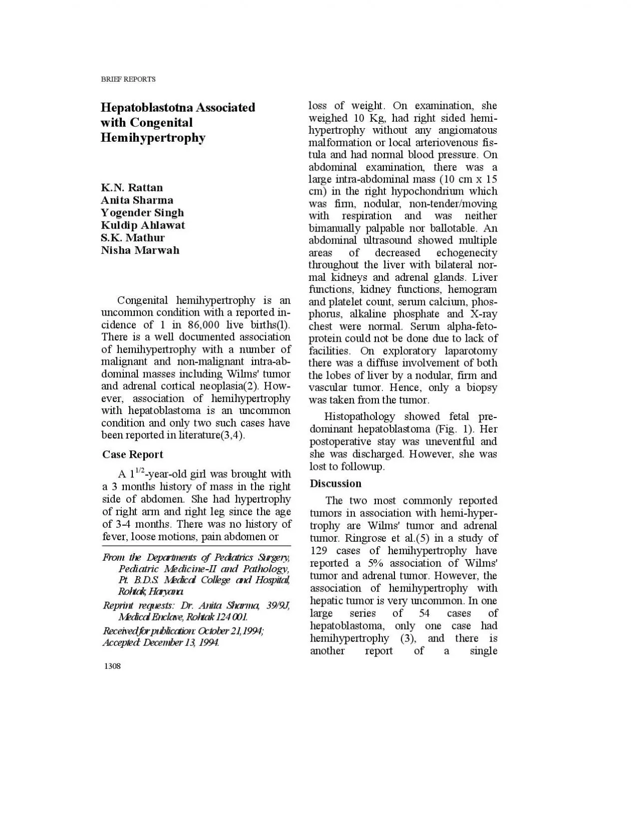
BRIEF REPORTS Hepatoblastotna Associated with Congenital Hemihypertrophy K.N. Rattan Anita Sharma Yogender Singh Kuldip Ahlawat S.K. Mathur Nisha Marwah Congenital hemihypertrophy is an cidence of 1 in 86,000 live births(l). There is a well documented association of hemihypertrophy with a number of malignant and non-malignant intra-ab- dominal masses including Wilms' tumor and adrenal cortical neoplasia(2). How- ever, association of hemihypertrophy with hepatoblastoma is an uncommon condition and only two such cases have been reported in literature(3,4). Case Report 1/2-year-old girl was brought with a 3 months history of mass in the right side of abdomen. She had hypertrophy of right arm and right leg since the age of 3-4 months. There was no history of fever, loose motions, pain abdomen or From the Departments of Pediatrics Surgery, Pediatric Medicine-II and Pathology, Pt. B.D.S. Rohtak, Haryana. Reprint requests: Dr. Anita Sharma, 39/9J, Medical Enclave, Rohtak 124 001. Received for publication: October 21,1994; Accepted: December 13, 1994. 1308 loss of weight. On examination, she hypertrophy without any angiomatous malformation or local arteriovenous fis- tula and had normal blood pressure. On abdominal examination, there was a large intra-abdominal mass (10 cm x 15 cm) in the right hypochondrium which was firm, nodular, non-tender/moving with respiration and was neither bimanually palpable nor ballotable. An abdominal ultrasound showed multiple throughout the liver with bilateral nor- mal kidneys and adrenal glands. Liver functions, kidney functions, hemogram and platelet count, serum calcium, phos- phorus, alkaline phosphate and X-ray chest were normal. Serum alpha-feto- protein could not be done due to lack of facilities. On exploratory laparotomy there was a diffuse involvement of both vascular tumor. Hence, only a biopsy was taken from the tumor. Histopathology showed fetal pre- dominant hepatoblastoma (Fig. 1). Her postoperative stay was uneventful and she was discharged. However, she was lost to followup. Discussion The two most commonly reported tumors in association with hemi-hyper- trophy are Wilms' tumor and adrenal 129 cases of hemihypertrophy have reported a 5% association of Wilms' tumor and adrenal tumor. However, the association of hemihypertrophy with hepatic tumor is very uncommon. In one large series of 5
4 cases of hepatoblastoma, only one case had hemihypertrophy (3), and there is another report of a single case(4). In yet another case report, the child had hemihypertrophy in association with hepatic hemangioendo- thelioma(6). Abdominal ultrasound is a, useful non-invasive modality for the detection of intra-abdominal masses in cases with hemihypertrophy. Raised alpha-feto- protein is a non-specific marker yet it is helpful in diagnosing hepatoblas- toma(3). Hepatoblastoma is the most common primary hepatic tumor in children under 5 years of age(3,7)- Besides hemihyper- trophy, hepatoblastoma can be associated with other congenital anomalies also; the exact incidence and cause of this as- sociation is unknown. In children, epi- thelial hepatoblastoma with predomi- nantly fetal pattern is the most common type and has better prognosis(3). Com- plete surgical resection remains the key in achieving long term survival(8). Radi- VOLUME 32-DECEMBER1995 ation or chemotherapy has little to offer as a primary treatment modality(3). Overall reported mortality is 76%(3). REFERENCES Holcombe GW, O'Neil JA, Mahboubi S, et al. Experience with hepatic hemangioendothelioma in infancy and childhood. J Pediatr 1988,23: 661-666. Faumeni JE, Ceiser CF, Mannin MD. Wilms' tumor and congenital hemihypertrophy : Report of five cases and review of literature. Pediatrics 1967,40: 886-889. Lack EE, Neave C. Vawter GF. Hepatoblastoma: A clinical and pathologic study of 54 cases. Am J Surg Pathol 1982, 6: 693-705. Geiser CG, Baez A, Schindler AM, Shih VE. Epithelial hepatoblastoma associ- ated with congenital hemihypertrophy and cystathionuria: Presentation of a case. Pediatrics 1970,45: 66-73. Ringrose RE, Jabbour JT, Keele DK. Hemihypertrophy. Pediatrics 1965, 36: 434-448. Davenport M, Mieli-Vegani G, Howard ER. Infantile hepatic heman- gioendothelioma: An association with hemihypertrophy. Pediatr Surg Int 1993,8: 505-506. Ishak KG, Glunz PR. Hepatoblastoma and hepatocellular carcinoma in infan- cy and childhood. Report of 47 cases. Cancer 1967,20: 396-422. Exelby PR, Filler RM, Grosfeld JL. Liv- er tumors in children with particular reference to hepatoblastoma and hepa- tocellular carcinoma: American Acade- my of Pediatrics Surgical Section Sur- vey 1974. J Pediatr Surg 1975, 10: 329- 337