
Munir Saadeddin FRCSEd Associate Professor amp Consultant College of Medicine King Saud University and King Khalid University Hospital Objectives of this Lecture To understand the importance of bony and soft tissues structure of Foot and Ankle ID: 1036091
Download Presentation The PPT/PDF document "Common Foot and ankle Disorders" is the property of its rightful owner. Permission is granted to download and print the materials on this web site for personal, non-commercial use only, and to display it on your personal computer provided you do not modify the materials and that you retain all copyright notices contained in the materials. By downloading content from our website, you accept the terms of this agreement.


1. Common Foot and ankle DisordersMunir Saadeddin ,FRCSEdAssociate Professor & ConsultantCollege of Medicine, King Saud University and King Khalid University Hospital
2. Objectives of this LectureTo understand the importance of bony and soft tissues structure of Foot and Ankle.To get a concise idea on common Foot and Ankle disorders.To differentiate from simple disorders and serious ones.To learn about initial management and prognosis.
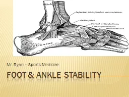
3. Importance of Foot and AnkleThey are the structures which are subject to most weight bearing (Loading) of the body.Have very important proprioception function.Their sensory role is very important.Their appearance or deformity is easily noticeable.Faulty or improper shoe wear can cause symptoms.With advancing age; deformity becomes more common.
4. Foot Skeleton: Medial Aspect
5. X-ray standing
6. Foot skeleton: Lateral aspect
7. Foot skeleton: Dorsal View
8. Common DisordersFlat Foot.Hallux Valgus.Heel Pain (Plantar Fasciitis).Ankle Sprains and Ankle Instability.Osteochondral lesions of Talus.Diabetic Foot.Charcot Foot.
9. Flat Foot Means reduced longitudinal arches of the foot.Most cases are developmental: i.e. arches do not develop normally.Usually is painless.Rarely acute flat foot can be encountered.Rigid flat foot can be the result of tarsal coalition( fibrous or bony cross union between bones of the foot )
10. Arches of The Foot
11. X-ray standing
12. Flat foot :Lat. Weight bearing X-rays
13. Flat Foot: rigid or flexibleRigid flat foot can be suspected by simple test: when patient is inspected from behind and asked to stand on tip-toes; the heel normally moves inward.In cases of rigid flat foot heel does not move inward.Also; on examination table: when ankle is held still and heel is moved sideways; it does not move in stiff heel as normally
14. Inspection of heels from behindNormally the heel is straight or minimally in valgus.This patient has normal appearance Right heel and excessive valgus Left heel.
15. Standing on tip-toesBoth heels correct their valgus and point medially in some varus:This is NOT rigid flat foot and there is no tarsal coalition or bony bar connecting tarsal bones.
16. Flat Foot managementUsually NO action is needed.Foot exercises is prescribed ; but its value is not confirmed.Orthotics and insoles are sometimes prescribed ; but its benefit is doubtful.However choosing correct and good type of shoes can be of benefit on the long run.Rigid flat foot may require surgical management.
17. Developmental Foot deformity: Hallux Valgus
18. Hallux ValgusMeans lateral deviation of big toe.Usually at the metatarsophalangeal joint.Often is associated with a bunion (swelling and protrusion at the medial aspect of big toe).Common at middle age and elderly, mainly females.Most cases are painless.When severe it interferes with shoe wear and may cause symptoms.
19. Hallux Valgus measurements (Normal foot)
20. Hallux Valgus MeasurementsHallux Valgus Angel: angle between line extending along 1st metatarsal and a line extending along proximal phalanx. Normal angles: <15 Mild HV: 16-25 Moderate HV: 26-35 Severe HV: > 35
21. Hallux Valgus Measurements1st intermetatarsal angle: Angle between 1st metatarsal long axis and 2nd metatarsal N < 10Hallux interphalangeus angle: Angle between long axis of proximal and distal phalanges N < 8
22. Hallux Valgus ManagementCorrect and suitable shoe wear.Avoidance of tight shoes.Protection to the bunions.Surgery is reserved for symptomatic and disturbing cases.Following surgery; patient has to continue proper shoe wear.
23. Hallux Valgus Pre Op
24. Hallux Valgus Post Op
25. Heel Pain: Plantar FasciitisCommon disorder at middle age and elderly.Insidious in onset; unilateral or bilateral.Vague pain at heel region.Localised tenderness to insertion of plantar fascia into calcaneum.Plain lateral X-ray of heel frequently shows calcaneal spur (prominence or ossification at the site of anterior calcaneum at plantar fascia insertion site)
26. X-ray: Bilateral calcaneal spur (Early)
27. Plantar fasciitisCommonly associated with flat feet.No visible heel swelling, no skin changes and no increase in local temperature.Inflammatory process is at site of pain; i.e. at plantar fascia insertion into calcaneum.Heel pain like stabbing pain when patient puts foot to the ground first thing in the morning; and gets less after some walking.
28. X-ray: Calcaneal Spur (Advanced)
29. Plantar Fasciitis managementAt present NO easy or simple management is available.Mainly conservative.Includes stretching exercises to plantar fascia: active and passive.Use of soft heel insoles (Silicone) may be helpful.Shock wave therapy (SWT) may be effective.Local steroid injections are helpful sometimes.
30. Ankle SprainsOne of most common injuries.Usually occurs during sports activities.But may occur at home or at street.Is the result of twisting injury.There is pain, swelling and local bruising.X-rays do not show fracture.The injury is partial or complete ligament rupture.
31. Ankle Ligaments (Lateral)
32. Ankle SprainMost commonly injured ligament is the Anterior Talo-Fibular Ligament.Ankle anterior drawer test is used to detect its rupture.Other ligaments are Posterior Talo-Fibular Ligament and Calcaneo-Fibular ligament.
33. Clinical picture of Ankle SprainsAlways there is a history of twisting injury.Pain, swelling and bruising at and around ankle.No tenderness of lateral malleolus; but tenderness anterior, posterior or inferior to it i.e. over ligaments.Dorsi-flection and plantar flexion possible; but inversion and eversion very painful. X-Rays : NO fracture.
34. Management of Ankle SprainRICE: Rest, Ice, compressors, Elevation.Used to apply Back-slab splints for few days.Rest should only be for few days.PRICES: recent view = Protection, relative Rest, Ice, Compression, Elevation and support.
35. Osteochondral Defects of talus (OCD)Very localised areas of joint damage; due to lack of blood supply.Lack of blood supply is often post traumatic, but occasionally No cause can be found.A local cartilage and varying depth of underneath bone are involved and may separate of main talus inside the ankle joint.Usually postero-medial part of dome of talus.Localised pain on weight bearing and even at rest may present.
36. Plain AP X-ray : lesion is suspected
37. CT Coronal view; lesion highly suspected
38. MRI: lesion is confirmed
39. Management of OCDDepends on how much symptoms and disturbance the patient suffers.Also when the OCD is large and Loose or almost loose.Arthroscopic debridement of the lesion and drilling of its crater (base).Rarely Fixation of a large defect which has significant bony part, by absorbable screws.
40. Diabetic FootLong term diabetes or failure to control diabetes adequately may result in Neuropathy.Neuropathy: is nerve damage.It can result in numbness, tingling and reduced sensation of the feet.Decreased circulation associated with neuropathy can result in small cuts on feet being overlooked and becoming infected.Infection in diabetic foot may result in Gangrene.
41. Care of Feet in DiabetesVery important as well as blood sugar control.Daily self inspection of feet is mandatory.If patient is unable to do self inspection (due to poor sight or hips and knees stiffness); a member of the family or assistant should do it.Regular inspections by healthcare personnel should be arranged A visit to a doctor should take place immediately whenever any complication occurs.
42. Surgery in Diabetic FootSkilled care of wounds and ulcers in diabetic foot is required.Wound debridement, antibiotics and repeated dressing should be done.Amputations may become necessary when there is Gangrene.Toe amputation or ray amputation, forefoot amputation, below or above knee amputation.
43. Charcot FootOccurs in people who have significant nerve damage to the foot.The bones of the foot become weak and the joints inflamed, swollen and lax. walking on the foot leads to disintegration and collapse of the joints and Deformity: such as Rocker- bottom deformity.
44. Charcot Foot CausesAny disorder which lead to Neuropathy.There is decreased sensation and decreased ability to feel temperature, pain and trauma.
45. Charcot Foot ClinicallyWarms of an area of foot or whole foot.May become red or dusky in colour.Swelling in the area.Pain or soreness.X-rays changes are important to detect and interpret, as early there is NO changes.Later: haziness, osteopenia, irregular joint destruction, subluxation or even dislocation.
46. Charcot Foot & Ankle
47. Diabetic foot 04/03/1428
48. (R)Diabetic foot : Early Charcot 1431
49. (R) Charcot Ankle 1431
50. (L) Side Early Charcot 1433
51. Advanced Case of Charcot
52. Diagnosis of Charcot FootGood history and clinical examination.Awareness.Exclusion of other causes which may give similar picture: like infection or tumour.MRI, bone scans, aspiration biopsies can help.
53. Non Surgical Management of Charcot FootImmobilisation:Custom Shoes and Bracing:Activity modification
54. Surgery in Charcot FootMay be indicated in certain cases
55. Post Op right Ankle 1435
56. Post Op Left foot 1435
57. Amputation in Charcot footMay be indicated as a last option.Mainly when there is severe instability which cannot be controlled by surgery or orthosis.Also when surgery fails to achieve stability.Presence of refractory infection increase the possibility of amputation.
58. Shoe wear
59. Importance of proper shoe wear
60. Spread of pressure during walking
61. How to reduce pressure during walking
62. Pressure during weight bearing