
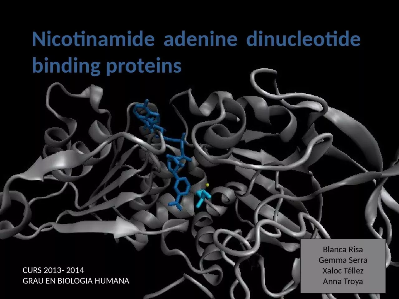
dinucleotide binding proteins Blanca Risa Gemma Serra Xaloc Téllez Anna Troya CURS 2013 2014 GRAU EN BIOLOGIA HUMANA Introduction Enzymes that bind nucleotides NADP NADPH ID: 1048222
Download Presentation The PPT/PDF document "Nicotinamide adenine" is the property of its rightful owner. Permission is granted to download and print the materials on this web site for personal, non-commercial use only, and to display it on your personal computer provided you do not modify the materials and that you retain all copyright notices contained in the materials. By downloading content from our website, you accept the terms of this agreement.


1. Nicotinamide adenine dinucleotide binding proteinsBlanca RisaGemma SerraXaloc TéllezAnna TroyaCURS 2013- 2014 GRAU EN BIOLOGIA HUMANA
2. IntroductionEnzymes that bind nucleotidesNAD(P) – NAD(P)HNAD-binding proteinsWhat do we study?Sequence identityStructure NAD-binding enzymes and classical Rossmann foldSuperimpositionsFingerprint coreFunctionCofactor interactionsCofactor orientationStereospecific transferConclusions Index
3. High-energy phosphate bonds in triphosphates: - ATP - GTPOxidation-reduction (redox): - Flavin: FAD and FMN - Nicotinamide: NAD and NADP Some enzymes require non-protein molecules called cofactors for activityNucleotides play a central role in cellular metabolismNucleotides can be involved in two different energy transfer processes:Introduction Enzymes that bind nucleotides
4. NAD molecule comprises:Nicotinamide ribose phosphate (NMN): H addition. Adenine ribose phosphate (AMP) Introduction Linked through a pyrophosphate bondNAD(P): oxidizing agentsNAD(P)H: reducing agents. NAD(P)-NAD(P)HNADP has additional phosphate groupNAD+NADP+NADHNADPH
5. Introduction OXIDATION reactionsREDUCTION reactionsReduction of NAD to NADHOxidation of NADH to NADketoneAlcoholketoneAlcohol++ 2e-- 2e-Chemical reactions
6. NAD(P)-Binding enzymesNAD(P)-binding proteins are ubiquitousThere are several distinct ways of binding NAD(P):NON CLASSICALAll-AlphaAll-BetaAlpha + BetaAlpha/BetaCLASSICALAlpha/betaBeta BarrelAldose reductaseMalate dehydrogenaseRossmann foldNAD(P)-binding enzymes
7. They can have different functions and catalyse similar or different reactions related to oxidoreductionFamilyProteinSpeciesPDB IDAlcohol dehydrogenase-likeAlcohol dehydrogenaseEquus caballus2OHXAlcohol dehydrogenaseRana perezi1P0FFormate/glycerate dehydrogenasesL-alanine dehydrogenasePhormidium lapideum1PJCFormate dehydrogenasePseudomonas sp.2NADLDH N-terminal domain-likeMalate dehydrogenaseEscherichia coli1EMDLactate dehydrogenaseThermotoga maritima1A5Z6-phosphogluconate dehydrogenase-like Prephenate dehydrogenaseSynechocystis sp. 2F1KSiroheme synthase Siroheme synthase CysGSalmonella typhimurium1PJSOrnithine cyclodeaminase-likeOrnithine cyclodeaminasePseudomonas putida1X7DTyrosine-dependent oxidoreductasesUridine diphosphogalactose-4-epimeraseHomo sapiens1EK5Amino acid dehydrogenase-like Glutamate dehydrogenasePyrobaculum islandicum1V9LGlyceraldehyde-3-phosphate dehydrogenase-likeGlyceraldehyde-3-phosphate dehydrogenaseEscherichia coli1GADTranscriptional repressor RexTranscriptional repressor RexThermus aquaticus1XCBCoA-binding domainSuccinyl-CoA synthetaseThermus thermophilus1OI7Potassium channel NAD-binding domainKtn Mja218Archaeon Methanococcus jannaschii1LSSWhat do we study?Class: Alpha and beta protein (α/β)Superfamily: NAD(P)-Binding Rossmann fold domains
8. Sequence identityMSA: Whole sequence MSA with whole sequence of 2OHX, 1P0F, 1PJC, 2NAD, 1PJS, 1EMD, 1A5Z, 2F1K, 1LSS, 1XCB, 1X7D, 1OI7, 1GAD, 1EK5, 1V9L
9. Sequence homologyMSA: Rossmann foldMSA with Rossmann fold of 2OHX, 1P0F, 1PJC, 2NAD, 1PJS, 1EMD, 1A5Z, 2F1K, 1LSS, 1XCB, 1X7D, 1OI7, 1GAD, 1EK5, 1V9L
10. Large protein molecules which can have several identical polypeptide chains Structure NAD(P)-binding enzymes Alcohol dehydrogenaseTwo separated domains (or more):Catalytic domains: binds to the substrate NAD(P)- Binding domainIn different regions of the polypeptide chainHave similar 3D structures.
11. Two Rossmann fold motifs (βαβαβ motifs) Crossover connection: α-helix (not always)Open parallel 6-stranded β-sheet (321456) with α-helices on both sidesTopological switch pointStructureNAD-binding classical fold
12. StructureSuperimposition of Rossmann fold(….)(….)(….)RMSD<2: 44>2 i <3: 61 Sc5.5-9.8: 22.5-5.5: 51< 2.5: 5233/105Superimposition of 2OHX, 1P0F, 1PJC, 2NAD, 1PJS, 1EMD, 1A5Z, 2F1K, 1LSS, 1XCB, 1X7D, 1OI7, 1GAD, 1EK5, 1V9L
13. StructureSuperimposition of Rossmann foldThe overall topologies of the NAD-binding domain show variations:Additional beta strandsDifferent helix length Different loopsCrossover region variable in structure (α-helix)Not all the 6 strands are essential to NAD- bindingRasmol display of the superimposition of 2OHX, 1P0F, 1PJC, 2NAD, 1PJS, 1EMD, 1A5Z, 2F1K, 1LSS, 1XCB, 1X7D, 1OI7, 1GAD, 1EK5, 1V9L
14. StructureDifferences in the Rossmann foldClassical Rossmann fold Alcohol dehydrogenase Malate dehydrogenaseLactate dehydrogenase Ktn Mja218
15. Siroheme synthase CysG L-alanine dehydrogenaseStructureDifferences in the Rossmann foldDifferent number of β-strands 123465123451234567
16. StructureOrnithine cyclodeaminase 123456Transcriptional repressor RexSuccinyl CoA synthetase123456123456Differences in the Rossmann fold
17. StructureGlyceraldehyde-3-phosphate dehydrogenase 1234, 5678Differences in the Rossmann foldSuperimposition of 1GAD (glyceraldehyde 3-phosphate dehydrogenase) and 2OHX (alcohol dehydrogenase)
18. StructureSuperimposition of Rossmann core (βαβαβ + β4)(….)Superimposition of 2OHX, 1P0F, 1PJC, 2NAD, 1PJS, 1EMD, 1A5Z, 2F1K, 1LSS, 1XCB, 1X7D, 1OI7, 1GAD, 1EK5, 1V9LRMSD<2: 78>2 i <3: 27 Sc5.5-9.8: 452.5-5.5: 39< 2.5: 2118/105
19. StructureSuperimposition of Rossmann core (βαβαβ + β4)Minimum structure conserved in most proteins: first motif (βαβαβ) + β4 (70 Aa)Rasmol display of superimposition of βαβαβ + β4, conserved in most proteins. Not included: 1XCB, 1OI7, 1GAD, 1EK5, 1V9L
20. StructureSuperimposition of fingerprint region βαβ(….)RMSD<2: 105>2 i <3: 0 Sc5.5-9.8: 1052.5-5.5: 0< 2.5: 0Any LOW SCORESuperimposition of 2OHX, 1P0F, 1PJC, 2NAD, 1PJS, 1EMD, 1A5Z, 2F1K, 1LSS, 1XCB, 1X7D, 1OI7, 1GAD, 1EK5, 1V9L
21. StructureSuperimposition of fingerprint region βαβSuperimposition of 2OHX, 1P0F, 1PJC, 2NAD, 1PJS, 1EMD, 1A5Z, 2F1K, 1LSS, 1XCB, 1X7D, 1OI7, 1GAD, 1EK5, 1V9LStructure is conserved in all proteins
22. StructureStructural aligment of fingerprint region βαβ30-35 amino-acids:Glycine-rich phosphate-binding sequence: GX1-2GXXGHydrophobic coreNegatively charged residue at C-t of the β2 (NAD, not NADP)Positively charged residue at the N-terminus of β1Glycine residuesHydrophobic residuesNegatively charged residuesPositively charged residues
23. StructureSuperimposition of fingerprint region βαβFormate dehydrogenase (2NAD)Ornithine cyclodeaminase (1X7D)Malate dehydrogenase (1EMD) Different glycine-rich phosphate-binding sequence: GX1-2GXXG2OHX1EMD2OHX2NAD2OHX1X7D
24. Fingerprint regionconserved characteristicsGX1-2GXXGHydrophobic coreNegative charged residuePositively charged residueWhy are they conserved?L-alanine dehydrogenase(NAD)Alcohol dehydrogenase(NADP) Which interactions are important in NAD binding? Which is their function?FunctionL-alanine dehydrogenase
25. Glycine-rich phosphate-binding loop There is a Cα –H· · ·O hydrogen bonds between the third and the first glycyl residueIn addition, Rossmann folds that bind NAD(P) also typically contain GXXXG or GXXXA motifs, both forming Van der Waals interactions with a valine or isoleucine residue located either seven or eight residues further back along the polypeptide chain.The first strictly conserved glycine allows for a tight turn of the main chain from the β -strand into the loop, which is important for positioning the second glycine. The second glycine allows for close contact to NAD(P) pyrophosphate The third glycine is important for the close packing of the helix with the β -strand. G X1-2 G X X GFunctionNAD
26. GX1-2GXXGXXXAGly 174Gly 176Gly 179Ile 172Ala 183Gly 203Gly 200Gly 198Ala 207Val 196NADNADPL-AlaDHADH8
27. Pyrophosphate Group Pyrophosphate group binds to the central region β sheet.Some residues form HB to the pyrophosphate groupThe second Glycine of GXGXXGResidues in the N-t of αAFunctionSer 133Val 177Gly 176Val 178L-alanine dehydrogenase
28. Hydrophobic Core The hydrophobic core of the fingerprint region, consisting of six positions occupied by small hydrophobic amino acids. These residues are necessary for the packing of the β strands against the α helix. Val 170Ile 1722222Ala 183Ala 186Ile 195Val 183NADFunctionL-alanine dehydrogenase
29. NADPNeutral residue small residue for the additional phosphate group NAD Conserved aspartate HB 2’OH adenosine riboseNegative ChargeAsp 197Gly 222FunctionL-AlaDHADH8
30. Positive Charge A conserved positively charged residue at the amino terminus of the β1 strand, usually Lys or Arg. Its role is not fully understood, but it appears to make a stabilizing interaction with the core elements β1 and β4. Lys 169NADFunctionL-alanine dehydrogenase
31. NAD(P) binding proteins accommodate water molecules, many of these form water-mediated HBConserved water molecule: HB between the Gycine-rich loop and pyrophosphate. This water typically makes 4 HB:Two invariant with pyrophosphate and conserved Gly. Two variant that involve the 1st or 2nd conserved Gly and a C-terminal residue of β4 Conserved water FunctionL-alanine dehydrogenase
32. Adenosine Group The adenine binds through an hydrophobic pocket.Mainly held in place by hydrophobic and Van der Waals interactions.The adenine can make one or several hydrogen bonds with the protein, some can be mediated by a bridging water molecule More interactionsFunctionSer 219Ile 198Val 238Leu 248Gly 174NADL-alanine dehydrogenase
33. Nicotinamide Ribose Group Nicotinamide ribose binds to the second motif curving down towards the interior of the sheet between β strands 4, 5, and 6.One side of the ring interacts with the structural framework of the NAD(P)-binding domain, and the other side faces the substrate binding site. 456NADGOLFunctionNADPGOLAlcohol dehydrogenase
34. NADGOLThe nicotinamide is held into place by up to six hydrogen bonds with the protein. Water molecules are occasionally used as ligands to the cofactor.FunctionNADH2ONADNADNADNADL-alanine dehydrogenaseNADH2OVal 297Val 266Ala 136Met 300Val 178
35. Function Most NAD molecules adopt an extended shapePosition of the pyrophosphate and adenosine moiety is quite constantThe nicotinamide ring is more variableConserved water molecule Cofactor orientationSuperimposition of 2OHX, 1PJC, 2NAD, 1EMD with cofactor
36. FunctionNAD must be positioned in the active site sufficiently close and in the correct orientation to allow electron transfer.Residues with aromatic rings are involved in better transferring of the HNAD binding is stabilized by interactions with protein: VDW and HB (direct or through water)Cofactor orientationNADGOLPhe 93Alcohol dehydrogenase with its substrate
37. Function Hydrogen transfer to NAD is stereospecific There are two different forms of NAD, which differ by a flip of the nicotinamide ring 180º around the glycosidic bond that links it to the ribose.The concavity that makes the protein and interactions with NAD define two classes:H is transferred to the position above the ringH is transferred to a position below the ring Class A Class B L-alanine dehydrogenaseGlyceraldehyde-3-phosphate dehydrogenase
38. Function180ºClass A Class B StereospecificityL-alanine dehydrogenaseGlyceraldehyde-3-phosphate dehydrogenase
39. EvolutionProtein Specy1DSSPanulirus versicolor (invertebrate)1NBOSpinaca oleracea2B4RPlasmodium falciparum1GADEscherichia Coli1U8FHomo sapiens Glyceraldehyde 3 phosphate dehydrogenase
40. Conclusions NAD-binding proteins show small sequence identity.The overall topologies of the NAD-binding domain show variations. Not all the 6 strands are essential to NAD- binding.There is a minimum structure conserved in most proteins: first motif (βαβαβ) and β4. β1 and β4 are located in the center of the NAD-binding domain and are involved in cofactor binding.The fingerprint region (βαβ) is conserved in all proteins and have several conserved residues important for its function. These residues are involved in NAD interactions with protein through VDW and HB (direct or through water). There is also a structurally water molecule conserved.Interactions between NAD and the protein allow the correct orientation of the cofactor and the electron transfer with the substrate. The transfer is stereospecific.It is difficult to decide if low seguence homology but structural preservation of NAD binding domains is due to:Convergent evolution from different ancestral genesDivergent evolution from a common ancestor (remote homologs)
41. ReferencesAnantharaman V, Koonin EV, Aravind L. Regulatory Potential, Phyletic Distribution and Evolution of Ancient, Intracellular Small-molecule- binding Domains. J. Mol. Biol. (2001) 307, 1271±1292.Auerbach G, Ostendorp R, Prade L, Korndörfer I, et al. Lactate dehydrogenase from the hyperthermophilic bacterium Thermotoga maritima: the crystal structure at 2.1 Å resolution reveals strategies for intrinsic protein stabilization. Structure 15 June 1998, 6:769–781.Baker P.J. Analysis of the structure and substrate binding of Phormidium lapideum alanine dehydrogenase. Nature structural biology. July 1998; 5:7.Bellamacina C.R. The nicotinamide dinucleotide binding motif: a comparison of nucleotide binding proteins. Faseb J. 1996 Sep;10(11):1257-69.Brandrn CI, Eklund H, et al . Structure of Liver Alcohol Dehydrogenase at 2.9-A Resolution. August 1973. Vol. 70, No. 8, pp. 2439-2442. Bottoms C, Smith P.E, Tanner J.A. Structurally conserved water molecule in Rossmann dinucleotide-binding domains. Protein Science. 2002. June 4.Bhuiya M, Sakuraba H, et al. The First Crystal Structure of Hyperthermostable NAD-dependent Glutamate Dehydrogenase from Pyrobaculum islandicum. J. Mol. Biol. (2005) 345, 325–337.Ghosh S, George S, Roy U. NAD: A master regulator of transcription. Biochimica et Biophysica Acta 1799 (2010) 681–693.Jessica L. Goodman J.L, Susan Wang S, et al. Ornithine Cyclodeaminase: Structure, Mechanism of Action, and Implications for the μ-Crystallin Famil. American Chemical Society. 2004. August 18.Kim SY, Hwang KY, et al. Structural Basis for Cold Adaptation: Sequence, biochemical properties, and crystal structure of malate dehydrogenase from a psychrophile aquaspirillium arcticum. J Biol. Chem. 1999, 274:11761-11767. Legrant P, Dumas R, et al. Biochemical Characterization and Crystal Structure of Synechocystis Arogenate Dehydrogenase Provide Insights into Catalytic Reaction. April 2006. Structure 14, 767–776. Mark A. Robien MA, Bosch J, Buckner F.S, et al. Crystal Structure of Glyceraldehyde-3-Phosphate Dehydrogenase from Plasmodium falciparum at 2.25 Å Resolution Reveals Intriguing Extra Electron Density in the Active Site. PROTEINS: Structure, Function, and Bioinformatics (2006); 62:570–577.
42. ReferencesRosell A, Valencia E, et al. Crystal Structure of the Vertebrate NADP(H)- dependent Alcohol Dehydrogenase (ADH8). J. Mol. Biol. (2003) 330, 75–85.Rubach J.K, Plapp B.V. Amino Acid Residues in the Nicotinamide Binding Site Contribute to Catalysis by Horse Liver Alcohol Dehydrogenase. Biochemistry 2003, 42, 2907-2915.Roosild T.P, Miller S, Booth IR, Choe S. A Mechanism of Regulating Transmembrane Potassium Flux through a Ligand-Mediated Conformational Switch. 2002, Cell, Vol. 109, 781–791, June 14. Rubach J.K, Plapp B.V. Amino Acid Residues in the Nicotinamide Binding Site Contribute to Catalysis by Horse Liver Alcohol Dehydrogenase. Biochemistry 2003, 42, 2907-2915.Song S, Xu Y, Lin Z, Tsou C. Structure of Active Site Carboxymethylated D-Glyceraldehyde-3-phosphate Dehydrogenase from Palinurus versicolor. J. Mol. Biol. (1999) 287, 719±725.Stroupe M.E, Leech H.K, Daniel D.S, Warren M.J, Getzoff E.D. CysG structure reveals tetrapyrrole-binding features and novel regulation of siroheme biosynthesis. Nat Struct Biol. 2003 Dec;10(12):1064-73. Sickmier EA, Brekasis D, Paranawithana D.S, Bonanno J.B, et al. X-Ray Structure of a Rex-Family Repressor/NADH Complex Insights into the Mechanism of Redox Sensing. Structure. January, 2005. Vol. 13, 43–54.Stockwell G.R, Thornton J.M. Conformational Diversity of Ligands Bound to Proteins. J. Mol. Biol. (2006) 356, 928–944.Peter Belenky, Katrina L. Bogan and Charles Brenner. NAD metabolism in health and disease. RENDS in Biochemical Sciences Vol.32 No.1.Tomita T, Fushinobu S, Kuzuyama T, Nishiyama M. Crystal structure of NAD-dependent malate dehydrogenase complexed with NADP(H). Biochemical and Biophysical Research Communications 334 (2005) 613–618.
43. ReferencesUchikoba H, Fushinobu S, Wakagi T, Konno M, Taguchi H, Matsuzawa H .Crystal Structure of Non-Allosteric L-Lactate Dehydrogenase From Lactobacillus pentosus at 2.3 Å Resolution: Specific Interactions at Subunit Interfaces. PROTEINS: Structure, Function, and Genetics. 2002;46:206–214.Michel G, Roszak W.A, Sauvé V, Maclean J, Matte A, Coggins J.R, Cygler M, Adrian. Structures of Shikimate Dehydrogenase AroE and Its Paralog YdiB: A common structural framework for different activities. J. Biol. Chem. 2003, 278:19463-19472.Stroupe M.E, Leech H.K, Daniel D.S, Warren M.J, Getzoff E.D. CysG structure reveals tetrapyrrole-binding features and novel regulation of siroheme biosynthesis. Nat Struct Biol. 2003 Dec;10(12):1064-73. Thoden J.B, Wohlers T.M, Fridovich-Keil J.L, Holden H.N. Crystallographic Evidence for Tyr 157 Functioning as the Active Site Base in Human UDP-Galactose 4-Epimerase. Biochemistry 2000, 39, 5691-5701. Lesk A.M. NAD-binding domains of dehydrogenases. University of Cambridge Clinical School, Cambridge; UK. Current Opinion in Structural Biology, 5: 775: 783. Kleiger G, Eisenberg D. GXXXG and GXXXA Motifs Stabilize FAD and NAD(P)-binding Rossmann Folds Through C – H· · ·O Hydrogen Bonds and van der Waals Interactions. J. Mol. Biol. (2002) 323, 69–76.
44. The classical NAD(P)-Binding fold domain is:Greek key B-sandwichRossmann folda and b4 helix bundleall answers are correct 2. Talking about the two Rossmann folds motifs that form a NAD(P)-Binding site domain:In the classical one, the two motifs form an open parallel six stranded B-sheet with alpha helices located on both side of the sheet.B-strands and a-helixes form a characteristic TIM Barrel in these motifsNAD is bounded in the topological switch pointB-strands are antiparallel 1, 2, 31, 32, 441, 2, 3, 4Questions
45. 3. NAD(P)-Binding enzymes Are involved in different redox reactions Require NAD or NADP as a cofactor to transfer electronsThe cofactor and the substrate must be nearly located to allow the electron transfer. Bind their substrates and NAD(P) in different domains a) 1, 2, 3b) 1, 3c) 2, 4d) 4e) 1, 2, 3, 4 4. Hydrogen transfer to NAD(P):Is stereospecificThere are two different classes of NAD: A and BThe two forms of NAD differ by a flip of the nicotinamide ring, 180º around the glycosilic bond that links it to the ribose What defines the two different classes is the concavity that makes the protein and the interactions with NAD.all the answers are correct 5. The fingerprint region of the Rossmann fold…It is a region of approximately 35 residuesIt has got some conserved residues which are important for its functiona and b are correctDoes not have any conserved residue involved in it is function.Is the most variable region in the Rossmann fold
46. 6. The fingerprint region of the Rossmann fold is characterized by…A glycine rich phosphate binding sequenceAn hydrophobic core of six residuesA negatively charged residue located in the C-terminus of the first B-strandA region of 20 consecutive leucine residues1, 2, 31, 32, 441, 2, 3, 4 7. As far as NAD(P) interaction is concerned…NAD(P) molecules adopt and extended shape similar to a boomerangNAD(P) binding is stabilized by interaction with the protein which involves hydrogen bonds and VDW contacts. A and b are correctNAD(P) pyrophosphate group does not have any interaction with the proteinAll the answers are correct8. NAD and NADP….Are oxidizing agents that can be reduced to NADH and NADPH, respectively. Are used as a cofactor to shuttle electrons between proteinsA and b are correctNADP has an additional phosphate group in 2’ of the adenosine riboseAll the answers are correct
47. 9. As far as NAD(P) interaction is concerned…Glycine residues are important to adopt a good conformation that allows a correct contact with the NAD(P) cofactorHydrogen bonds are involved in the interaction between NAD(P) and the proteinThe pyrophosphate group interacts with an structurally conserve water moleculeWater molecules are not involved in forming hydrogen bonds 1, 2, 31, 32, 441, 2, 3, 4 10. As far as interaction between NAD(P) is concerned… Residues with aromatic rings are involved in the hydrogen transferA negatively charged residue at the C-terminus end of the second b-strand is important to discriminate between NAD and NADPa and b are correctThere is not any difference between binding NAD or NADP cofactors. All the answers are correct
48.
49. Glyceraldehyde 3 phosphate dehydrogenase
50. Glyceraldehyde 3 phosphate dehydrogenase