
Unit 1 Principles of Anatomy amp Physiology in Sport The Skeletal System Consists of 206 bones Bones living tissue constantly adapting to stresses placed on them Split into Axial amp ID: 928434
Download Presentation The PPT/PDF document "P1 – Describe the Structure & Func..." is the property of its rightful owner. Permission is granted to download and print the materials on this web site for personal, non-commercial use only, and to display it on your personal computer provided you do not modify the materials and that you retain all copyright notices contained in the materials. By downloading content from our website, you accept the terms of this agreement.


Slide1
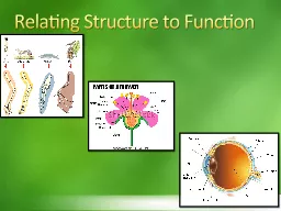
P1 – Describe the Structure & Function of the Skeletal System
Unit 1 – Principles of Anatomy & Physiology in Sport
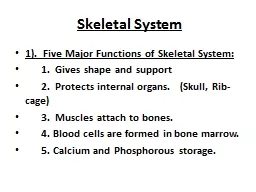
Slide2The Skeletal System
Consists of 206 bones
Bones = living tissue, constantly adapting to stresses placed on them
Split into Axial &
Appendicular
skeleton
Has different types of bones
Has a number of functions
Bones meet together at joints
Slide3Cranium
Mandible
Sternum
Rib
Vertebral Column
Pelvis
Sacrum
Coccyx
Clavicle
Scapula
Humerus
Ulna
Radius
Carpals
Metacarpals
Phalanges
Femur
Patella
Tibia
Fibula
Tarsals
Metatarsals
Phalanges
Ischium
Slide4Cranium – box like structure
brain sits inside
protects the brain
made up of a number of different bones
bones fuse together between 1-3 yrs old
Mandible – jaw bone
Sternum – breast bone long, flat bone centre of chest
Slide5Ribs
– long, flat bones
12 pairs
1
st
7 pairs attached to sternum other 5 pairs called false ribsPelvis – made up of 2 sets of 3 bones
Ilium – upper wing like bones, provides the sockets for the hip bones, upper edge = iliac crest Pubis
– pubic bone, front of the pelvis Ischium – lower, posterior bones, sitting bones
Slide6Clavicle – collar bone
long, thin bone
makes up the anterior (front) of the
shoulder girdle
is a strong, mobile attachment for arms
Scapula – large, triangle shape
back (posterior) part of shoulder girdleHumerus – long bone, upper arm proximal head = shoulder joint
distal end = elbow joint
Slide7Ulna
– Forearm
little finger side
Radius
– forearm
thumb side moves around ulna when turning handCarpals – 8 short bones
wrist 2 rows of 4Metacarpals – 5 long bones palm of the hand
run from the carpals to each finger & thumb
Slide8Phalanges
– small long bones
fingers & toes
3 in each finger & toes
2 in each thumb & big toe
Femur – thigh bone longest & strongest
top (proximal end) sits in socket of pelvis bottom (distal end) forms the knee jointPatella – kneecap
large, triangle shaped sesamoid bone sits inside the quadriceps tendon protects knee joint
Slide9Tibia – shin bone
medial, thicker bone
proximal end = knee joint with femur
distal end = ankle joint
Fibula
– lateral, thinner bone non weight bearing
distal end = ankle jointTarsals – 7 bones (ankle)Metatarsals – 5 foot bones
Slide10Skeletal System Information
Appendicular
System
126 bones
Axial System
80 bones
Slide11Appendicular
System
126 bones
Upper body Lower body
Clavicle =
collar bone Pelvis = 2 x 3 bones
Scapula = Shoulder blade Femur = Thigh
Humerus = upper arm Patella = Kneecap
Radius = to the thumb Tibia = Shin
Ulna = to the little finger Fibular = Outside/ankle
Carpals = wrist
Tarsals
= under ankleMetacarpals = hand Metatarsals = FootPhalanges = fingers Phalanges = Toes
Axial System
80 bones
Skull = 28 bones
Hyoid
Sternum
Ribs = 12 pairsVertebral Column = 33 vertebrae
Slide12Vertebral Column
33 Bones – 5 Sections
C 1 - 7
T 1 - 12
L 1 - 5
S 1 - 5
Coccyx 1 - 4
Slide13Vertebral Column
Spine
33 irregular bones
Approx 40% of your height
Very small movement between each vertebrae
Lots of movement of spine altogether
Slide14Bones
Living tissue =
grow during childhood
Bleed & hurt if damaged
Able to repair if broken
Ossification – a hardening process that takes place when bones mature. Makes bones solid
structures that can withstand a lot of pressure/stresses.Collagen = resilienceCalcium = strength
Slide15Types of BonesF.L.I.S.S.
F
L
I
S
S
lat
Protection
Scapula, sternum, ribs, pelvis
ong
Movement
Femur, humorus, tibia
rregular
Shape
Vertebrae, facial bones
hort
Cube shaped
Carpals, tarsals
esamoid
Inside a tendon
Patella