
Anatomy and physiology Function The digestive system is the first organ system to develop in animals This system allows animals to take in nutrients that provide it with energy and materials necessary to grow and maintain their bodies ID: 206198
Download Presentation The PPT/PDF document "Digestive System" is the property of its rightful owner. Permission is granted to download and print the materials on this web site for personal, non-commercial use only, and to display it on your personal computer provided you do not modify the materials and that you retain all copyright notices contained in the materials. By downloading content from our website, you accept the terms of this agreement.

Slide1
Digestive System
Anatomy and physiology Slide2
Function
The digestive system is the first organ system to develop in animals.
This system allows animals to take in nutrients that provide it with energy and materials necessary to grow and maintain their bodies.
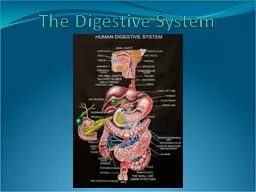
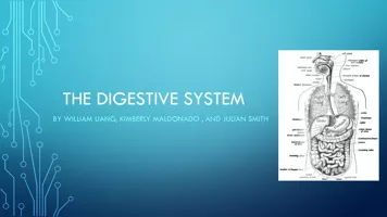
The digestive system is at it’s core a tube that starts with the mouth and ends with the anus. This tube is called the alimentary canal, also known as the gastrointestinal tract (GI tract).The GI tract travels through the pharynx, esophagus, stomach, small intestine, and large intestine.Accessory organs such as the liver, pancreas, gallbladder, and salivary glands, secrete substances that help in the chemical breakdown and absorption of food.Slide3
matchingSlide4
Six Steps of Digestion
1. Ingestion
2. propulsion
3. mechanical breakdown4. chemical breakdown5. absorption6. defecationAnimationSlide5
Ingestion
, the first step in the digestion process. The teeth and tongue mechanically break down the food. Increasing it’s surface area. Saliva begins the foods chemical breakdown.
Propulsion
, swallowing and peristalsis – muscles in the esophagus contract to push food down. This delivers food to the stomach.Mechanical breakdown, occurs in the stomach, the churning actions therein further reduce the food to smaller particles.Chemical breakdown, in the stomach and small intestine requires enzymes secreted by the secondary digestive organs. The food then becomes chemically less complex, and easy to absorb. Absorption, the small intestine and the large intestine absorbe
the chemical nutrients and send it to the bloodstream so that these resources can be sent to the rest of the body.Defecation, lastly, depleted of nutrients, the waste is removed from the body via the rectum and anus.Slide6
The layers of the Alimentary Canal
Mucosa:
is the innermost layer, and composed of mucus covered epithelial cells. It lines the inner walls of the alimentary canal.
Submucosa: layer of irregular dense connective tissue, contains blood vessels, lymphatic vessels, and nerves. Lymphatic vessels and glands in the submucosa secrete substances that aid in the digestion and absorption Muscularis Externa: composed of two layers of smooth muscle this layer propels food though the canal using peristalsis. Nerves run between the two muscle layers.Serosa: A thin slippery layer that surrounds the alimentary canal and reduces friction between organs in the abdominal cavity. Slide7
Layers of the Alimentary Canal
The cavity, empty space inside the alimentary canal is called the lumen.Slide8
Organs of the Digestive System
Oral cavity
:
Ingestion of foodMechanical breakdown of food (chewing)Chemical breakdown of foodPropulsion of food.Slide9
Organs of the Digestive System
Nasal Cavity:
Separated from the oral cavity via the palate. The uvula which hags down from the soft palate prevents food from entering the nasal cavity.Slide10
Organs of the Digestive System
Teeth and gums:
The gums are known as the
gingiva, a soft tissue that covers the necks of the teeth and the upper jaw and mandible.Children have 20 deciduous teethAdults have 32 permanent teeth. Slide11
Anatomy of the ToothSlide12
Organs of the Digestive System
Salivary Glands:
Parotid: largest salivary glands, just in front of the ears.
Submandibular: near the lower lawSublingual: under the tongueSecrete saliva: Mostly water, contains mucus, antibodies, and several enzymes. The enzymes help break down starches into simpler sugars and begin the breakdown of fats.Slide13
Organs of the Digestive System
Pharynx:
The throat
3 Parts: nasopharynx, oropharynx, laryngopharynxEpiglottis: moves down to prevent food passing into the trachea durring swallowing. Propels food down into the esophagus
Swallowing x-ray
Esophagus:
Posterior to the trachea and heart
Passes through a hole in the diaphragm
Uses a muscular contraction known as
peristalsis
to propel foodSlide14
Major Organs of the Digestive System:
Stomach
MatchingSlide15
Stomach
4
major regions: cardia, fundus, body, and pyloric region
The stomach twists and contracts to churn food and break it down both physically and chemically before passing through the pyloric sphincter into the duodenum of the small intestine.3 muscle layersLongitudinal muscle layer (up and down) – top layerCircular muscle layer (around the stomach) – middle layerOblique muscle layer (diagonally) – Inner layerThe rugae (folds) can flatten out increasing the stomach’s volume.
A full stomach can hold about 2 liters or more.the stomach churning food (ramen)
Artificial stomach
The mixture of gastric juice and food is known as
chymeSlide16
Stomach lining
Stomach lining
Epithelia cells
at the surface, covered in mucus (mucus prevents our digestive juices from eating into our own tissues)Gastric juice is secreted from gastric pits – controlled by the parasympathetic nervous systemThe cells surround gastric pits that lead down into the gastric glands.Mucus-secreting cellsParietal cells secrete hydrochloric acid (HCl
) and intrinsic factor (helps the body absorb B12
)
Chief cells
secrete pepsinogen – activates protein-digesting enzymes
Enteroendocrine
cells
– produce gastrin (hormone) that stimulates the production of more gastric juice.Slide17
Small Intestine
Named for it’s diameter not its length.
Longest segment of the GI tract at 6-7 meters
Most chemical breakdown of food occurs in the small intestineParts of the small intestineDuadenum – secretions from the liver, gallbladder, and pancreas enter the duadenum from the duadenal ampullaJejunum (8 feet)Ileum (12-13 feet)
Chemical digestion, absorption, and propulsion via peristalsis occurs in all 3 regions.Slide18
Lining of the Small
Intestine
Circular
folds, villi, and microvilli all greatly increase the surface area of the lining of the small intestine.Intestinal crypts are similar to gastric pits.Slide19
Chemical Breakdown in the Small Intestine.
When
chyme
enters the duodenum, secretions from the pancreas and bile from the gallbladder are secreted, to help break down the food. Bile is important in the emulsification of fats.The pancrease secretes many chemicalsBicarbanate: to neutralize the acidic chymePancreatic amylase: breaks down starches into disaccharides (2-sugar molecules)
Pancreatic lipase: chemically breaks down lipids and fats. The emulsification caused by the bile salts increases the surface area of the lipid, allowing more of the pancreatic enzyme to work on the lipids at one time.
Pancreatic proteases
: Breaks down protein, stays inactive until it enters the small intestine. This protects the pancreas from being digested by it’s own enzymes.Slide20
Absorbtion
Epithelial cells in the villi freely absorb monosaccharaides and amino acids and transports them into the blood vessels in the villus. This nutrient rich blood is collected and sent to the liver.
Free fatty acids also enter the epithelial cells and get repackaged for transport.
B12 binds to intrinsic factor which will bind to receptors in the ileum. This will allow the celss to absorb the large molecule.Vitamins A, D, E, and K are absorbed with fats.Slide21
Major Organs of Digestion: Large Intestine
Large diameter, shorter length.
Mostly propels waste and absorbs water.
Contains colonies of bacteria that live with us symbiotically. They help us with the synthesis of some B vitamins and vitamin K.The cecum: where the ileum empties into the large intestine. The ileocecal valve opens in response to gastrin produced by the stomach.The appendix protects the body from infectious organisms.