
immunopathology concerns disorders caused by alterations of the immune response 3 main categories of alterations excessive response hypersensitivity reactions inappropriate response ID: 914392
Download Presentation The PPT/PDF document "Immunopatology As a branch of Immunology..." is the property of its rightful owner. Permission is granted to download and print the materials on this web site for personal, non-commercial use only, and to display it on your personal computer provided you do not modify the materials and that you retain all copyright notices contained in the materials. By downloading content from our website, you accept the terms of this agreement.


Slide1
Immunopatology
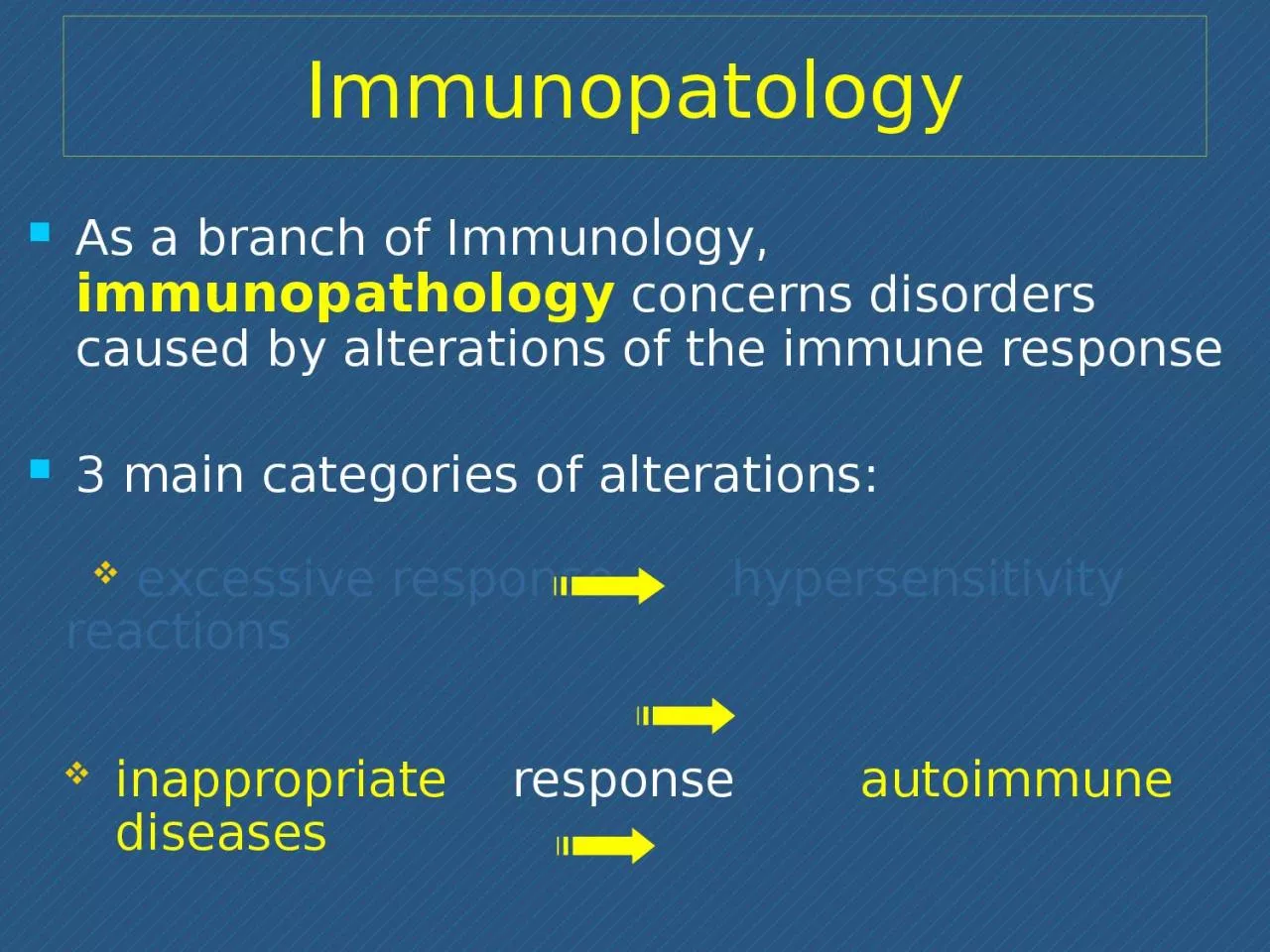
As a branch of Immunology, immunopathology concerns disorders caused by alterations of the immune response3 main categories of alterations: excessive response hypersensitivity reactionsinappropriate response autoimmune diseases defective response immunodeficiencies
Slide2Factors involved in the aetiopathogenesis of autoimmune diseases
Slide3Immune reactions to self antigens (i.e., autoimmunity) are the underlying cause of numerous human diseases
Autoimmune diseases are estimated to affect 2% to 5% of the population in developed countries, and appear to be increasing in incidenceThe evidence that some human diseases are indeed the result of autoimmune reactions is more persuasive for some than for others in which the role of autoimmunity is suspected but not provedAutoimmunity
Slide4Slide5In many autoimmune disorders, multiple high-affinity
autoantibodies have been identified, and in some cases these antibodies are known to cause pathologic abnormalitiesSimilarly, in some of these diseases, there is growing evidence for the activation of pathogenic self-reactive T cellsExperimental models in rodents have proved very informative, providing circumstantial evidence in support of an autoimmune etiology of selected pathologiesEvidence for the autoimmune nature of some human diseases
Slide6Introducing autoimmunity
Immunological tolerance (central and peripheral)Negative selectionRole of regulatory T cells (Treg)Physiological unresponsiveness to self- antigens based on
Slide7Immunological tolerance
Billions of different antigen receptors are randomly generated in developing T and B lymphocytesIt is not surprising that during this process, receptors that recognize self antigens are producedSince these antigens cannot all be concealed from the immune system, there must be means of eliminating or controlling self-reactive lymphocytes [tolerance]
The
breakdown of tolerance is the basis of autoimmunity
Slide8Where can lymphocytes encounter self antigens?
Central and peripheral tolerancethymus, bone marrow
T and B precursorsBTreg
Slide9s
ome immature lymphos (both B and T) are killed by apoptosis (negative selection)in the bone marrow, some self-reactive B lymphos switch to new antigen receptors that are not self-reactive (receptor editing)in the thymus, some T lymphos differentiate into regulatory T cells (Treg)Central tolerance: thymus and bone marrow
The interaction of lymphoid precursors with self antigens in the central lymphoid organs may have three main outcomes:
Slide10Where can lymphocytes encounter self antigens?
Central and peripheral tolerancethymus, bone marrowperipheral lymphoid tissues
Slide11Mechanisms of peripheral
toleranceSelf-reactive mature lymphos which gain access to peripheral tissues may undergo negative selection (apoptosis) or functional inhibition (anergy)T cells are made anergic in the absence of costimulatory signals by APCB cells become anergic if they encounter antigen in the absence of specific helper T cellsCurrent investigations have not yet completely disclosed the molecular mechanisms underlying anergy
Slide12Peripheral tolerance of T lymphos: role of T regulatory cells (
Treg)The responses of T lymphos to self antigens may be actively suppressed by Treg, a subtype of T cells (CD4+) generated in the thymus by self antigen recognition (high IL-2 receptor number; role for IL-2?)Main mechanisms proposed to explain how Treg control immune responses:secretion of immunosuppressive cytokines (e.g., IL-10, TGF-β) which down-regulate T cell responsesblocking of costimulatory signals by APC leads to inhibition of T cell activation (direct contact ? undefined mechanism)
Slide13In a normal response, T cells recognize antigen, proliferate and differentiate into effector cells
Tregs generated by self antigen recognition inhibit the development or functions of effector T cells (negative control of autoreactive T cells)TregIL-10, TGF-
β
Slide14Postulated failures of Treg-mediated regulation of autoreactive T cells
Inadequate number of Treg cells owing to their inadequate development, proliferation or survivalDefects in Treg cell function (poor production of immune suppressive cytokines, e.g. IL-10, TGFβ)
Resistance of pathogenic effector T cells
to suppression by Treg cells: increased production of cytokines which impede Treg function, such as TNF-α, IL-4, IL-6
intrinsic defects of Treg
Slide15Essential
steps in the pathogenesis of autoimmunitySusceptibility genesEnvironmental trigger(e.g. infections, tissue injury)
Failure ofself-tolerance
Activation ofself-reactive lymphocytesImmune responses against self tissues
Persistence of functional
self-reactive lymphocytes
?
Slide16Major factors involved in the pathogenesis of autoimmunity
Autoimmunity arises from:
(1)
the
inheritance of susceptibility genes
, that may interfere with selftolerance;
(2)
association with
environmental triggers
(infection, tissue injury, inflammation) that:
alter the display of self antigens
promote
entry of self-reactive
lymphocyte
into tissues
enhance their activation
Slide17Genetic susceptibility to autoimmunity (1)
Most autoimmune diseases are polygenic; affected individuals inherit multiple genetic polymorphisms that contribute to disease susceptibilityThe products of many of these polymorphic genes influence the development of self-tolerance:some are believed to influence negative selection of self-reactive T cells (central tolerance)others control T cell anergy to self antigens (peripheral tolerance)The mechanistic links between susceptibility genes and failure of tolerance are not yet conclusively established
Slide18Genetic susceptibility to autoimmunity (2)
Among the genes linked to autoimmunity, the strongest associations are with MHC (human HLA) genes, especially class II MHC genesClass II MHC genes are involved in the selection and activation of CD4+ T cells that regulate both humoral and cell-mediated immune response to protein antigensIn many autoimmune diseases, the disease-associated HLA molecules differ in their peptide binding cleft from HLA molecules that are not disease-associatedDisease-associated HLA molecules favour the binding of particular self-peptides that will ultimately be recognized by self-reactive T lymphos
Slide19Role of infections in autoimmunity (1)
Viral and bacterial infections may contribute to the development and exacerbation of autoimmunityTwo main mechanisms:Microbes may activate the APC to express costimulatory molecules for T lymphos; when these APC present self antigens, the self-reactive T cells are activated Some microbial antigens may cross-react with self antigens (molecular mimicry); immune response initiated by the microbes may activate T cell specific for self antigens
Slide20Role of infections in autoimmunity (2)
Example of molecular mimicry:Rheumatic fever: after streptococcal infection, anti-streptococcus antibodies cross-react with miocardial proteins; onset of inflammatory response (myocarditis)Myocarditis also takes place due to homologies between myocardial protein antigens and some antigens of Chlamydia and Trypanosoma cruzi Lyme artrhritis: homologies between a surface molecule of
Borrelia burgdorferi and a lymphocyte antigen (LFA-1, lymphocyte function antigen-1)
Slide21Other factors involved in the development of autoimmunity
Anatomic alterations in tissues, possibly induced by inflammation, ischemic injury or trauma, may lead to exposure of self antigens normally concealed from the immune systemIntra-ocular antigens (post traumatic uveitis)Sperm proteins (orchitis after vasectomy)Hormonal influences: many autoimmune diseases have a higher incidence in females than in malesSystemic lupus erythematosus affects women about 10 times as frequently as men
Slide22Organ-specific
Autoimmune attack vs. self-antigens of a given organ
It results in a damage of organ structure and functionTreatment is focused on the replacement of organ function
Organ-specific vs systemic autoimmune diseases
Systemic
Targets are widespread self-antigens
Damage affects structures as blood vessels, cell nuclei, etc.
Treatment is aimed to inhibit excessive activation of the immune system
Slide23Organ-specific
Hashimoto thyroiditis (thyroid destruction)Grave’s disease (hypertyroidism)
Addison’s disease (adrenal g. failure)Juvenile diabetes mellitusMultiple sclerosisAtrophic gastritisMyasthenia gravisExamples of organ-specific
and systemic autoimmune diseases
Systemic
Systemic lupus (SLE)
Rheumatoid arthritis
Scleroderma
Dermatomyositis
Slide24Pernicious
anemia
Atrophic gastritis in pernicious anemia: loss of stomach parietal cells is due to autoimmune reaction mediated by auto-antibodies against parietal cells and intrinsic factor
normal mucosa
atrophic mucosa
Intrinsic factor
: glycoprotein secreted by gastric mucosa; favours absorption of
iron
and
vitamin B12
(
essential
cofactors for
erythropoiesis
)
Slide25Organ-specific
Hashimoto thyroiditis (thyroid destruction)Grave’s disease (hypertyroidism)
Addison’s disease (adrenal g. failure)Juvenile diabetes mellitusMultiple sclerosisAtrophic gastritisMyasthenia gravisExamples of organ-specific
and systemic autoimmune diseases
Systemic
Systemic lupus (SLE)
Rheumatoid arthritis
Scleroderma
Dermatomyositis
Slide26Antireceptor antibodies disturb the normal function of receptors
Graves diseaseAuto-antibodies against the thyroid-stimulating hormone (TSH) receptor activate thyroid cells: hyperthyroidism
Genetic susceptibilityBacterial and/or viral infections (?)
Myastenia gravisAuto-antibodies against ACh receptor impair neuromuscular transmission: muscle weaknessGenetic susceptibilityTreg dysfuntion (low levels of transcription factor FOXP3, crucial for Treg function)