
Kaleb Decker Harsh Moolani The tongue is a muscular organ in the mouth that is covered in moist pink tissue called mucosa Tiny bumps called papillae give the tongue its rough texture Thousands of tiny nervelike cells called taste buds cover the surface of the papillae ID: 909746
Download Presentation The PPT/PDF document "THE DIGESTIVE SYSTEM By Chance Meeks" is the property of its rightful owner. Permission is granted to download and print the materials on this web site for personal, non-commercial use only, and to display it on your personal computer provided you do not modify the materials and that you retain all copyright notices contained in the materials. By downloading content from our website, you accept the terms of this agreement.

Slide1
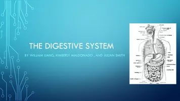
THE DIGESTIVE SYSTEM
By
Chance Meeks
Kaleb Decker
Harsh
Moolani
Slide2Slide3.The tongue is a muscular organ in the mouth that is covered in moist, pink tissue called
mucosa
.Tiny bumps called
papillae
give the tongue its rough texture .Thousands of tiny nerve-like cells called, taste buds, cover the surface of the papillae.The tongue is vital for chewing, swallowing food, and speech
Tongue
Slide4Slide5.The pharynx is a
fibromuscular
tube which extends from the base of the skull to the lower border of the cricoid cartilage
.The muscular walls of the pharynx are compromised of an outer layer made up of 3
circulary disposed muscles, the CONSTRICTERSDuring swallowing successive contraction of the constricters
helps to propel the ball of food down the esophagus
Pharynx
Slide6Slide7.The glands are found in and around your mouth and throat. We call the major salivary glands, the parotid, the submandibular and sublingual glands
.They all secrete saliva into your mouth, the parotid through tubes that drain saliva, called salivary ducts, near your upper teeth submandibular under your tongue and the sublingual through many ducts in the floor of your mouth
Salivary Glands
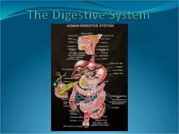
Slide8Slide9.The small intestine is part of the gastrointestinal tract (gut) following the stomach and followed by the large intestine
.It is where the vast majority of digestion and absorption of food takes place
.The small intestine in an adult human measures about 7 meters in length and 2.5-3cm in diameters
.Made up of 3 structural parts:
Duodenom, Jejunum, and Ileum.
Small intestine
Slide10Slide11.Stomach is a muscular organ located on the left side of the upper abdomen
.As food reaches the end of the esophagus, it enters the stomach through a muscular valve called the lower esophageal sphincter
.Stomach secretes acids and enzymes that digest food. Stomach muscles contract periodically, churning food to enhance digestion
.Ridges of muscle tissue called
rugae line the stomach.
It’s 12 inches long and 6 inches wide
Stomach
Slide12Slide13.Teeth are hardest substances in the human body
.Besides being essential for chewing, they play an important role for speech
.Parts of the teeth include: Enamel, Dentin, Pulp,
Cementum
, and the Periodical Ligament.The normal adult mouth has 32 teeth.Crown of each tooth projects into the mouth while the root descends into the gum line, into the jaw.
Teeth
Slide14Slide15. The liver is a large, meaty organ that sits on the right side of the belly. Weighing about 3 pounds, the liver is reddish-brown in color and feels rubbery to the touch. Normally you can't feel the liver, because it's protected by the rib cage
.The liver has two large sections, called the right and the left lobes. The gallbladder sits under the liver, along with parts of the pancreas and intestines. The liver and these organs work together to digest, absorb, and process food.
. The liver's main job is to filter the blood coming from the digestive tract
.
Liver
Slide16Slide17. The pancreas is about 6 inches long and sits across the back of the abdomen, behind the stomach.
. The head of the pancreas is on the right side of the abdomen and is connected to the duodenum (the first section of the small intestine) through a small tube called the pancreatic duct
.The main jobs of the pancreas is to produce digestive juices and produce hormones
.
Pancreas
Slide18Slide19. The gallbladder is a small pouch that sits just under the liver. The gallbladder stores bile produced by the liver. After meals, the gallbladder is empty and flat, like a deflated balloon. Before a meal, the gallbladder may be full of bile and about the size of a small pear
. The gallbladder squeezes stored bile into the small intestine through a series of tubes called ducts. Bile helps digest fats, but the gallbladder itself is not essential.
.
Gall Bladder
Slide20Slide21. The esophagus is a muscular tube connecting the throat (pharynx) with the stomach. The esophagus is about 8 inches long, and is lined by moist pink tissue called mucosa.
. The upper esophageal sphincter (UES) is a bundle of muscles at the top of the esophagus. The muscles of the UES are under conscious control, used when breathing, eating, belching, and vomiting. They keep food and secretions from going down the windpipe
.When the lower esophageal sphincter is closed it prevents acids and stomach contents from traveling backwards
.
Esophagus
Slide22Slide23. The colon is also called the large intestine. The ileum (last part of the small intestine) connects to the
cecum
(first part of the colon) in the lower right abdomen.
.The colon is divided into 4 parts: The ascending colon, the transverse colon, the descending colon, and the sigmoid colon
. The colon removes water, salt, and some nutrients forming stool. Muscles line the colon's walls, squeezing its contents along. Billions of bacteria coat the colon and its contents, living in a healthy balance with the body.
.
Colon (Large Intestine)
Slide24Slide25. The lower opening of the digestive tract. It is associated with the anal sphincter and lies in the cleft between the buttocks, through which fecal matter is extruded.
Anus
Slide26Slide27.http://www.webmd.com/oral-health/picture-of-the-tongue
.http://www.emory.edu/ANATOMY/AnatomyManual/pharynx.html
.http://www.entnet.org/HealthInformation/salivaryGlands.cfm
.
http://www.news-medical.net/health/What-Does-the-Small-Intestine-Do.aspx. http://www.news-medical.net/health/What-Does-the-Small-Intestine-Do.aspx
.
http://www.webmd.com/oral-health/picture-of-the-teeth
.
http://www.webmd.com/digestive-disorders/picture-of-the-liver
.
http://www.webmd.com/digestive-disorders/picture-of-the-pancreas
.
http://www.webmd.com/digestive-disorders/picture-of-the-gallbladder
.
http://www.webmd.com/digestive-disorders/picture-of-the-esophagus
.
http://www.webmd.com/digestive-disorders/picture-of-the-colon
. http://dictionary.webmd.com/terms/anus
.
http://www.mamashealth.com/organs/stomach.asp
Citation Page