
MOI Direct blow to the anterior shin Etiology A direct blow causes bleeding into the narrow space housing the nerves and blood vessels The tight surrounding fascia allows very limited expansion ID: 908898
Download Presentation The PPT/PDF document "Conditions of the Foot, Ankle, and Lower..." is the property of its rightful owner. Permission is granted to download and print the materials on this web site for personal, non-commercial use only, and to display it on your personal computer provided you do not modify the materials and that you retain all copyright notices contained in the materials. By downloading content from our website, you accept the terms of this agreement.

Slide1
Conditions of the Foot, Ankle, and Lower Leg (FALL)
Slide2MOI
:
Direct blow to the anterior shinEtiology: A direct blow causes bleeding into the narrow space housing the nerves and blood vessels. The tight surrounding fascia allows very limited expansion. Pressure increases on the tibial artery and the deep peroneal nerve which cuts off blood flow and feeling.Medical Emergency: Intramuscular pressure and neurovascular compromise leads to ischemia (lack of oxygen) and necrosis (tissue pain)Signs and Symptoms: Severe throbbing pain, decreased strength with dorsiflexion and great toe extension, decreased sensation on the dorsum (top) of the foot, decreased circulation at the dorsal pedis pulse, glossy red tight skinTreatment: Emergency Fasciotomy
Acute Anterior Compartment Syndrome
Slide3Acute Anterior Compartment Syndrome
Slide4MOI:
Develops gradually with athletic activity
Etiology: Gradual increase in pressure in the anterior compartment due to swelling and muscle hypertrophySigns and Symptoms: Weak dorsiflexion and great toe extension, anterior shin pain, decreased sensation on the dorsum of the foot, decreased circulation.Evaluation: Have athlete exercise to the point when symptoms occur. Once symptoms occur, test strength, sensation, and circulation before symptoms decreaseTreatment: Ice and evaluation, BUT NO COMPRESSION, monitor neurological status and girth measurements of the affected area, crutches for loss of sensation, and referralChronic Anterior Compartment Syndrome
Slide5Chronic Anterior
Compartment Syndrome
Slide6MOI:
Direct blow to the lower leg or a rotational force from planting or turning on a fixed foot
Etiology: Direct blow or rotational force that results in a transverse or spiral oblique fracture of the tibia/fibulaSigns and Symptoms: Obvious deformity, swelling, ecchymosis (discoloration), crepitus(noise), and inability to bear weight especially when the tibia is involvedTreatment: Immobilization, PRICES, refer to physicianSpecial Test: Percussion /Bump TestAnd Compression/Squeeze TestFracture(s) of the Tibia/Fibula
Slide7Fractures to the Tibia/Fibula
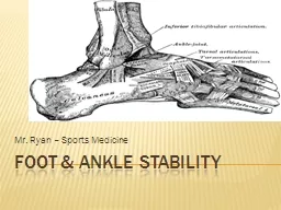
Slide8Special Tests for Stability
of the Tibia and Fibula
Percussion or Bump Test-helps determine a fractureCompression or Squeeze Test-helps determine a fracture or anterior/posterior tibiofibular sprain
Slide9MOI
: Overuse/Repetitive mechanical stress
Etiology: The bone cannot adapt to repetitive loading and attempts to adapt with increasing the breakdown of bone. In return, the laying of new bone follow. Once the breakdown of bone exceeds the laying down of new bone, the bone begins to weaken and a stress fracture formsSigns and Symptoms: Pain during weight bearing activity and eventually non-weight bearing activity, localized swelling and point tenderness over the affected areaTreatment: Refer for x-rays two weeks after injury; bone scan or MRI may be necessary; treat symptomatically for pain and return to activity when the asymptomatic (normally 4 -6 weeks); REST is the best treatment.Stress Fracture(s) of the Tibia/Fibula
Slide10Stress Fractures of Tibia/Fibula
Slide11MOI: Overuse/Repetitive stress
Etiology:
Overuse/Repetitive stress forces acting on the proximal insertions of the Flexor Digitorum Longus, Tibialis Posterior, and /or Tibialis Anterior. Inflammation present along muscle attachments to tibia and insertions on the plantar surfaceSigns and Symptoms: Aching pain in shin that is aggravated with push-off phase and shock absorption during heel strike; athlete may excessively pronate(have flat feet)Treatment: Ice cup, taping or orthotics for foot mechanics, stretch routine, and rule out stress fracture. Active Rest!Shin Splints
Slide12Shin Splints
Slide13MOI:
Strong force during resistive forceful plantarflexion of the ankle with the knee extended or forceful dorsiflexion of the ankle with contraction of the Gastrocnemius/Soleus
Etiology: Partial/complete rupture of the Achilles tendonSigns and Symptoms: Athlete report a sensation of being kicked or shot in the calf, audible snap, and painful loss of function. Observable gap with a window shade affect, palpable gap, bunching of the tendon proximally, and massive swellingSpecial Tests: Thompson TestAchilles Tendon Rupture
Slide14Achilles Tendon Rupture
Slide15Grades of Achilles Tendon Strain
Slide16Thompson Test-for Achilles’
T
endon Ruptures
Slide17MOI: Overuse injury
Etiology:
Microtrauma/Inflammation and possible thickening of the Achilles tendonPredisposing Factors: Tight achilles Tendon, Extremely High Arches, Excessively Flat FeetSigns and Symptoms: Pain proximal to calcaneal insertion, morning pain/stiffness, clicking sensation when walking, palpable crepitus, and swelling and thickening of the tendon. Functional Tests: loss of full dorsiflexion, painful and active and resistive plantarflexionTreatment: Ice cup and Ultrasound, heel lifts(both shoes), stretching, strengthening, and tapingAchilles Tendon Tendonitis
Slide18Achilles Tendon Tendonitis
Slide19MOI for Ankle Sprains
Slide20MOI
:
Inversion (inversion/plantarflexion)Etiology: Partial/complete rupture of the Anterior Talofibular Ligament, Calcaneofibular Ligament, and/or Posterior Talofibular Ligament, avulsion fracture of the malleolus, chip fracture of the medial malleolusSigns and Symptoms: Pain/Point tenderness over lateral ligamentous structures, as well as, the medial and lateral malleoli. Inability to bear weight without limp, swelling laterally and posteriorly with swelling migrating to the toes and possible crepitus. Special Tests: +Anterior Drawer, +Talar TiltTreatment: PRICES, Crutches, RehabilitationLateral Ankle Sprains
Slide21Lateral Ankle Sprain
Slide22Special Tests for Lateral Ankle Sprains
Talar
Tilt Test-tests for the Calcaneofibular ligamentAnterior Drawer (Ankle)-tests for the Anterior Talofibular ligament
Slide23MOI:
Eversion (Eversion/Dorsiflexion)
Etiology: Partial/Complete rupture of the deltoid ligament, avulsion fracture of the medial malleolus, chip fracture of the lateral malleolus, spiral oblique fracture of the shaft of the tibia/fibulaSigns and Symptoms: Pain/Point tenderness over medial ligamentous structures, as well as, the medial and lateral malleoli. Inability to bear weight without limp, swelling medially and posteriorly with additional swelling migrating to the toes and possibly crepitus.Special Tests: + Kleiger TestMedial Ankle Sprains
Slide24Medial Ankle Sprains
Slide25Kleiger Test-determines a Medial Ankle Sprain and injury to the DELTOID ligament
Slide261
st
DEGREE: Mild stretching of the ligament with mild swelling, minor point tenderness, and no instability.2nd DEGREE: Partial tear of the ligament with moderate swelling, pain, moderate point tenderness, ecchymosis, and instability with firm end field.3rd DEGREE: Complete rupture of the ligament with severe swelling, ecchymosis, pain, and gross instability with a soft end field.Classification of Ankle Sprains
Slide27MOI:
Direct blow to the heel
during extreme weight bearingEtiology: Contusion to the heel area with irritation of the fat pad, calcaneus, and surrounding nervesSigns and Symptoms: Pain during weight bearing especially on the lateral side during heel strike, swelling in the heel area, ecchymosis, and palpable mass of blood.Treatment: Ice, padding, taping, crutchesHeel Contusion (Heel Bruise)
Slide28Heel Bruise (Heel Contusion)
Slide29MOI: Overuse injury,
caused by friction and pressure
Signs and Symptoms: Redness, swelling, warmth, callus formation, and point tenderness at the inserting of the Achilles TendonTreatment: PaddingRetro-calcaneal bursitis (Pump Bump)
Slide30MOI: Overuse
Etiology:
The plantar fascia becomes inflames due to repetitive forces. Tightness of the plantar fascia is a common cause. Having a tight gastrocnemius/soleus complex as well as hamstrings may contribute.Signs and Symptoms: Pain and point tenderness at mid plantar surface and migrating to the calcaneus. Pain in the morning and during the push off phase of gait. Athlete may walk on the lateral side of foot to decrease their pain.Treatment: Ice cup and Ultrasound, Anti-inflammatory drugs, orthotics tape, stretching.Plantar Fasciaitis
Slide31Plantar
Fasciaitis
Slide32MOI:
Forces hyperextension or hyperflexion of the Metatarsalphalangeal Joint of the Great Toe.
It is often seen when athletes wear flexible shoes on a hard surface.Etiology: Sprain of the plantar or dorsal ligaments of the MP joint of the great toe.Signs and Symptoms: Pain during push-off phase of gait, pain and swelling at the MP Joint, pain with passive extension or flexion and side to side motions. Treatment: Ice, tape, rigid shoesSprain of the Metatarsalphalangeal Joint of the Great Toe – “Turf-Toe”
Slide33Metatarsophalangeal Joint Sprain “Turf Toe”
Slide34MOI: Direct blow
Signs and Symptoms:
Pain with gait, swelling, ecchymosisTreatment: Refer to physician for x-raysToe Fractures
Slide35Sometimes called a Jones Fracture
MOI:
Forced inversionEtiology: Fracture occurs between the peroneus brevis and peroneus tertius. May disrupt blood supply-risk of non-union. It is very hard to heal!Signs and Symptoms: Swelling, ecchymosis, point tenderness at the base of the 5th metatarsalTreatment: Refer for x-raysBase of the 5th Metatarsal Fractures
Slide365
th
Metatarsal Fractures
Slide37MOI: Inversion
Etiology:
The inversion mechanism causes the muscles (peroneus brevis and peroneus tertius) to pull off a piece of the bone at the base of the 5th metatarsal boneSigns and Symptoms: Swelling, ecchymosis, point tenderness at the base of the 5th.Treatment: Refer for x-rays.Avulsion Fractures
Slide38Avulsion Fractures
Slide39MOI:
Friction
Etiology: As the layers of skin rub together, friction causes a seperation. The body responds with fluid formation in this seperation. Signs and Symptoms: Pain, fluid formation, open wound is possible Treatment: Padding, Lubrication, Prevent infection at all timesBlisters