
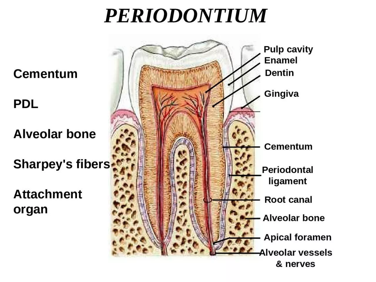
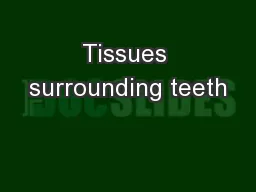
Alveolar bone Sharpeys fibers Attachment organ Cementum Periodontal ligament Alveolar bone Apical foramen Pulp cavity Enamel Dentin Gingiva Root canal Alveolar vessels amp nerves TEETH INSITU ID: 918460
Download Presentation The PPT/PDF document "PERIODONTIUM Cementum PDL" is the property of its rightful owner. Permission is granted to download and print the materials on this web site for personal, non-commercial use only, and to display it on your personal computer provided you do not modify the materials and that you retain all copyright notices contained in the materials. By downloading content from our website, you accept the terms of this agreement.


Slide1
PERIODONTIUM
Cementum
PDL
Alveolar boneSharpey's fibersAttachmentorgan
Cementum
Periodontalligament
Alveolar bone
Apical foramen
Pulp cavity
Enamel
Dentin
Gingiva
Root canal
Alveolar vessels& nerves
Slide2TEETH IN-SITU
Slide3PeriodontiumGingiva(fibrous CT)Periodontal Ligament(fibrous CT)Alveolar bone(minerlaized CT)Cement(minerlaized CT)
Slide4Slide5CementumIt is a hard avascular connective tissue that covers the roots of teeth
Slide6Role of CementumIt covers and protects the root dentin (covers the opening of dentinal tubules)2) It provides attachment to the periodontal fibers3) It compensates for tooth resorption
Slide7Varies in thickness:
thickest in the apex andin the inter-radicular areas of multirootedteeth, and thinnest in the cervical area10 to 15 m in the cervical areas to50 to 200 m (can exceed > 600 m) apically
Slide8Cementum simulates bone Organic fibrous framework, ground substance, crystal type, development Lacunae Canaliculi Cellular component Incremental lines (also known as “resting” lines; they are produced by continuous but phasic, deposition of cementum)
Slide9Differences between cementum and bone Not vascularized – a reason for it being resistant to resorption Minor ability to remodel
More resistant to resorption compared to bone Lacks neural component – so no pain 70% of bone is made by inorganic salts (cementum only 45-50%)
Slide10Clinical Correlation
Cementum is more resistant to resorption: Important in permitting
orthodontic tooth movement
Slide11Development of Cementum
Cementum formation occurs along the
entire tooth
Hertwig’s epithelial root sheath (HERS) –Extension of the inner and outer enamelepitheliumHERS sends inductive signal to ectomesen-chymal pulp cells to secrete predentin bydifferentiating into odontoblastsHERS becomes interruptedEctomesenchymal cells from the inner portionof the dental follicle come in with predentin bydifferentiating into cementoblastsCementoblasts lay down cementum
Slide12CementoblastsDerive from dental follicle
Slide13Slide14Properties of Cementum
Physical:
Cementum is pale yellow with a dull surface Cementum is more permeable than other dental tissues
Relative softness and the thinness at the cervical portion means that cementum is readily removed by the abrasion when gingival recession exposes the root surface to the oral environment
Slide15Chemical Composition of Cementum
45% to 50% hydroxyapatite (inorganic)
50% to 55% collagenous and noncollagenous matrix proteins(organic)
Slide16Collagenous componentTYPE ITYPE IIITYPE XIITYPE VTYPE XIV
Slide17Classification of CementumPresence or absence of cellsOrigin of collagenous fibers of the matrixPrefunctional and functional
Slide18Cellular and Acellular Cementum
Acellular cementum
: covers the root
adjacent to dentin whereas cellularcementum is found in the apical areaCellular: apical area and overlyingacellular cementum. Also common ininterradicular areas
Cellular: Has cellsAcellular: No cells and has no structureAcellular cementum (primary cementum)Cellular Cementum (secondary cementum
Slide19A: Acellular cementum
B: Hyaline layer of Hopwell-SmithC: Granular layer of TomesD: Root dentin
Cellular cementum usually overlies acellular cementum
Slide20Acellular
Cellular
Variations also noted where acellular and cellular reverse in position
and also alternate
Slide21Dentin
GT
Lacuna of cementocyte
Canaliculus
CEMENTUM
Acellular cementum
Cellular cementum
Hyaline layer (of Hopewell Smith)Granular layer of tomes
Dentin with tubules
Slide22Cementoblast and cementocyte
Cementocytes in lacunae and the channels that their processes extend arecalled the canaliculiCementoid: Young matrix that becomes secondarily mineralized
Cementum is deposited in increments similar to bone and dentin
Slide23Classification Based on the Nature and Origin of Collagen Fibers
Organic matrix derived form 2 sources:
Periodontal ligament (Sharpey’s fibers)CementoblastsExtrinsic fibers if derived from PDL. These are in the same
direction of the PDL principal fibers i.e. perpendicular oroblique to the root surface Intrinsic fibers if derived from cementoblasts. Run parallel tothe root surface and at right angles to the extrinsic fibers The area where both extrinsic and intrinsic fibers is calledmixed fiber cementum
Slide24Combined classificationAcellular Extrinsic Fiber Cementum (AEFC-Primary Cementum) Located in cervical half of the root and constitutes the bulk of cementum The collagen fibers derived from Sharpey’s fibers and ground substance from cementoblasts Covers 2/3rds of root corresponding with the distribution of primary acellular cementum
Principal tissue of attachment Function in anchoring of tooth Fibers are well mineralized
Slide25Slide26Slide27Slide28Distribution of Cementum on the RootAcellular afibrillar: cervical enamelAcellular extrinsic: Cervix to practically the whole root (incisors, canines) increasing in thickness towards the apical portion 50200μmCellular: Apical third, furcations
Slide29Cemento-enamel junction(CEJ) Cementum overlaps enamel 60% Cementum just meets enamel 30% Small gap between cementum and enamel 10%
Slide30Slide31Slide32Aging of Cementum
Smooth surface becomes irregular
due
to calcification of ligament fiber bundles where they are attached to cementumContinues deposition of cementum occurs with age in the apical area. [Good: maintains tooth length; bad: obstructs the foramen]Cementum resorption. Active for a period of time and then stops for cementum deposition creating reversal linesResorption of root dentin occurs with aging which is covered by cemental repair
Slide335.Cementicles
Calcified ovoid or round nodule found in the PDL Single or multiple near the cemental surface Free in ligament; attached or embedded in cementum
Aging and at sites of trauma