
Ingestion taking fooddrink into mouth Propulsion movement through alimentary canal swallowing peristalsis Mechanical Digestion Physical breakdown of food chewing churning ID: 1036836
Download Presentation The PPT/PDF document "The Digestive System Digestive Process..." is the property of its rightful owner. Permission is granted to download and print the materials on this web site for personal, non-commercial use only, and to display it on your personal computer provided you do not modify the materials and that you retain all copyright notices contained in the materials. By downloading content from our website, you accept the terms of this agreement.

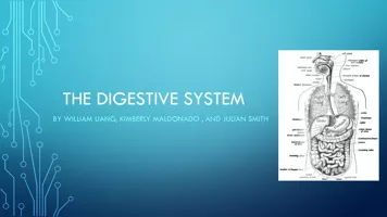
1. The Digestive System
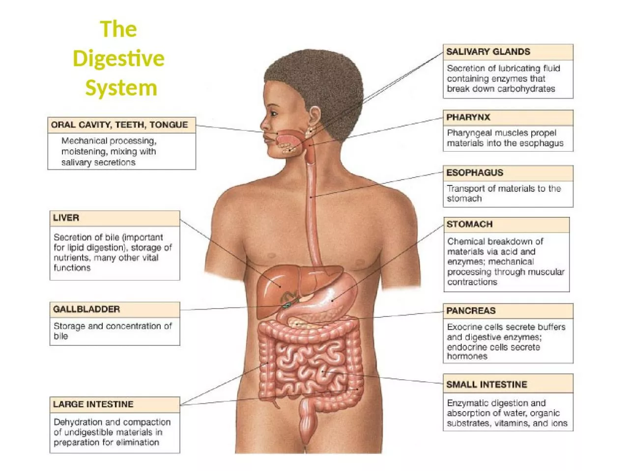
2. Digestive ProcessesIngestion – taking food/drink into mouth.Propulsion – movement through alimentary canal (swallowing, peristalsis).Mechanical Digestion – Physical breakdown of food (chewing, churning).
3. Absorption – transport of digested products from lumen of G.I. tract to blood and lymph (inside body).Defecation – elimination of indigestable and waste products from body (feces).Chemical Digestion – Enzymatic breakdown of food (from complex to simple building blocks).
4.
5.
6. Lingual amylase (breaks down starch).Lysozyme – antibacterial agent in saliva.Salivary Glands Intrinsic (inside oral cavity) e.g., lips & cheeks.Extrinsic (outside oral cavity):1) Parotid (largest) - a serous gland.2) Submandibular - a serous gland.3) Sublingual (smallest) - a mucus gland“Mumps”
7. The Oral CavityTypical Adult Teeth (in one quadrant)Incisors 2Canines 1Premolars 2Molars 3 = 8Times 4 quadrants:Total: 4 x 8 = 32
8.
9. – Mucous glands in tela submucosa (layer) to lubricate bolus.The Esophagus - is a Muscular tube.– ~ 10 inches long.– Transitions from skeletal to smooth muscle.(hence voluntary to involuntary)– Outermost layer is Adventitia or Serosa.outsideperitonealcavityinsideperitonealcavity
10. The Stomach - an acidic storage area.
11. Only Absorption of alcohol and aspirin.Enzymatic Digestion of proteins occurs here (Pepsin breaks down proteins).Stomach – acidic (pH 2) storage of chyme. Mechanical Digestion continued (churning).Rugae allows for expansion when volume of contents increase.Has 3 muscle layers, for churning.
12. Can you identify the 4 Tunics?Unique in the GI tract,the stomach has 3 musclelayers:
13. Chief cells: make Pepsinogen, which is cleaved to pepsin (↑HCl), to digest proteins.Production and secretion of gastric juices controlled by CNS.e.g., Vagus nerveParietal cells: make Hydrocholic acid (HCl) in gastric glands.Gastric Gland
14. Gastric Bypass (Malabsorptive) Surgery
15. Lacteals absorption lipidsSmall Intestine: Chemical Digestion/AbsorptionDuodenumJejunumIleumIncrease Surface Area for AbsorptionIntestinal glands Goblet cellsStem cells1) Plicae Circulares2) Villi (Intestinal)3) Microvilli
16. The Small Intestine
17. Vascular Arcade (from superior mesenteric a.)Mesentery Proper - is a double layered serous membrane attached to the small intestine.Roles:- Supports branches of blood vessels.- Supports lymphatics of the jejunum and ileum.- Supports nerves of the jejunum and ileum.Villi of Small Intestine
18. The Mesentery Proper of the Small IntestineThe Greater Omentum
19. Distinguishing features of each region of the Small Intestine
20. Begins as pouch inferior to end of ileumEnds at anus.The Cecum:Contains the Ileocecal valve and connected to appendix.The Large Intestine1) Reabsorb water and compact feces.Functions of Large Intestine:3) Stores fecal matter.2) Absorb vitamins (helps make Vit K) electrolytes.The Colon: – Ascending, Transverse, Descending, Sigmoid.
21.
22. The ColonLack of villiAbundance of goblet cellsAbundance of mucous-secreting intestinal glands
23. The Rectum and Anus
24. The Liver
25.
26. Diagrammatic view of Liver lobular organization.
27. Anatomy between the Liver (makes Bile), the Gallbladder (stores and concentrates bile), and the Pancreas (makes/releases pancreatic juices) into the Duodenum.
28. The roles of the Greater and Lesser Duodenal Papillae
29. Histology of the PancreasExocrine Gland:Pancreatic JuicesAmylasesProteasesLipases Endocrine Gland:Makes HormonesInsulinGlucagonSomatostatinGastrin
30. Histology of the G.I. Tract
31. Lines entire digestive tractA Mucous MembraneMoistened by glandular secretionsSimple or stratified depending on area of tractPleated for expansion (↑Surface Area)
32. is a Mucous Membrane (wet, absorbs, protects) 1) Epithelium 2) Lamina propria 3) Muscularis mucosae2. Tela Submucosa1. Tunica MucosaAreolar Connective TissueContains:Submucosal plexus (nervous innervation)Blood and Lymphatic VesselsThese are the 4 Layers!
33. 3. Tunica Muscularis Externa4. Tunica Serosa (or Adventitia*)Smooth muscle layers1) Inner Circular Layer 2) Outer Longitudinal LayerMyenteric PlexusSerous membrane – aka visceral peritoneum* Name depends on location:Inside peritoneal cavity = serosaOutside peritoneal cavity = adventitiaWhat are some exceptions?
34.
35. Layers: How are they different in each diagram above?1. _______________2. ______________3. _______________4. _______________
36. The Peritoneum: Two layersVisceral peritoneum (a.k.a. serosa)Parietal peritoneum - Lines inner surfaces of body wallGreater omentumLesser omentumMesentery properMesocolonMesenteries: – Fused double sheets of peritoneal membrane to suspend portions of digestive tract:e.g.
37. Retroperitoneal Structures – these are attached to posterior abdominal wall, and are not in the peritoneal cavity. - Most of the Duodenum - Ascending colon - Descending colon - Pancrease.g.
38. Horizontal section through the upper abdomen showing the position of the liver relative to other visceral organs.