
Function Dr Baghbanian M Gastroenterologist Shaheed Sadoughi hospital 2012 liver tests 1 detect the presence of liver disease 2 distinguish different types of liver ID: 333137
Download Presentation The PPT/PDF document "Evaluation of Liver" is the property of its rightful owner. Permission is granted to download and print the materials on this web site for personal, non-commercial use only, and to display it on your personal computer provided you do not modify the materials and that you retain all copyright notices contained in the materials. By downloading content from our website, you accept the terms of this agreement.


Slide1
Evaluation of Liver Function
Dr.
Baghbanian
M.
Gastroenterologist
Shaheed
Sadoughi
hospital / 2012Slide2
liver tests
(
1) detect the presence of liver
disease
(2)
distinguish
different types of liver
disorders
(3)
extent of liver damage
(
4) follow the response to
treatmentSlide3
Liver tests
Can
be normal in
serious
liver
disease
Can be abnormal in non hepatic diseases
Rarely
suggest a specific
diagnosis
They
suggest a general category of liver disease, such as
hepatocellula
r
or
cholestatic
Slide4
liver carries out thousands of biochemical functions
most
cannot be easily measured by blood tests.
Laboratory
tests measure only a limited number of these functions
.Slide5
Aminotransferases /Alkaline
phosphatase
do not measure liver function at all.
Rather, they detect:
liver cell damage
interference with bile flow.
Thus, no one test FOR assess the liver's total functional capacity.Slide6
Liver test
Bilirubin
aminotransferases
alkaline
phosphatase
albumin
prothrombin
time tests. Slide7
USE MULTIPLE TEST for detection of liver disease
probability of liver disease is high When :
more than one of these tests are abnormal
tests persistently abnormal on serial determinations
probability of liver disease is
lowWhen
:
all test results are normalSlide8
Tests Based on Detoxification and Excretory Functions
Serum
Bilirubin
Blood Ammonia
Serum EnzymesSlide9
Serum Bilirubin
breakdown product of the
porphyrin
of
heme
-containing
proteins
two
fractions:
Conjugated = direct
water soluble
excreted by the kidney.
Unconjugated
=
indirect
insoluble
in water
bound to albumin in the blood. Slide10
Normal Serum Bilirubin
Total = 1 -
1.5
mg/
dL
.
Direct <15
% of the
total → considered indirect
upper
limit of normal for conjugated
=
0.3 mg/
dL
.Slide11
Isolated unconjugated
hyperbilirubinemia
is
rarely due to liver
disease
Causes:
hemolytic
disorders
genetic conditions such as
:
Crigler-Najjar
Gilbert's syndromesSlide12
bilirubin elevated but <15% direct
should prompt a workup for
hemolysis
In the
absence of
hemolysis
, an isolated,
unconjugated
hyperbilirubinemia
in an otherwise healthy patient can be attributed to
Gilbert's
syndrome, and
no further evaluation is required.Slide13
conjugated hyperbilirubinemia
always implies liver or
biliary
tract disease.
In
most liver diseases, both conjugated and
unconjugated
fractions of the
bilirubin
tend to be
elevatedSlide14
rate-limiting step in bilirubin metabolism
transport
of conjugated
bilirubin
into the bile
canaliculi
not conjugation Slide15
Fractionation of the bilirubin
rarely helpful in determining the
cause
of jaundice
Except :
purely
unconjugated
hyperbilirubinemia
,.Slide16
Degree of elevation of bilirubin
not
as a prognostic
marker
But is important
in
:
viral
hepatitis:
higher
bilirubin
→ greater
hepatocellular
damage.
alcoholic hepatitis:
Total
serum
bilirubin
correlates with poor
outcomes
component
of the Model for
End stage
Liver Disease (
MELD
)
drug-induced liver disease:
elevated
total serum
bilirubin
indicates
more severe injury.Slide17
Urine Bilirubin
Unconjugated
bilirubin
binds to albumin in the serum and is not filtered by the kidney.
any
bilirubin
in
urine is conjugated
bilirubin
;
the
presence of
bilirubinuria
implies the presence of liver disease
.
In
patients recovering from jaundice, the urine
bilirubin
clears prior to the serum
bilirubin
.Slide18
Blood Ammonia
is produced
during
normal protein metabolism
intestinal
bacteriain
the colon.
liver plays
: detoxification
of ammonia by converting it to
urea→ excreted
by the
kidneys
Striated muscle
→detoxification of ammonia(combination with
glutamic
acid ) Slide19
Elevated ammonia levels
Has very
poor correlation
with:
presence or severity of
acute encephalopathy
hepatic function
.Slide20
Elevated ammonia levels
occasionally useful for
occult liver disease
in mental changes.
correlate with outcome in
fulminant
hepatic failure.
in severe portal hypertension and
shunting
around the liver
even in normal or near-normal hepatic function.Slide21
Serum Enzymes
The
liver contains
thousands
of
enzymes
These
enzymes have
no known function
probably
cleared by
r
eticuloendothelial
cells
liver cells damage → entrance of Enzymes into serumSlide22
3 type of LIVER enzyme tests
enzymes whose elevation reflects
damage to
hepatocytes
2) enzymes whose elevation reflects
cholestasis
3) enzyme tests that do
not fit
either pattern.Slide23
Enzymes that Reflect Damage to Hepatocytes
include:
aspartate
aminotransferase
(AST) =
serum
glutamic
oxaloacetic
transaminase
(SGPT)
alanine
aminotransferase
(ALT) =
serum
glutamic
pyruvic
transaminase
(SGPT)
sensitive
indicators of liver cell injury
most
helpful in recognizing acute
hepatocellular
diseases
(hepatitis) Slide24
AST is found in
Liver
cardiac muscle
skeletal muscle
kidneys
brain
pancreas
lungs
leukocytes, and erythrocytesSlide25
ALT is found primarily in the liver and is more specific for liver injury.
The
aminotransferases
are normally present in the serum in low concentrations.Slide26
Aminotransferases
damage
to the liver cell → enzymes release into blood
Liver cell necrosis is not required
poor correlation with degree of liver cell damage
not prognostic in acute
hepatocellular
disorders.Slide27
Levels of aminotransferases
normal : 10-40
U/L.
<300
U/L are nonspecific and may be found in any type of liver disorder.
Minimal
ALT elevations
in asymptomatic blood donors rarely indicate severe liver disease;
fatty liver
is
the most
cause.Slide28
Aminotransferases >1000 U/L
Extensive
hepatocellular
injury such:
viral
hepatitis
ischemic
liver injury (prolonged hypotension or acute heart failure)
toxin- or drug-induced
liver injury.Slide29
The pattern of the aminotransferase
acute
hepatocellular
disorders: ALT ≥ AST.
chronic
viral hepatitis
: ALT ≥ AST
cirrhosis : AST ≥ ALT
Slide30
Alcoholic liver disease
AST/ALT >2:1 is suggestive
AST/ALT >3:1 is highly suggestive
The AST is rarely >300 U/L
ALT is often normal.
A low level of ALT in the serum is due to an alcohol-induced
deficiency of
pyridoxal
phosphate.Slide31
Aproach to asymptomatic elevation of serum
aminotransferaseSlide32
Obstructive jaundice
Aminotransferases
not
greatly elevated
Exception:
passage of a
gallstone
into the common bile duct → acute
biliary
obstruction
→
aminotransferases
1000–2000
→ decrease quickly → liver-function
tests rapidly evolve
typical
of
cholestasis
.Slide33
Aproach to isolated elevation of bilirubinSlide34
Enzymes that Reflect Cholestasis
Are usually elevated in
cholestasis
Alkaline
phosphatase
5
'-
nucleotidase
Gama
glutamyl
transpeptidase
(GGT
) Slide35
Gama glutamyl
transpeptidase
(GGT)
GGT is more diffuse in liver
→
is
less specific
for
cholestasis
than alkaline
phosphatase
or 5'-nucleotidase.
GGT
in
occult alcohol use?
lack of specificity
/ questionable.Slide36
Serum alkaline phosphatase
found
in
:
Liver
Bone
Placenta
Small intestineSlide37
ALKP non pathologically elevated
Age > 60
Blood types O and B after fatty meal (influx of
intestinal
ALKP into the blood.)
Children and adolescents undergoing rapid bone growth, (
bone)
Late in
normal pregnancies
(influx of
placental
)Slide38
Elevation of liver-derived alkaline phosphatase
N
ot specific
for
cholestasis
< 3 fold
occur in :
any
type of liver disease
.
>4 fold
occur in:
cholestatic
liver
disorders
infiltrative
liver diseases such as
cancer
and
amyloidosisSlide39
If an elevated ALKP is only finding
First
aproach
: ALKP
electrophoresis
.
S
econd
approach
:
inactivation
by
heat
heat-stable : placenta
or a tumor is the
source.
heat –unstable: intestinal, liver
, and
bone
measurement of serum 5'-nucleotidase or GGTSlide40
In the absence of jaundice or elevated aminotransferases, an elevated ALKP of liver origin
Often
:
early
cholestasis
less often
:
hepatic infiltration by
tumor
or
granulomata
.Slide41
isolated elevations of the alkaline phosphatase
Hodgkin's disease
diabetes
hyperthyroidism
congestive heart failure
amyloidosis
inflammatory bowel disease.Slide42
Level of ALKP IS
NOT helpful in distinguishing
between
intrahepatic
and
extrahepatic
cholestasis
obstructive
jaundice due to cancer, common duct stone,
sclerosing
cholangitis
, or bile duct stricture. Slide43
Alkaline phosphatase
increased in :
intrahepatic
cholestasis
due to drug-induced hepatitis
primary
biliary
cirrhosis
rejection of transplanted livers
rarely, alcohol-induced
steatohepatitis
.Slide44
Serum alkaline phosphatase
Greatly elevated in
hepatobiliary
disorders in
AIDS
AIDS
cholangiopathy
due to cytomegalovirus or
cryptosporidial
infection
tuberculosis with hepatic involvementSlide45
Aproach to isolated elevation of ALKPSlide46
Serum Albumin
S
ynthesized
exclusively by
hepatocytes
.
L
ong
half-life: 18–20
days
Not a good indicator of
acute
or
mild
hepatic dysfunction (slow turnover
)Slide47
Hypoalbuminemia
C
ommon in
chronic liver disorders
such as cirrhosis than in acute liver disease
R
eflects
severe liver damage
and decreased albumin synthesis.
is not specific for liver
disease
and
occur in:
protein malnutrition
protein-losing
enteropathies
nephrotic
syndrome
chronic infections that inhibit albumin synthesis. Slide48
Serum Globulins
Immunoglobulins
produced by
B lymphocytes
Globulins are increased in chronic hepatitis and cirrhosis. Slide49
increased Serum Globulins
In cirrhosis: due to the increased synthesis of
antibodies
against intestinal bacteria.
Cause : cirrhotic liver fails to clear bacterial antigens that normally reach through the hepatic circulation.Slide50
Specific globulins are helpful in recognition of certain liver diseases
Diffuse polyclonal
IgG
↑ in
autoimmune hepatitis
IgM
↑in
primary
biliary
cirrhosis
IgA
↑
in
alcoholic liver disease.Slide51
Coagulation Factors
Are made
exclusively in
hepatocytes
.
Exception:
factor VIII
,
(
which is produced by vascular endothelial cells
) Slide52
Coagulation Factors
Half-lives are shorter
than albumin
6 h for factor VII to 5 days for fibrinogen.
Single best acute measure
of hepatic synthetic function FOR diagnosis and assessing the prognosis of acute
parenchymal
liver disease.Slide53
Coagulation Factors
Prothrombin
time
: measures factors II, V, VII, and X.
(25710)
Depends on vitamin K: synthesis of factors II, VII, IX, and X
(29710)Slide54
Prothrombin time
May be elevated in :
hepatitis
cirrhosis
vitamin K deficiency
obstructive jaundice
fat
malabsorption
Slide55
prothrombin time >5 s above control
If not corrected by
parenteral
vitamin K
is a poor prognostic sign in acute viral hepatitis and other acute and chronic liver diseases. Slide56
MELD (model of end stage liver disease)
Allocate for liver transplantation.
Has 3 component:
INR,
Total serum
bilirubin
CreatinineSlide57
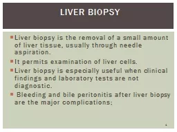
Percutaneous Liver Biopsy
Is a safe procedure
Easily performed at the bedside
With local anesthesia and ultrasound guidance. Slide58
Percutaneous Liver Biopsy Indication
Hepatocellular
disease of uncertain cause
Prolonged hepatitis (chronic active hepatitis)
(3) Unexplained
hepatomegaly
(4) Unexplained
splenomegaly
(5) Hepatic filling defects IN imaging
(6) Fever of unknown origin
(7) Staging of malignant lymphomaSlide59
Liver biopsy
is most accurate in disorders causing diffuse changes IN liver
Sampling error in focal infiltrative disorders such as hepatic metastases.
Should not be the initial procedure in
cholestasis
. Slide60
Liver biopsy Contraindications
Significant
ascites
Prolonged INR
Under these circumstances, the biopsy can be performed via the
transjugular
approachSlide61
Ultrasonography
First diagnostic test in
cholestasis
:
dilated
intrahepatic
extrahepatic
biliary
tree
gallstones.
space-occupying lesions IN
liver, →distinguish between cystic and solid masses, and helps direct
percutaneous
biopsies. Slide62
Ultrasound with Doppler
Detect the patency of the :
portal vein
hepatic artery
hepatic veins
First test in patients suspected Budd-
Chiari
syndrome.Slide63
Abnormal in...
Liver Test
Cirrhosis, severe hepatocellular injury
Albumin
Cholestasis, hepatocellular enzyme induction, canalicular injury, children during bone growth, bone disease, pregnancy (placenta origin)
Alkaline phosphatase
Hepatocellular injury (ethanol, drug-induced hepatitis, hepatitis B and C, ischemic injury, chronic liver disease, NAFLD, chronic viral hepatitis, alcoholism, nonspecific viral injury, and cholestatic or replacement disease); acute biliary obstruction; rarely in hyperthyroidism, celiac disease, skeletal muscle disease
Aminotransferases (AST, ALT)
Any acute or chronic liver disease; congenital disorders of bilirubin metabolism.
Bilirubin
Cholestasis
5′ nucleotidase
Cholestasis; medications, ethanol; rarely anorexia nervosa, hyperthyroidism, myotonic dystrophy
GGT
Impaired synthesis of vitamin K-dependent coagulation factors
INR
Ischemic injury, Epstein-Barr virus infection, hemolysis, solid tumor
Lactate dehydrogenase
Alcohol consumption, gout
Uric acidSlide64
Aproach to cholestatic
liver enzyme elevationsSlide65